

Research Article
CBCT Three-Dimensional Cephalometry of the Temporomandibular Joint in Post-Treatment Patients with Articular Pathology
Jorge A. Learreta1* and Esteban E. Barrientos2
1Residence program in TMJ, Catholic University of Salta, Buenos Aires, Argentina
2Orthodontic Specialist of the Argentinian Orthodontic Association, Post-Graduate Orthodontic Society of Corrientes, Corrientes, Argentina
*Address for Correspondence: Jorge A. Learreta, Residence program in TMJ, Catholic University of Salta, Buenos Aires, Argentina, E-mail: jorgelearreta@fibertel.com.ar
Dates: 20 April 2017; Approved: 14 May 2017; Published: 19 May 2017
Citation this article: Learreta JA, Barrientos EE. CBCT Three-Dimensional Cephalometry of the Temporomandibular Joint in Post-Treatment Patients with Articular Pathology. Sci J Res Dentistry. 2017;1(1): 010-016.
Copyright: © Learreta JA, et al. This is an open access article distributed under the Creative Commons Attribution License, which permits unrestricted use, distribution, and reproduction in any medium, provided the original work is properly cited.
Abstract
The temporomandibular joint is a fundamental articulation for professionals performing orthodontic treatments. Different etiologic agents may alter hard and soft tissues. Treatment of TMJ pathologies through Electronic Mandibular Deprogramming, validated by surface electromyography, allows a muscle position to be used as a correct benchmark of the mandible to initiate an orthodontic treatment. Although these parameters offer better results and more stability, doubts about condyle position in the mandibular fossa always remained. The objective was to determine the three-dimensional condylar position in the mandibular fossa of post-treatment patients with TMJ disorders based on Cone Beam Computed Tomography (CBCT) images.
CBCT images of 19 patients treated for TMJ pathologies were studied and analyzed through high-resolution image software (Dolphin Imaging 11.7) for diagnosis. A total of 38 articulations were analyzed through sagittal and coronal cephalograms. Results showed that, in the sagittal plane, the anteroposterior position of the condylar axis was in mesial relationship to the center of the mandibular fossa, while in the coronal plane, statistical evidence enabled validation of the condylar centric position. Cephalometry allowed determination of the three-dimensional condylar position in patients treated for articular pathology. The present study provides information that may be considered as a criterion to complement the orthodontic diagnosis.
Introduction
The temporomandibular joint is a fundamental articulation for professionals performing orthodontic treatments. It is made up of osseous tissues (mandibular condyle and fossa) and soft tissues (articular disc, ligaments, capsule and surrounding tissues). Knowledge of these anatomical structures is fundamental for diagnosing TMJ tissue pathologies.
Sectional studies in sagittal, frontal and horizontal planes of the temporomandibular joint allowed a three-dimensional analysis of the TMJ and the joint capsule in relation with surrounding structures. Posteromedial sagittal section cuts of the mandibular condyle show the superior lamina strongly attached to the petrotympanic fissure, where some fibers extend through it, their insertion terminating in the anterior ligament of the hammer. The thinner inferior lamina is attached to the medial region of the pterygoid muscle fascia. Laterally, it inserts into the neck of the mandible and into the mandibular condyle. The retrodiscal area presents loose connective tissue, with lobules of fatty tissue, and it contains a venous plexus and numerous nerve fibers [1]. It is vascularized by atrial, anterior tympanic and superficial temporal arteries, and innervated by the temporal auricular nerve [2]. It is the underlying posterior nutritional pole, given the existence of vascular plexuses that allow nutrition and hydration of the articulation, where a large number of cells constituting synovial fluid elements are also found [3].
Hard and soft tissues [4], may be altered by different etiological agents. The mandibular condyle may present shape and position alterations [5]. Position alterations, such as posterior disc displacement, have been correlated as causal factors of temporomandibular joint symptomatology and pathology [6-11].
Different imaging methods are employed to study hard structures: transcranial radiographs [12,13]. Laminographies [14,15]. Linear computed tomography [16,17] and Cone Beam Computed Tomography (CBCT) [18-20]. High resolution CBTC multiplanar images provide essential information for diagnosing TMJ pathologies with substantially lower radiation doses compared to helical computed tomography, allowing the evaluation of TMJ anatomy without overlapping or distortion of morphology and the articular space. CBCT is effective in evaluating bone structures of the TMJ and their pathological alterations [18].
TMJ cephalometry allowed positional three-dimensional study of the mandibular condyle in relation to the mandibular fossa, in sagittal [6,14,16,17,21-26] and coronal planes [21,27-31].
The treatment of TMJ pathologies through the use of Electronic Mandibular Deprogramming, validated by Surface Electromyography, allows the use of a muscular position as a correct benchmark of the mandible to approach orthodontic treatment [32-34].
Although these parameters provided better results and more stability, doubts about condyle position in the mandibular fossa always remained.
The present research aims to determine the three-dimensional condylar position within the mandibular fossa in post-treatment patients with TMJ disorders based on CBCT images.
Method and Materials
Nineteen consecutive patients previously treated for their temporomandibular pathologies were studied by CBCT images and Dolphin Imaging Software 11.7, permitting the Second Phase of the treatment [Figure 1,2].
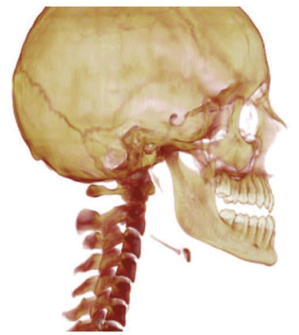 Figure 1: Right hemi cranium view obtained by reconstruction
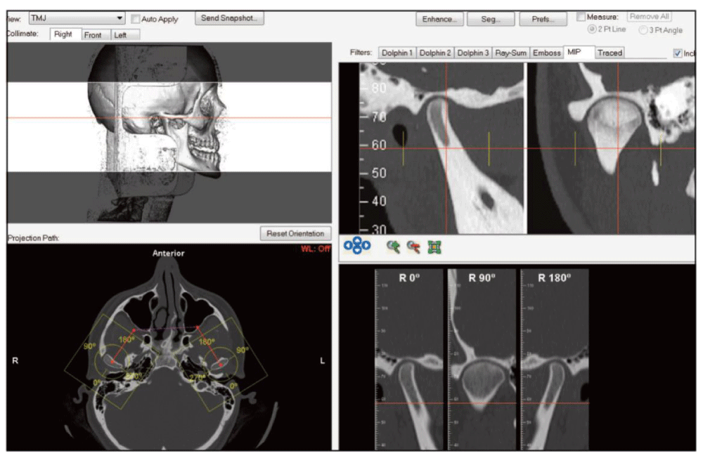 Figure 2: Screen printing of the software image used as a planning and diagnostic tool, in which images similar to those used to conduct research, were observed.
TMJ pre-orthodontic, cephalometric and routine tests were indicated. Patients were informed about procedures and consented to the present study. In the differential diagnosis of temporomandibular joint pathology, age and sex were not significant for this investigation. A total of 38 joints were evaluated as independent units [Figure 3a,b,c].
 Figure 3a-c: CBCT images analyzed with Dolphin Imaging 11.7 Software. Sagittal cut 1 - a, coronal cuts1 - b and 1 - c
CBTC images were obtained in patients with their Intraoral Devices (IODs) at maximum occlusion. The dimensions and location of the IODs were established after evaluation by Electronic Deprogramming and Surface Electromyograph.
Figure 3a-c: CBCT images analyzed with Dolphin Imaging 11.7 Software. Sagittal cut 1 - a, coronal cuts1 - b and 1 - c
CBTC images were obtained in patients with their Intraoral Devices (IODs) at maximum occlusion. The dimensions and location of the IODs were established after evaluation by Electronic Deprogramming and Surface Electromyograph.
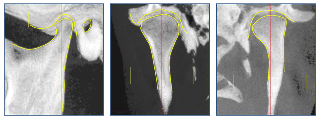 Figure 4a-c: Hard structures. Sagittal view a. Coronal views b - c
Taking as a reference the major condylar axis in a horizontal plane, a sagittal cut was reconstructed, which indicates the following sections: external auditory meatus, articular eminence, mandibular condyle, fossa and 2/3 of the mandibular ramus (Figure 4a). Acoronal cut shows the mandibular condyle, neck, fossa, and 2/3 of the mandibular ramus (Figure 4 b-c).
Figure 4a-c: Hard structures. Sagittal view a. Coronal views b - c
Taking as a reference the major condylar axis in a horizontal plane, a sagittal cut was reconstructed, which indicates the following sections: external auditory meatus, articular eminence, mandibular condyle, fossa and 2/3 of the mandibular ramus (Figure 4a). Acoronal cut shows the mandibular condyle, neck, fossa, and 2/3 of the mandibular ramus (Figure 4 b-c).
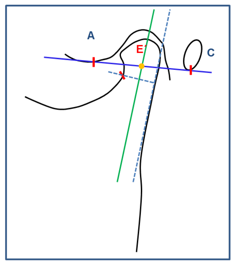 Figure 5: Sagittal Cephalogram construction: Reference plane extending from point A - C, and mandibular condyle axis can be observed. Point E´, intersection of the reference plane and condylar head axis.
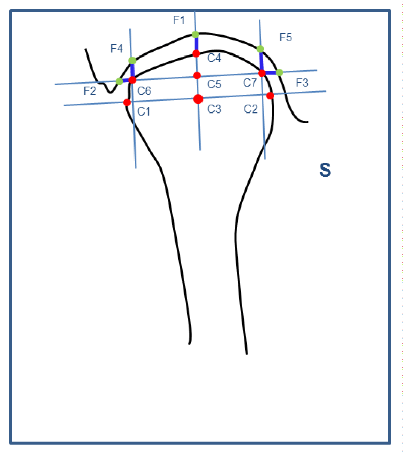 Figure 6: Construction of the coronal Cephalogram to study the articular space in sagittal and horizontal planes. Note the condyle centric position within the mandibular fossa.
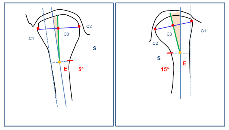 Figure 7a,b: Coronal direction analysis of the condyle. Note the difference in the degree of internal rotation in articulation b, compared to articulation c.
Sagittal Cephalogram
In a previous research a cephalometric tracing was described through the TMJ sagittal plane [15-35]. For the present research two factors were applied to determine the condylar relationship (Figure 3a).
Vertical relationship: defined as the distance between two parallels to the reference plane. It extends from the most inferior point of the external auditory meatus (point C), to the most inferior point of the articular eminence (point A), tangent to the uppermost point of the mandibular fossa and condylar head.
Anteroposterior relationship of the axis: described as the relation between the linear distance from point A to point E ‘and the linear distance from point A to point C on the reference plane, determining a percentage relationship postulated in the following formula:
Point E’ anteroposterior relationship = (distance A _ E’ / distance A _C) * 100 = %
Coronal cuts were analyzed on a new tracing:
Coronal Cephalogram
1. Analysis of the Articular Space (Figure 6).
Cephalogram construction
1. Point C1, external pole, the outermost point of the mandibular condyle
2. Point C2, internal pole, the innermost point of the mandibular condyle
3. Coronal axis of the condylar head, plane which cuts the external and internal poles of the condyle
4. Point C3, condylar center, point equidistant to the external and internal poles, traced on the coronal plane
5. Plane perpendicular to point C3, perpendicular to the condylar coronal plane
6. Point C4 superior point of the condyle, intersection of the plane perpendicular to point C3, on the superior border of the condyle
7. Point F1 superior point of the mandibular fossa, plane intersection perpendicular to point C3 and the border of the mandibular fossa
8. Point C5, equidistant to point C3, center of the condyle and the superior point of the mandibular fossa, point C5 traced on the perpendicular plane
9. Plane parallel to the coronal plane of the condyle, which cuts C5
10. Point C6, intersection of the parallel plane and the superior external border of the condyle
11. Point C7, intersection of the parallel plane and the superior internal border of the condyle
12. Perpendicular external to the coronal plane, intersection with point C6
13. Perpendicular internal to the coronal plane, intersection with point C7
14. Point F2, intersection from the parallel to the coronal plane and the external border of the mandibular fossa
15. Point F3, plane intersection parallel to the coronal plane and the internal border of the mandibular fossa
16. Point F4, intersection of the external perpendicular and the superior border of the mandibular fossa
17. Point F5, intersection of the internal perpendicular and the superior border of the mandibular fossa
Cephalometric analysis
Vertical Relationship:
1. Superior external articular space, linear distance between points F4 andC6.
2. Superior medial articular space, linear distance between points C4 and F1.
3. Superior internal articular space, linear distance between points C7 and F5.
Horizontal Relationship:
1. External articular space, linear distance between points C6 and F2.
2. Internal articular space, linear distance between points C7 and F3.
Direction analysis of themandibular condyle: Construction of the mandibular condyle neck axis: Figure 7-a-b
1. External plane, plane adapted to the external border of the neck and the superior mandibular ramus.
2. Point S, point on the internal border of the condylar neck at its upper and narrower portion, as it changes from straight to oblique, in an upward and inward direction.
3. Perpendicular to the external plane, which cuts point S.
4. Point E, equidistant to Point S and the plane external of the neck and ramus.
5. Neck axis of the condylar head, a plane parallel to the external plane which cuts point E.
Construction of the condylar axis: Condylar axis, a plane which cuts points C3 (condylar center) and E.
Cephalometric analysis
1. Direction angle of the condyle, the angle formed by the neck and condylar axes. In the coronal cut, each section of articular space analysis was defined as a population:
2. Superior external articular space
3. Superior medial articular space
4. Superior internal articular space
5. External articular space
6. Internal articular space.
Statistics
A possibility of retaining the mean equality hypothesis between populations was analyzed: (a) superior external articular space - superior medial articular space. (b) Superior medial articular space - internal medial articular space. (c) External articular space - internal articular space. The null hypothesis is postulated, that is, the absence of difference in the articular space between sections of the coronal plane.
The hypothesis test was carried out using the Student’s t-test of equality in means. A 5% error probability level was set by rejecting the null hypothesis, (p < 0.05).
Results
The cephalometric results of 19 patients treated for their temporomandibular joint pathologies are presented. A total of 38 joints were studied through CBCT images and analyzed with Dolphin Imaging Software 11.7, utilizing sagittal and coronal cephalograms. (Tables 1 and 2)
| Table 1: Results of cephalometric factors in sagittal plane. | ||||
| Sagittal Cephalogram | ||||
| Variable | Number of observations | Mean | Variance | Standard deviation |
| Anteroposterior axis relationship | 38 | 47.6 % | 12.4 | 3.6 % |
| Vertical relationship | 38 | 2.7 mm | 0.9 | 0.9 |
| Table 2: Results of cephalometric factors in coronal plane. | ||||
| Coronal Cephalogram | ||||
| Variable | Number of Observations | Mean | Variance | Standard Deviation |
| External Articular Space | 35 | 2.2 mm | 0.8 | 0.9 |
| Superior External Articular Space | 38 | 2.3 mm | 0.6 | 0.8 |
| Superior Medial Articular Space | 38 | 2.7 mm | 0.9 | 1 |
| Superior Internal Articular Space | 38 | 2.7 mm | 1.3 | 1.1 |
| Internal Articular Space | 38 | 2.6 mm | 1 | 1 |
| Condylar Head Direction | 38 | 8.7º | 32.9 | 5.7 |
Since the external wall tracing of the mandibular fossa was absent, Point F2 could not be traced, therefore a total of 35 external articular spaces were studied. |
||||
Results of the hypothesis test allow the following conclusions:
• Statistical evidence does not allow retention of the null hypothesis that the mean of the articular space is different for the superior articular space to the superior external articular space, p> 0.05 (not significant).
• Statistical evidence does not allow the retention of the null hypothesis that the mean of the articular space is different for the superior articular space to the superior internal articular space, p> 0.05 (not significant).
• Statistical evidence does not support the hypothesis that the mean of the articular space is different for the external articular space to the internal articular space, p > 0.05 (not significant).
Discussion
For years, dentistry used occlusal positions as a gold standard for the correct mandibular position in orthodontic diagnoses, although dental alterations may lead to anomalous mandibular positions [36-38]. Studies to determine the condylar position through records of centric relation, showed that the condyles moved to a superior and posterior position within the mandibular fossa, this being a non-physiological position and a misconception [39-41].
Nineteen patients treated for TMJ pathologies were studied with the use of Electronic Mandibular Deprogramming and validated by Surface Electromyography, which allowed the use of a muscular position as a correct benchmark of the mandible to approach orthodontic treatment [32-34].
CBCT images were studied and analyzed with high-resolution image software (Dolphin Imaging 11.7) for diagnosis. A total of 38 articulations were analyzed through sagittal and coronal cephalograms.
CBTC high quality images allow diagnosis of condylar shape alterations, osteophytes, erosions, fractures, ankylosis, developmental abnormalities and condylar position changes within the fossa. The examination time is shorter and the patient dose is lower than with conventional CT. It may therefore be considered as the imaging technique of choice when investigation of bone changes of the TMJ is the task at hand [19].
Honda K, et al. [42], compared the diagnostic reliability of CBCT and helical computed tomography (helical CT) for the detection of osseous abnormalities of the mandibular condyle, using macroscopic observations. Twenty-one temporomandibular joint autopsy specimens underwent imaging with CBCT and helical CT. The specimens were macroscopically evaluated for cortical erosion, osteophytosis and sclerosis. Macroscopic observations and imaging findings show that CBCT is a dose-effective and a cost- effective alternative to helical CT for the diagnostic evaluation of TMJ osseous abnormalities.
The morphology of the mandibular fossa can be studied through CBTC images. The inclination of the articular eminence, height and thickness of the mandibular fossa roof were analyzed according to age, gender and bone findings of osteoarthritis and condylar shape.
The inclination of the articular eminence and the height of the fossa were greater in males. The thickness of the mandibular fossa was greater with the presence of osteophytes and erosion in the condyle [43]. Moreover, the inclination tends to be higher in patients without TMJ dysfunction [44].
Pathological alterations of the condylar head, such as surface alterations, degenerative changes, bifid condyle and fractures can be accurately diagnosed through CBTC images [45-48]. Surface changes were common in patients with TMJ disorders and significantly associated with age [48].
The description of independently studied axes of the mandibular condyle and ramusin coronal planes, allowed study of three-dimensional changes in the condyle and ramus, after vertical ramus osteotomy [28]. In a coronal plane, perpendicular to the Frankfort Horizontal (FH) plane, the following angles were constructed:
1 - Coronal condylar angle: the angle between the FH plane and the long condylar axis (the line between the most medial and lateral points). 2- Coronal ramus angle: the angle formed between the FH plane and the line tangentto the lateral border of the ramus.
In the proposed coronal cephalogram, the direction angle of the mandibular condyle allowed study of its morphology. The angle is formed between the condylar axis and the ramus. The cephalogram interpretation allows study of the degree of internal or external inclination of the condyle with the mandibular ramus. The values obtained were: 8.7° mean 5.74° s / d and 32.9 variance.
In a previous study, a cephalogram was developed in sagittal plane that allowed analysis of the mandibular condyle position, with references distant from the articular surface, since the mandibular condyle may suffer shape alterations [15]. The following factors were utilized: Point E’ anteroposterior proportion is indicative of the anteroposterior position of the condyle in the mandibular fossa. A 50% value was considered as center of the mandibular fossa in an anteroposterior sense, and described as the point equidistant between the inferior point of the articular eminence and the inferior point of the external auditory meatus.
In a previous study, a cephalogram was developed in sagittal plane that allowed analysis of the mandibular condyle position, with references distant from the articular surface, since the mandibular condyle may suffer shape alterations [15]. The following factors were utilized: Point E’ anteroposterior proportion is indicative of the anteroposterior position of the condyle in the mandibular fossa. A 50% value was considered as center of the mandibular fossa in an anteroposterior sense, and described as the point equidistant between the inferior point of the articular eminence and the inferior point of the external auditory meatus.
The vertical relationship is a linear measurement from the inferior point of the mandibular fossa to the uppermost section of the condylar head, these values are indicative of the superior articular space, which was 2.7 mm media, 0.93 s/ d.
The coronal cephalogram developed in the present study allowed the analysis of the superior articular space in three sections: the superior external, superior middle, and superior internal articular spaces, these were: 2.3 mm 0.8 s/ d, 2.7mm 0.95 s/ d and 2.7 mm 1.1 s/ d. In a horizontal relationship, the external and internal articular spaces, had values of 2.2 mm 0.9 s/ d and 2.6 mm 1 s/ d, respectively.
The statistical evidence allowed retention of the mean equality hypothesis of the articular space in a coronal sense, showing that the position of the condyle was centered within the mandibular fossa. These values may be referential to determine the position of the mandibular condyle in patients who initiate orthodontic treatments.
Owen [13], states that “In the ideal situation, every orthodontic case would be finished with optimal skeletal and soft tissue balance, with adequate anterior guidance, and with the condyles positioned in a physiologically acceptable range”. For the author, the concentric position is ideal for the majority of patients. The treatment would be completed when the condyles are located in a therapeutic area, being Owen’s description their posterior limit, and Geld’s [49,50] their anterior limit.
Ricketts refers to the correct functional position as the physiological centric positions i.e., when maximum inter cuspation occurs with the centric condylar position: the centric condylar position and centric occlusion coincide [51].
The results of our investigation are validated by Dalili Z [29], who studied the three-dimensional condyle position in CBCT images of 40 patients with normal temporomandibular joint and class I skeletal pattern. The condylar position was analyzed in the sagittal plane, studying the superior, anterior and posterior articular space. In a coronal plane, the medial and lateral articular space was analyzed. Research has shown that the centric condylar position in the fossa is the most common position in patients with normal function. Alterations of the condylar position in sagittal and coronal planes are interpreted as pathologies.
Mazzetto MO, et al. [52], analyzed the condylar position within the fossa in patients with signs and symptoms of temporomandibular disorders through CBCT. He studied the position of the condyle in a sagittal view, making linear measurements of the anterior, superior and posterior articular spaces. Results showed that there was a great variation in the condylar position, with a predominance of the posterior position in patients with signs and symptoms of temporomandibular pathology.
Ikeda K, et al. [30], confirmed that changes in disc position are represented by alterations in the condylar position, in both sagittal and coronal planes. The magnitude and direction of the disc displacement can be estimated from the distance of the articular space displayed in CBCT.
Conclusion
The use of cephalometry in sagittal and coronal planes, applied to a population of patients treated for temporomandibular pathologies, allowed a definition of the three-dimensional condylar position within the mandibular fossa. Results showed that in a sagittal plane the condylar axis was in mesial relation to the fossa, and in a coronal plane, statistical evidence confirms the centric condylar position within the fossa. The present study provides information that may be considered as a criterion to complement the orthodontic diagnosis.
References
- Cordula Schmolke. The relationship between the temporomandibular joint capsule, articular disc and jaw muscles. Anatomical Institute, University of Bonn, Germany. J Anat. 1994; 184: 335-345. https://goo.gl/qxPd5B
- Siéssere S, Vitt M, Sousa LG, Semprini M, Hallak SC. Bilaminar Zone: Anatomical aspect, irrigation and innervation. Braz. J. morphol. Sci. 2004; 21: 217-220. https://goo.gl/ZqqyHt
- Learreta JA. [Anatomy of the temporomandibular joint: update of the temporomandibular joint anatomy: an update]. Rev. Soc. Odontol. Silver. 1997; 10: 17 -26. https://goo.gl/isfSZ6
- Learreta JA: Current diagnosis of temporomandibular pathologies. J Craniomandib Pract. 2009; 27: 125-133.
- https://goo.gl/v0gTUQ
- Learreta JA. [Compendium on diagnosis of pathologies of the TMJ]. San Pablo: Artes Médicas; 2004. p. 89-96.
- Owen AH 3rd. Orthodontic/orthopedic treatment of craniomandibular pain dysfunction. Part 2: posterior condylar displacement. J Craniomandibular Pract. 1984; 2: 333-49. https://goo.gl/m1J4LR
- Paknahad M, Shahidi S. Association between mandibular condylar position and clinical dysfunction index. J Craniomaxillofac Surg. 2015; 43: 432-6. https://goo.gl/gp5q8K
- Al-Rawi NH, Uthman AT, Sodeify SM. Spatial analysis of mandibular condyles in patients with temporomandibular disorders and normal controls using cone beam computed tomography. European Journal of Dentistry. 2017;11(1):99-105. doi:10.4103/ejd.ejd_202_16. https://goo.gl/JHV94S
- Pullinger AG, Solberg WK,Hollender L, Guichet D. Tomographic analysis of mandibular condyle position in diagnostic subgroups of temporomandibular disorders. J Prosthet Dent. 1986; 55: 723-9. https://goo.gl/nyo7to
- Imanimoghaddam M, Madani AS, Mahdavi P, Bagherpour A, Darijani M, Ebrahimnejad H. Evaluation of condylar positions in patients with temporomandibular disorders: A cone-beam computed tomographic study. Imaging Science in Dentistry. 2016; 46: 127-131. doi:10.5624/isd.2016.46.2.127. https://goo.gl/bahXKv
- Gateno J, Anderson PB, Xia JJ, Horng JC,Teichgraeber JF, Liebschner MA. A comparative assessment of mandibular condylar position in patients with anterior disc displacement of the temporomandibular joint. J Oral Maxillofac Surg. 2004; 62: 39-43. https://goo.gl/Vrtdk5
- Weinberg, L.A.: An Evaluation of Duplicability of Temporomandibular Joint Radiographs, J. PROSTHET. DENT. 1970; 24: 512-541. https://goo.gl/TqRl9d
- Owen, A. H.: Orthodontic/Orthopedic Treatment of Craniomandibular Pain Dysfunction Part1: Diagnosis with Transcranial Radiographs. J Craniomand Prac1984; 2: 238-249. https://goo.gl/QFxuXu
- Ricketts RM. Variations of the temporomandibular joint as revealed by cephalometric laminagraphy. Am J Orthod. 1950; 36: 877-98. https://goo.gl/XBouCp
- Learreta JA, Barrientos EE. Temporomandibular Joint Method to Study the Morphology and Relationship of the Hard Structures. Cranio. 2010; 28. https://goo.gl/LpYfDT
- Blaschke DD, Blaschke TJ. A method for quantitatively determining temporomandibular joint bony relationships. J Dent Res. 1981; 60: 35-43. https://goo.gl/hRWqf3
- Pullinger A, Hollender L. Variation in condyle-fossa relationships according to different methods of evaluation in tomograms. Oral Surg Oral Med Oral Pathol. 1986; 62: 719-27. https://goo.gl/nXtv9s
- Barghan S, Tetradis S, Mallya SM. Application of cone beam computed tomography for assessment of the temporomandibular joints. Australian Dental Journal. 2012; 57: 109-118. https://goo.gl/jK7DC6
- Tsiklakis K, Syriopoulos K, Stamatakis HC. Radiographic examination of the temporomandibular joint using cone beam computed tomography. Dentomaxillofacial Radiology. 2004; 33: 196–201. https://goo.gl/pao0Cn
- Silvia Caruso, Ennio Storti, Alessandro Nota, Shideh Ehsani, Roberto Gatto, “Temporomandibular Joint Anatomy Assessed by CBCT Images,” BioMed Research International. 2017; 2017: 1-10. https://goo.gl/Bptk8N
- Christiansen E. L, Thomsom J. R, Zimmerman G, Roberts D, Hasso A. N, Hinshaw D. B and Kopp S.: Computed tomography of condylar and disk positions within the temporomandibular joint. Oral Surg Oral Med Oral Pathol. 1987; 64: 757-767. https://goo.gl/EKSGoL
- Owen AH 3rd. Orthodontic/orthopedic treatment of craniomandibular pain dysfunction. Part 1:diagnosis with transcranial radiographs. J Craniomandibular Pract. 1984; 2: 238-49. https://goo.gl/QBiktn
- Dumas AL, Moaddab MB,Willis HB, Homayoun NM. A tomographic study of the condyle/fossa relationship in patients with TMJ dysfunction. J Craniomandibular Pract. 1984; 2: 315-25. https://goo.gl/4JtSzB
- Cohlmia JT, Ghosh J, Sinha PK, Nanda RS, Currier GF. Tomographic assessment of temporomandibular joints in patients with malocclusion. Angle Orthod. 1996; 66: 27-35. https://goo.gl/6t1lvL
- Pandis N,Karpac J,Trevino R,Williams B. A radiographic study of condyle position at various depths of cut in dry skulls with axially corrected lateral tomograms. Am J Orthod Dentofacial Orthop. 1991;100: 116-22. https://goo.gl/DDMnYk
- Katzberg RW, Keith DA, Ten Eick WR, Guralnick WC. Internal derangements of the temporomandibular joint: an assessment of condylar position in centric occlusion. J Prosthet Dent. 1983; 49: 250-4. https://goo.gl/rgB1Qb
- Ueki K, Hashiba Y, Marukawa K, Nakagawa K, Alam S, Okabe K, Yamamoto E. The effects of changing position and angle of the proximal segment after intraoral vertical ramus osteotomy. International Journal of Oral and Maxillofacial Surgery. 2009; 38: 1041-1047. https://goo.gl/qzUPTq
- Ueki K, Moroi A, Sotobori M, Ishihara Y, Marukawa K, Yoshizawa K, et al. Changes in temporomandibular joint and ramus after sagittal split ramus osteotomy in mandibular prognathism patients with and without asymmetry. J Craniomaxillofac Surg. 2012; 40: 821-827. https://goo.gl/ICpud8
- Dalili Z, Khaki N, Kia SJ, Salamat F. Assessing articular space and condylar position in the people with normal function of temporomandibular joint with cone-beam computed tomography. Dent Res J (Isfahan). 2012; 9: 607-612.
- https://goo.gl/fSNsVN
- Ikeda K, Kawamura A. Disc displacement and changes in condylar position. Dentomaxillofac Radiol. 2013; 42: 84227642. https://goo.gl/4DLYYr
- Ikeda, K., Kawamura, A. and Ikeda, R. (2011), Assessment of Optimal Condylar Position in the Coronal and Axial Planes with Limited Cone-Beam Computed Tomography. Journal of Prosthodontics. 20: 432-438. doi:10.1111/j.1532-849X.2011.00730. https://goo.gl/pWn7B3
- Learreta JA, Beas J, Bono A. Aumento del espacio libre interoclusal como factor etiológico de las clase III. Caso Clínico. Revista Iberoamericana de Ortodoncia. 1998; 17: 51-58. https://goo.gl/YrQ0RY
- Learreta JA, Bono A. A Importancia da Deporgramacao Mandibular no Dianóstico Ortodontico. Jornal Brasileiro de Ortodontia & Ortopedia Facial 1998; 18:72-77.
- Learreta JA, Moses AJ. Cephalometric Variation in Patients with and Without Intraoral Neuromuscular Repositioning Appliance. J Gen Orthod. 1999; 10: 14-21. https://goo.gl/ooEyoA
- Learreta JA, Barrientos EE. Temporomandibular Application of a Cephalometric Method to the Temporomandibular Joint in Patients With or Without Alteration in the Orientation of the Mandibular Condyle Axis. Cranio. 2013; 31: 46-55. https://goo.gl/92iTfE
- AlKofide EA, AlNamankani E. The association between posture of the head and malocclusion in Saudi subjects. CRANIO. 2007; 25. https://goo.gl/zPazry
- D'Attilio M, Caputi S, Epifania E, Festa F, Tecco S. Evaluation of cervical posture of children in skeletal Class I, II, and III. Cranio; 2005; 23: 219-228. https://goo.gl/5Gtfce
- Quintana Espinosa MT, Martínez Brito I. Interferencias oclusales y su relación con las maloclusiones funcionales en niños con dentición mixta. Rev méd electrón. 2010; 32. https://goo.gl/T1Q907
- Ismail YH, Rokni A. Radiographic study of condylar position in centric relation and centric occlusion. J Prosthet Dent. 1980; 43: 327-30. https://goo.gl/QKrFB4
- Weinberg LA. An evaluation of occlusal factors in TMJ dysfunction-pain syndrome. J Prosthet Dent. 1979; 41: 198-208. https://goo.gl/8xNMNz
- Williamson EH. Laminagraphic study of mandibular condyle position when recording centric relation. J Prosthet Dent. 1978; 39: 561-4 https://goo.gl/KACGBr
- Honda K, Larheim TA, Maruhashi K, Matsumoto K, K Iwai K. Osseous abnormalities of the mandibular condyle: diagnostic reliability of cone beam computed tomography compared with helical computed tomography based on an autopsy material. Dentomaxillofacial Radiology. 2006; 35: 152–157. https://goo.gl/GHN5o8
- Elgüy D, Elgüy M, FiGekçioLlu E, DölekoLlu S, Ersan N. Articular Eminence Inclination, Height, and Condyle Morphology on Cone Beam Computed Tomography. Hindawi Publishing Corporation. The Scientific World Journal. 2014; 2014: 1-6. https://goo.gl/PtD9BN
- Sümbüllu MA, Aglayan FC, Akgül HM, Yilmaz AB. Radiological examination of the articular eminence morphology using cone beam CT. Dentomaxillofacial Radiology. 2012; 41: 234-240. https://goo.gl/ZyP0ao
- Anjos Pontual ML, Freire JSL, Barbosa JMN, Frazao MAG, Anjos Pontu A, Fonseca da Silveira MM. Evaluation of bone changes in the temporomandibular joint using cone beam CT. Dentomaxillofacial Radiology. 2012; 41: 24-29. doi: 10.1259/dmfr/17815139. https://goo.gl/tWBC0Z
- Shintaku WH,.Venturin JA, Azevedo B, Noujeim M. Applications of cone-beam computed tomography in fractures of the maxillofacial complex. Dent Traumatol. 2009; 25: 358-366. https://goo.gl/l68bRT
- Cho BH, Jung YH. Nontraumatic bifid mandibular condyles in asymptomatic and symptomatic temporomandibular joint subjects. Imaging SCI Dent. 2013; 43: 25-30. https://goo.gl/L08EdV
- Alves N, Quezada SA, Villalobos GA, Lara S, SJ, Deana NF. Riveros PC. Morphological Characteristics of the Temporomandibular Joint Articular Surfaces in Patients with Temporomandibular Disorders. Int. J. Morphol. 2013; 31: 1317-1321. https://goo.gl/Wi5fty
- Gelb, H. Clinical management of head, neck and TMJ pain and dysfunction. Philadelphia, Penn: W.B. Saunders; 1977. p.109. https://goo.gl/gjT3d3
- Gelb H. New concepts in craniomandibular and chronic pain management. Mosby: Wolfe; 1995; 55: 373. https://goo.gl/cPir6E
- Ricketts RM. Provocations and perceptions in cranio-facial orthopedics. Library of Congress Catalogue. 1989; 671-672. https://goo.gl/HpiAC5
- Mazzetto MO, Veneziam GC, Magri LV, Nasr MK, Paiva AF, Paiva G.Evaluation of the condylar position in subjects with signs and symptoms of functional disorders of the temporomandibular joint through images made with cone beam computed tomography on the sagittal plane. Braz Dent Sci. 2014; 17: 77-82. https://goo.gl/oTzHxr
Authors submit all Proposals and manuscripts via Electronic Form!