
Da Silva J1, Gil M2, Nagai M2, Chen G3, Sun J3, Stock S4 and Ishikawa-Nagai S1*
1Department of Restorative Dentistry & Biomaterials Sciences, Harvard School of Dental Medicine, Boston, MA, USA
2Department of Oral Medicine, Infection, and Immunity, Harvard School of Dental Medicine, Boston, MA, USA
3DMD graduate Class of 2017, Harvard School of Dental Medicine, Boston, MA, USA
4Department of Mathematics and Computer Science, College of the Holy Cross, Worcester, MA, USA
*Address for Correspondence: Da Silva J, Gil M, Nagai M, Chen G, Ishikawa-Nagai S, et al. Near Infrared Fluorescence Imaging For Early Caries Detection. Sci J Res Dentistry. 2017;1(2): 027-032.
Dates: 08 August 2017; Approved: 16 August 2017; Published: 17 August 2017
Citation this article: Da Silva J, Gil M, Nagai M, Chen G, Ishikawa-Nagai S, et al. Near Infrared Fluorescence Imaging For Early Caries Detection. Sci J Res Dentistry. 2017;1(2): 027-032.
Copyright: © Ishikawa-Nagai S, et al. This is an open access article distributed under the Creative Commons Attribution License, which permits unrestricted use, distribution, and reproduction in any medium, provided the original work is properly cited.
Keywords: Biomaterial(S); NIR Fluorescence Imaging; Caries Detection; Imaging Agents
Abstract
Detection of early stage carious lesions is critical for active prevention. Near-Infrared Fluorescence (NIRF) is an investigational imaging modality. The purpose of this study was to investigate the feasibility and validity of NIRF imaging using an imaging agent in the detection of early occlusal and interproximal caries on extracted human teeth.
Method: Infiltration rate of OsteoSense750 (OS750, bisphosphonate derivative) into enamel carious lesions was measured using a florescence microscope, and the correlation between infiltration and lesion depth was determined. Stability of OS750 fluorescence signal intensity was examined as a factor of dilution ratio and time of excitation illumination exposure. The correlation between fluorescence intensity and carious lesion depth measured by high resolution 3D X-ray microscope was assessed. Finally, diagnostic performance (sensitivity and specificity) of NIRF imaging was compared to ICDAS for occlusal caries and to bitewing radiographs for interproximal caries using bootstrap resampling methods.
Results: The mean infiltration rate of OS750 into enamel carious lesions was 93% and there was little correlation between infiltration and lesion depth (R= -0.08987). OS750 maintained 100% of fluorescence intensity for 15 minutes with 1:10 dilution and 80% intensity for 5 minutes with 1:100 dilution. The correlation between OS750 fluorescence intensity and lesion depth was 0.64745. The sensitivity and specificity for occlusal caries with OS750 were 1.0 and 0.66 versus 0.81 and 0.8 for ICDAS. Interproximal caries values were 0.86 and 0.8 using OS750 and 0.34 and 0.9 by bitewing.
Conclusion: NIRF imaging using OS750 showed excellent properties in infiltration rate and intensity representative of the magnitude of the carious lesion. Diagnostic measures demonstrated much higher sensitivity and NPV than conventional clinical methods supporting the potential of NIR fluorescence imaging with OS750 for early stage caries detection.
Introduction
Near-infrared fluorescence (NIRF) imaging has been introduced as an investigational imaging modality that can address previously unmet clinical needs [1-3]. It has been used for imaging in many medical fields from measuring cancer and protease activity, tissue perfusion, and diseased vasculature, to assessing hydroxyapatite quality [4-6]. The requirements for NIRF imaging are 1) excitation light penetration into the target tissue and selective excitation of the NIRF imaging agent; 2) ability of NIRF imaging agents to selectively target disease markers safely; 3) use of emission filters and optical lenses to capture fluorescence by blocking scattered emission; 4) efficient detection of fluorescence emitted from targets [6]. Dental applications of various optical imaging techniques investigated to date have shown near-infrared (NIR, 700 ~ 2500nm) fluorescence imaging to be of particular interest. NIR noninvasive in vivo imaging has relatively low tissue absorption, scatter, and minimal auto fluorescence of tissues in the NIR range [7-12]. NIR has the potential to detect both primary caries and recurrent carries [9].
In 1958, ICG (Indocyanine green) was submitted for approval to the FDA for use in indicator-dilution studies in humans, and has been used clinically for decades as a contrast agent in measuring hepatic clearance and cardiovascular function [4]. There are several principal advantages of ICG: absorption maximum around 800nm, low toxicity (LD50 of 50-80 mg/kg in mice, rats and rabbits), and rapid excretion [13]. Recent applications of NIRF imaging with ICG involve vascular mapping and tissue perfusion, inflammation, atherosclerosis, protease activity, cell death, tumor cell surface targeting, and hydroxyapatite [4]. Zaheer and colleagues synthesized a NIRF version of the hydroxyapatite-binding molecule pamidronate [14]. The pamidronate was conjugated to a tetra-sulfonated heptamethine indocyanine (IRDye78-NHS). The NIRF derivative OsteoSense750EX (PerkinElmer, Waltham, USA) is a commercially available fluorescence in vivo bisphosphonate imaging agent [14]. OsteoSense750EX has been used for imaging of osteoblastic activity [14], optical detection of bone mineral [15], and detection of breast cancer micro calcification [16,17]. Bisphosphonates are synthetic phosphonate derivatives in which the canonical P-O-P bond is replaced by a nonhydrolyzable P-C-P bond [14]. Bisphosphonates have a very high affinity for bone mineral because they bind to hydroxyapatite crystals [18], and inhibit hydroxyapatite breakdown, thereby effectively suppressing bone resorption. Bisphosphonate therapy is a clinical intervention for postmenopausal osteoporosis due to the ability of bisphosphonates to selectively suppress osteoclast activity thereby retarding bone resorption [18]. Bisphosphonates can offer substantial clinical benefits in diseases such as osteoporosis, where an imbalance between osteoblast-mediated bone formation and osteoclast-mediated bone resorption exists. In addition, since tooth enamel contains a high percentage of hydroxyapatite (96%), enamel caries with demineralized hydroxyapatite may be great binding site for bisphosphonates.
The purpose of this study was to investigate the feasibility and validity of NIRF imaging in the detection of early occlusal and interproximal caries on extracted human teeth.
Materials and Methods
OsteoSense750EX® (OS750, PerkinElmer, USA), a targeted fluorescent in-vivo imaging agent was used as NIRF imaging probe for caries detection. OS750 excitation occurs at 749 nm and emission at 770nm which rarely overlaps with tooth auto fluorescence [Figure. 1A]. OS750 is comprised of bisphosphonate and ICG as a contrast agent [Figure 1B] [14, 16]. First the infiltration of OS750 into a carious lesion was assessed. The fluorescence signal stability of OS750 was compared to other NIR fluorescence imaging probes. The correlation between fluorescence intensity of OS750 and carious lesion depth was assessed. Finally, diagnostic measures including specificity and sensitivity of early stage of occlusal and interproximal caries by OS750 were compared to International Caries Diagnosis and Assessment System (ICDAS) and bite-wing radiographs. Extracted human premolars and molars without restorations or cavitation were used in this study. Institutional Review Board of Harvard University approved the study.
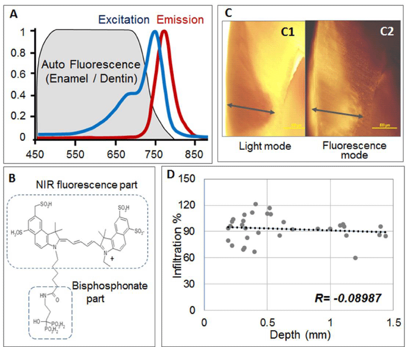 Figure 1: Property of OsteoSense750 (OS750).
A: Wavelength for excitation (749nm) and emission (770nm) of OS750.
B: Chemical formula of OS750.
C: Images of fluorescence of OS750 infiltrated into the caries lesion on the flat surface. C-1) Light microscopic image, C-2) Fluorescence microscopic image.
D: Correlation diagram of infiltration percentage and caries depth.
Figure 1: Property of OsteoSense750 (OS750).
A: Wavelength for excitation (749nm) and emission (770nm) of OS750.
B: Chemical formula of OS750.
C: Images of fluorescence of OS750 infiltrated into the caries lesion on the flat surface. C-1) Light microscopic image, C-2) Fluorescence microscopic image.
D: Correlation diagram of infiltration percentage and caries depth.
Infiltration of OS750 into the carious lesion
Thirty–three white spot lesions on the flat surface (interproximal, facial or lingual) of 24 extracted human teeth were selected and randomly numbered. One microliter of 1:100 dilution of the stock solution of OS750 at 200 µM in PBS was applied to lesions for 30seconds, then rinsed with a distilled water for 10 seconds and allowed to dry. Each tooth was abraded from the buccal-lingual or mesial-distal surface to a 3 mm section that included the area of interest using Buehler Vector Powerhead (Isomet 1000: Buehler, USA), then further polished to approximately 1.0 mm section. A fluorescence microscope BX51 (Olympus America, Philadelphia, USA) was used to capture two different images of each specimen: a histological image under light microscope setting of 4X power and a fluorescent image with the fluorescence microscope under 4X power. Samples were excited with a 750 nm filter and an image was captured with a 770 nm emission filter [Figure 1C]. The depth of the caries was measured from the histological picture captured under the light microscope. In the same manner, the extent of the fluorescent probe infiltration was measured from the fluorescence image taken by the fluorescence microscope [Figure 1C]. Percentage of infiltration was calculated (percentage=fluorescence-microscope/ light-microscope x 100) and Spearman’s correlation coefficient between infiltration percentage and caries lesion depth was analyzed.
Stability of emitted fluorescence signal of OS750
The fluorescence signal intensity of OS750 was compared to other 3 NIRF probes IR-820, CFTM 750 and Cardio Green (Sigma-Aldrich, Natick, MA) over time. Twenty-four square areas (approximately 5mm x 5mm) on the flat surfaces (lingual and buccal) of 12 intact teeth were etched with 38% phosphoric acid for 30 seconds, rinsed for 10 seconds and allowed to dry. Four NIR fluorescence probes were tested at 2 dilution concentrations (2 µM at 1:100 dilution and 20 µM at 1:10 dilution). One microliter of the each concentration was applied to the etched and surrounding area, and rinsed and dried. The fluorescence imaging system [Figure 2A], was used to detect fluorescence. Each tooth was placed on the stage [Figure. 2A-1], facing the Charge-Coupled Device (CCD) camera (MC285SPD-L0B0: Texas Instrument, USA) [Figure2A-2]. The NIR illumination light (750nm, Xenon light source MAX 301 Asahi Spector, Japan) was excited over the specimens at 45 degree angle and a fixed distance [Figure 2A-3]. Emission fluorescence was captured by the CCD camera and real-time images of the fluorescence were recorded. The square area (3mm x 3mm) in the center of each etched area (fluorescent area) was selected and the intensity of the fluorescence signal at 5 time points (baseline, 5, 10, 15 and 20 minutes) were quantified using the Photoshop Histogram Analysis and was plotted using Microsoft Excel [19,20]. Each measurement was repeated 3 times and the average readings were used for the analysis.
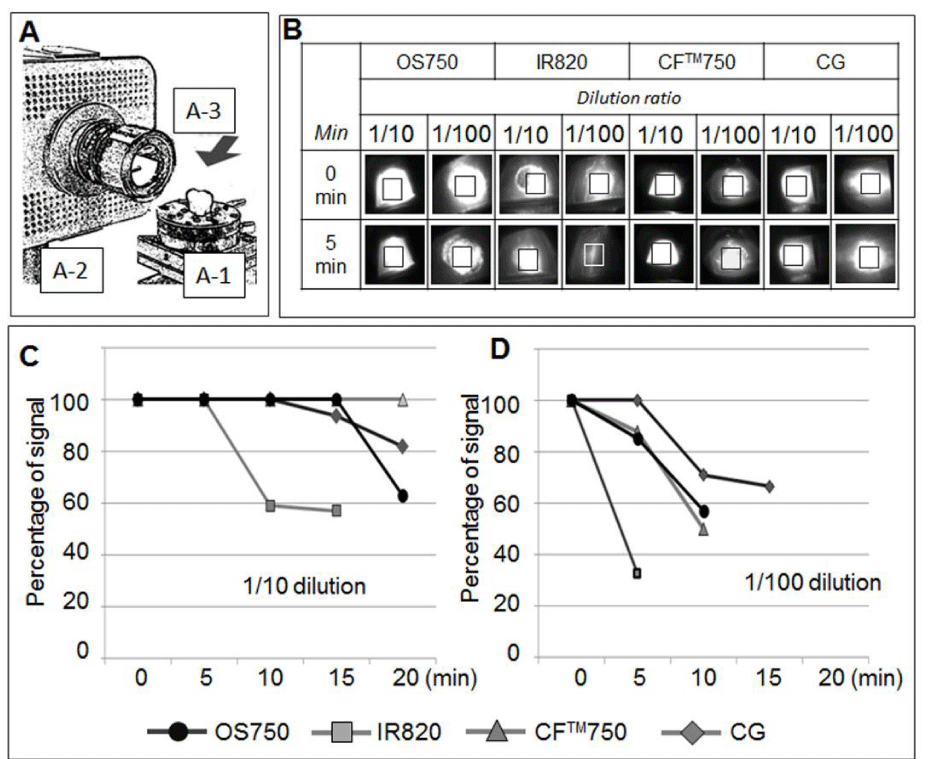 Figure 2: Property of OS750 fluorescence signal intensity.
A: Fluorescence detection system used. Distance and angle between ROI and camera and illumination was standardized. A-1) Precision stage to hold specimens. A- 2) Charged Couple Device (CCD) camera (MC285SPD-L0B0 with emission filter (770nm). A-3) The NIR illumination light (Xenon light source MAX 301 Asahi Spector) with excitation 750nm.
B: Fluorescence images of 4 NIR fluorescence agents (OS750, IR820, CFTM750 and CG-Cardio Green) tested in 2 different dilution ratios (1/10 and 1/100) in 0 minute-baseline and 5 minutes.
C: Change of fluorescence intensity over the time with 1/10 dilution. Intensity was stable in 15 minutes except for IR20.
D: Change of fluorescence intensity over the time with 1/100 dilution. Intensity of IR820 decreased drastically in 5 minutes, but other 3 agents kept over 80% of initial intensity.
Figure 2: Property of OS750 fluorescence signal intensity.
A: Fluorescence detection system used. Distance and angle between ROI and camera and illumination was standardized. A-1) Precision stage to hold specimens. A- 2) Charged Couple Device (CCD) camera (MC285SPD-L0B0 with emission filter (770nm). A-3) The NIR illumination light (Xenon light source MAX 301 Asahi Spector) with excitation 750nm.
B: Fluorescence images of 4 NIR fluorescence agents (OS750, IR820, CFTM750 and CG-Cardio Green) tested in 2 different dilution ratios (1/10 and 1/100) in 0 minute-baseline and 5 minutes.
C: Change of fluorescence intensity over the time with 1/10 dilution. Intensity was stable in 15 minutes except for IR20.
D: Change of fluorescence intensity over the time with 1/100 dilution. Intensity of IR820 decreased drastically in 5 minutes, but other 3 agents kept over 80% of initial intensity.
Correlation between fluorescence signal intensity and carious lesion depth
Seventeen interproximal white spot carious lesions (7 premolars and 10 molars) and 17 occlusal enamel caries without cavitation (7 premolars and 10 molars, ICDAS 1 or 2) were used to evaluate correlation between lesion depth and fluorescence intensity. One microliter of OS750 (1:100 dilution) was applied to the carious lesion and allowed to air dry. Fluorescence signals were captured and recorded with the aforementioned manner. Fluorescence signal intensity at the center of each lesion (approximately 2x2mm) was quantified. The lesion depth was measured by a high resolution 3D X-ray microscope (Xradia MicroXCT-200, Zeiss). All teeth were scanned using the same filter, magnification, exposure time, kV and voltage. Following reconstruction, the images were analyzed using the Xradia viewing software [Figure 3A, B]. Three images of the same tooth were carefully observed and corresponding section of carious lesion was identified. Each lesion depth was measured 3 times and the average of the readings was used in the analysis. Spearman’s correlation coefficient between lesion depth and fluorescence intensity was calculated.
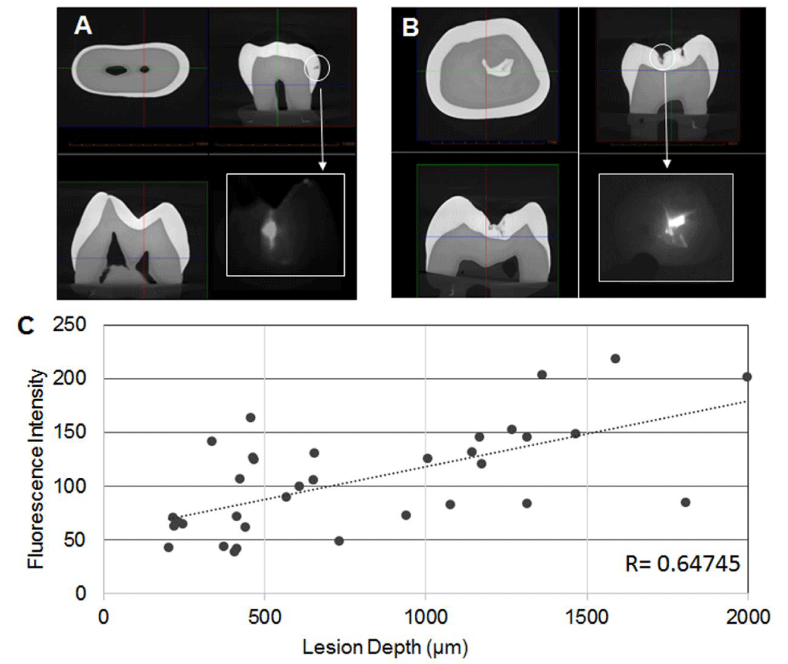 Figure 3: Correlation between fluorescence intensity and caries lesion depth measured by a high resolution 3D X-ray microscope.
A: Representative reconstructed images of the premolar by Xraida and fluorescence image of interproximal lesion.
B: Representative reconstructed images of the molar by Xraida and fluorescence image of occlusal lesion.
C: Correlation diagram of fluorescence intensity and lesion depth.
Figure 3: Correlation between fluorescence intensity and caries lesion depth measured by a high resolution 3D X-ray microscope.
A: Representative reconstructed images of the premolar by Xraida and fluorescence image of interproximal lesion.
B: Representative reconstructed images of the molar by Xraida and fluorescence image of occlusal lesion.
C: Correlation diagram of fluorescence intensity and lesion depth.
Diagnostic measures of carious detection performance by NIRF imaging, radiograph bitewings and ICDAS
Occlusal caries: Fifty-eight occlusal areas including central fossa, mesial and distal pits without any cavitation were selected as area of interest (ROI) and assessed by ICDAS and NIR fluorescence imaging. Three ICDAS calibrated examiners scored each ROI as “sound” or “carious (ICDAS 1 or 2)”. Each ROI on an occlusal surface was treated by 2~3µl of OS750 (1:100 dilution) in the same manner. The tooth was then examined with a fluorescence imaging system as previously described [Figure 4A-1]. Fluorescence images were captured with the CCD camera from the occlusal [Figure 4A-2]. Three trained examiners who did not participate in the ICDAS assessed images as “no fluorescence signal observed” or “fluorescence signal observed” [Figure 4A-3].
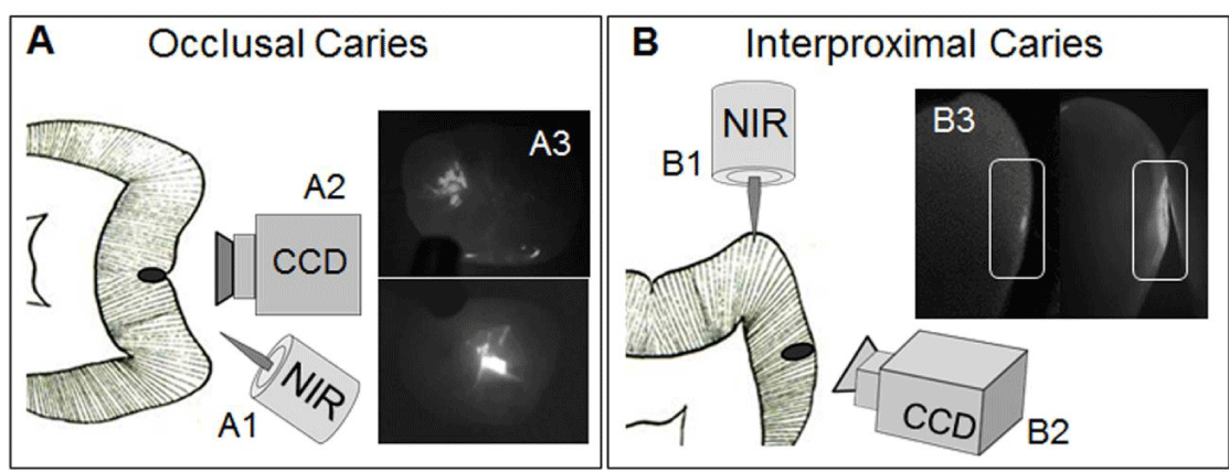 Figure 4: Scheme of caries imaging by OS750 using ex-vivo setting.
A: Optical geometry and fluorescence image of occlusal caries. Illumination light was shined from the occlusal surface (A-1) and emission signal was captured directly from occlusal surface by CCD camera (A-2). Representative fluorescence images captured (A-3).
B: Optical geometry and fluorescence image of interproximal caries. Illumination light was shined from the marginal ridge (B-1). Emission fluorescence signal from caries was captured indirectly from the buccal side using CCD camera (B-2). Representative fluorescence images captured (A-3).
Figure 4: Scheme of caries imaging by OS750 using ex-vivo setting.
A: Optical geometry and fluorescence image of occlusal caries. Illumination light was shined from the occlusal surface (A-1) and emission signal was captured directly from occlusal surface by CCD camera (A-2). Representative fluorescence images captured (A-3).
B: Optical geometry and fluorescence image of interproximal caries. Illumination light was shined from the marginal ridge (B-1). Emission fluorescence signal from caries was captured indirectly from the buccal side using CCD camera (B-2). Representative fluorescence images captured (A-3).
Interproximal caries: Thirty-three interproximal surfaces without cavitation were selected and assessed with bitewing radiographs and OS750 NIR fluorescence imaging. Each ROI was treated by 2~3µl of OS750 (1:100 dilution). The tooth was placed on the stage of the fluorescence imaging system with buccal side facing to the camera [Figure 4B]. The NIR illumination light was on the marginal ridge of the interproximal area [Figure 4B-1]. Real-time images of the fluorescence signal coming through the buccal surface were recorded by the CCD camera [Figure 4B-2]. Three trained examiners assessed images as “no fluorescence signal observed” or “fluorescence signal observed” [Figure 4B-3]. A conventional bitewing radiograph using a short cone technique was obtained for each tooth. Radiographs were taken using F speed film and the x-ray tube set at 70 kvp, 7 mA, and 22 pulses. Three examiners who did not assess the NIR images evaluated radiographs and scored each ROI as “no lesion” or “lesion”.
Statistical analysis: The majority score for each method was used in the statistical analysis. The gold standard, 3D X-ray microscope was used to evaluate occlusal caries and presence or absence of lesion was determined by an experienced examiner. For interproximal caries, histologic analysis by light microscope was used and the examiner determined presence or absence of a lesion.
The overall sensitivity and specificity for each detection method was calculated. Standard errors were estimated using bootstrap resampling. Statistical analyses were performed using the SAS version 9.1 statistical package (SAS Institute, NC, USA).
Results
Properties of OS750 on the carious lesion
Average infiltration rate was 93.72 ± 14.45% (Max: 121.5%, Min: 59.8%) and Spearman’s correlation coefficient between infiltration rate and lesion depth was -0.08987, little correlation [Figure 1-C]. Fluorescence intensity of NIR probes at 1:10 dilution was stable in 5 minutes, and about 100% of intensity was kept for 15 minutes on OS750, CFTM750 and CG [Figure 2B, C]. At 1:100 dilution, 80% of intensity was kept for 5 minutes except for IR820, but intensity decreased dramatically in 10 minutes [Figure 2B, D]. Lesion depth of teeth tested varied 203.7 µm ~1999.0 µm, fluorescence signal intensity measured were 38.39 ~ 218.24. There was a moderate positive correlation (Spearman’s R=0.64745) between lesion depth and fluorescence intensity.
Diagnostic measures of caries detection performance of NIR florescence imaging
Carious lesions were found in 43 of the 58 occlusal ROIs, and in 23 of the 33 of the interproximal ROIs. The values of sensitivity and specificity, and the corresponding 95% confidence interval for each detection method are shown in [Table 1]. For occlusal caries detection, NIRF imaging was in agreement with the gold standard. ICDAS missed 8 of 43 lesions. For absent caries, ICDAS misdiagnosed only 3 of 15 teeth and NIRF imaging misdiagnosed 5 of 15. Although sensitivity of the NIR method was 1.0, specificity was lower (0.667) compared to ICDAS (0.8). For the interproximal carious detection, NIRF imaging missed 3 lesions of 23. By contrast, bitewing missed 15 of 23 lesions. When carious lesions were not present, NIRF imaging had 2 false positive out of 10 and bitewings had 1 false positive. Therefore, sensitivity was substantially higher for NIR compared to bitewings (0.87 versus 0.35).
| Table 1: Summary of sensitivity, specificity, PPV, and NPV and corresponding 95% confidence interval. Occlusal caries by NIR fluorescence imaging and ICDAS. Interproximal caries by NIR fluorescence imaging and Radiograph Bitewing. | ||||||
| Occlusal caries | ||||||
| NIR Fluorescence Occlusal View | ICDAS | |||||
| Condition | Totals | Condition | Totals | |||
| Absent | Present | Absent | Present | |||
| Test Positive | 5 | 43 | 48 | 3 | 35 | 38 |
| Test Negative | 10 | 0 | 10 | 12 | 8 | 20 |
| Totals | 15 | 43 | 58 | 15 | 43 | 58 |
| Lower limit | Upper limit | Lower limit | Upper limit | |||
| Sensitivity | 1.0 | 0.897857 | 1.0 | 0.813953 | 0.660821 | 0.91078 |
| Specificity | 0.66667 | 0.386866 | 0.897857 | 0.8 | 0.513732 | 0.910 |
| PPV | 0.895833 | 0.76557 | 0.76557 | 0.921053 | 0.775159 | 0.979387 |
| NPV | 1.0 | 0.655464 | 1.0 | 0.6 | 0.364117 | 0.800229 |
| Interproximal caries | ||||||
| NIR Fluorescence Buccal View | Radiograph BiteWing | |||||
| Condition | Totals | Condition | Totals | |||
| Absent | Present | Absent | Present | |||
| Test Positive | 2 | 20 | 24 | 1 | 8 | 9 |
| Test Negative | 8 | 3 | 9 | 9 | 15 | 14 |
| Totals | 10 | 23 | 33 | 10 | 23 | 33 |
| Lower limit | Upper limit | Lower limit | Upper limit | |||
| Sensitivity | 0.869565 | 0.653332 | 0.965692 | 0.347826 | 0.171896 | 0.571788 |
| Specificity | 0.8 | 0.442182 | 0.964573 | 0.9 | 0.541155 | 0.994758 |
| PPV | 0.909091 | 0.693756 | 0.984096 | 0.888889 | 0.506703 | 0.994172 |
| NPV | 0.727273 | 0.393167 | 0.926723 | 0.375 | 0.195502 | 0.592424 |
Discussion
NIRF with OS750 has been widely used for diagnostic imaging of osteoblastic activity in animals, but has never been applied in dentistry, especially for diagnostic imaging of caries. This study confirmed NIRF as a promising method for caries imaging. More importantly, regardless of lesion size and depth, OS750 was shown to selectively target enamel caries with 93% infiltration rate which was an advantage for NIRF detection and imaging of early caries. In a clinical application with OS750 as an NIRF probe, the half-life of fluorescence intensity is an important characteristic. OS750 at 1:10 dilution gives 15min without any decrease in emission intensity, and at 1:100 dilution there is 90% retention of the fluorescence intensity for 5 minutes. This is a sufficient duration for clinical application.
We hypothesized that the degree of fluorescence intensity will vary depending on the amount of OS750 bound to the lesion which is in turn related to the volume of the lesion. There is a positive correlation (0.64745) between fluorescence signal intensity and lesion depth which is an advantage of NIRF imaging. Strong fluorescence intensity can be interpreted as representing a deep carious lesion. This characteristic can be explained by the excellent penetration of OS750 into carious lesions. This unique phenomenon of OS750 is the result of the bisphosphonate moiety.
A known side effect of systemic bisphosphonate use is an increased risk of pathologic fractures, Osteonecrosis of the Jaw (ONJ) and atrial fibrillation [18]. However, there are important differences in the use of bisphosphonate in this application. OS750 is applied topically with rinsing for dental diagnostic imaging while oral systemic therapy for osteoporosis requires more frequent and systemic use at higher doses compared to caries detection. Furthermore, although bisphosphonate interferes in normal bone cell metabolism thus causing ONJ, tooth enamel is acellular tissue that bisphosphonates do not affect when carious diagnostic imaging is performed.
A sensitive test is usually positive in the presence of disease and should be chosen when there is a serious consequence for missing a disease. For example, when there is reason to suspect a dangerous but treatable condition, sensitive tests are helpful during the early stages of a diagnostic workup [21]. The caries process, if allowed to progress, will become irreversible and need surgical intervention that many require invasive tooth preparation and can ultimately lead to tooth loss. Therefore, a sensitive test is desired for the detection of early stage caries. The sensitivity of NIRF imaging, the new method, was much higher than that of ICDAS and bite wing radiographs demonstrating improved efficacy of this method as a potential early stage caries detection tool. NIRF imaging could be much more likely to reveal the presence of disease early on. Although the sensitivity of NIRF imaging is superior to that of ICDAS and bite wing radiographs, further improvement would be desirable to reduce false positives and false negatives. One potential solution to false positives on occlusal caries is reducing the strength of the NIR light. Occlusal caries illumination light excites ROIs directly. Therefore, reducing strength of the NIR light could reduce the false positive rate. One possible way to reduce false negatives for interproximal caries is the geometry used in the technique. The method used in this study had the NIR light indirectly excite the area of interest from the occlusal marginal ridge. Therefore, NIR light intensity decreased as it traveled through the occlusal enamel to the probes in the area of interest. Altering the intensity of the excitation light source may change the sensitivity of the technique. Possible geometry would be direct illumination of excitation light by using laser fiber optics of very small diameter that can reach into the interproximal space. Another alternative would be using a probe with a higher wavelength of excitation/emission that has no overlap with the auto fluorescence of enamel and dentin. The emission would scatter less, enabling the emitted fluorescent signal to penetrate teeth more completely without compromising the contrast of the image.
This in vitro study supports our hypothesis that NIRF imaging using OS750 has the ability to detect early enamel carious lesions on occlusal and interproximal surfaces. Although the strong affinity of the bisphosphonate moiety to hydroxyapatite crystals is an important characteristic for caries imaging, a limitation of this study is the uncertainty of the impact of bisphosphonate on demineralization and remineralization. It has been reported that bisphosphonate derivatives, such as MHDP (methane-hydroxy-diphosphonate) and EHDP (ethane-1-hydroxy-1,1-diphosphonate), were effective in inhibiting dissolution of enamel [22-24]. Our preliminary study indicated that topical application of etidronate which is a first generation bisphosphonate led to significant reduction in lesion depth progression compared to the fluoride application [25]. This suggests efficacy in inhibiting enamel caries progression, which may explained by the ability of bisphosphonate to bind with high affinity to hydroxyapatite and penetrate deeply into enamel. This property of bisphosphonate has never been fully investigated in dentistry. The interaction of bisphosphonate in the demineralization and remineralization process needs to be clarified in a future study.
Conclusions
NIRF imaging using OS750 showed excellent infiltration into carious lesions with fluorescence intensity being representative of the magnitude of the carious lesion. Diagnostic measures of NIRF imaging demonstrated higher sensitivity than conventional clinical methods. These results support the goal of diagnosing early stage caries and preventing caries progression prophylactically.
Acknowledgements
We thank Dr. Shahrzad Khorashadi and Dr. Jennifer Bai for the technical support on NIR image acquisition, and Mrs. Karen Cox for the technical support for the Xradia micro-CT. We also thank the Olympus Corporation Japan for providing NIRF imaging system and fluorescence microscope under the Harvard Medical School Sponsored Program.
References
- Gao M, Yu F, Lv C, Choo J, Che L. Fluorescent chemical probes for accurate tumor. 2017. https://goo.gl/C9yhbL
- Yu F, Gao M, Li M, Che L. A dual response near-infrared fluorescent probe for hydrogen. 2015. https://goo.gl/LDRZta
- Han X, Yu F, Songa X, Chen L. Quantification of cysteine hydropersulfide with a ratio metric near-infrared fluorescent probe based on selenium–sulfur exchange reaction. Chem Sci.7:5098. https://goo.gl/R43MdW
- Frangioni JF. In vivo near-infrared fluorescence imaging. Current Opinion in Chemistry Biology. 2003; 7: 626-634. https://goo.gl/UsXCWX
- Luo S, Zhang E, Su Y, Cheng T, Shi C. A review of NIR dyes in cancer targeting and imaging. Biomaterials. 2011; 32: 7127-38. https://goo.gl/a9dVhD
- Sevick-Muraca EM. Translation of near-infrared fluorescence imaging technologies: emerging clinical applications. Annu Rev Med. 2012; 63: 217-31. https://goo.gl/3LkZGV
- Hall A, Gerkin JM. A Review of Potential New Diagnostic Modalities for Caries Lesions. J Dent Res. 2004; 83: 89-94. https://goo.gl/yzKSqV
- Lee RC, Darling CL, Staninec M, Ragadio A, Fried D. Activity assessment of root caries lesions with thermal and near-IR imaging methods. J Biophotonics. 2016; 10: 433-445. https://goo.gl/sRdf1N
- Simon JC, Darling CL, Fried D. A system for simultaneous near-infrared reflectance and trans illumination imaging of occlusal carious lesions. Proc SPIE Int Soc Opt Eng. 2016; 13: 9692. https://goo.gl/XWEkD2
- Tom H, Simon JC, Chan KH, Darling CL, Fried D. Near-infrared imaging of demineralization under sealants. J Biomed Opt. 2014; 19: 77003. https://goo.gl/QgAQc7
- Simon JC, Lucas S, Lee RC, Darling CL, Staninec M, Vaderhobli R, et al. Near-infrared imaging of secondary caries lesions around composite restorations at wavelengths from 1300-1700-nm. Dent Mater. 2016; 32: 587-95. https://goo.gl/4sEkaj
- Kang H, Darling CL, Fried D. Enhancement of OCT images with vinyl polysiloxane (VPS). Proc SPIE Int Soc Opt Eng. 2016; 13: 9692. https://goo.gl/RfNx2L
- Alander JT, Kaartinen I, Laakso A, Pätilä T, Spillmann T, Tuchin VV, et al. A review of indocyanine green fluorescent imaging in surgery. Int J Biomed Imaging. 2012; 2012: 940585. https://goo.gl/rjvb6m
- Zaheer A, Lenkinski RE, Mahmood A, Jones AG, Cantley LC, Frangioni JV. In vivo near-infrared fluorescence imaging of osteoblastic activity. Nature Biotechnology. 2001; 19: 1148–54. https://goo.gl/WVTHLE
- Kozloff KM, Weissleder R, Mahmood U. Noninvasive optical detection of bone mineral. J Bone Miner Res. 2007; 22: 1208-16. https://goo.gl/C1nHRU
- Felix DD, Gore JC, Yankeelov TE, Peterson TE, Barnes S, Whisenant J, et al. Detection of breast cancer micro calcification using (99m) Tc-MDP SPECT or Osteosense 750EX FMT imaging. Nucl Med Biol. 2015; 42: 269-73. https://goo.gl/unRPn3
- Bhushan KR, Misra P, Liu F, Mathur S, Lenkinski RE, Frangioni JV. Detection of breast cancer micro calcifications using a dual-modality SPECT/NIR fluorescent probe. J Am Chem Soc. 2008; 130: 17648-9. https://goo.gl/bHvi4P
- Drake MT, Clarke BL, Khosla S. Bisphosphonates: mechanism of action and role in clinical practice. Mayo Clin Proc. 2008; 83: 1032-45. https://goo.gl/8RZLtK
- Deng Z, Chen JF, Wang DZ. Transgenic overexpression of miR-133a in skeletal muscle. BMC Musculoskelet Disord. 2011; 12: 115. https://goo.gl/sMMXrp
- Xing W, Zhang TC, Cao D, Wang Z, Antos CL, Li S, et al. Myocardin induces cardiomyocyte hypertrophy. Circ Res. 2006; 98: 1089-97. https://goo.gl/KMAVtU
- Fletcher and Fletcher. Clinical Epidemiology, Chapter 3: Diagnosis. Lippincott. 2005. https://goo.gl/rnNLBJ
- De Rooij JF, Nancollas GH. The Formation and Remineralization of Artificial White Spot Lesions: A Constant Composition Approach. J Dent Res. 1984; 63: 864-867. https://goo.gl/Crf86e
- Christoffersen J, Christoffersen MR, Ruben J, Arends J. The Effect of EHDP Concentration on Enamel Demineralization in vitro. J Dent Res. 1991; 70: 123-126. https://goo.gl/1728Wc
- Budz JA, Lo Re M, Nancollas GH. The Influence of High- and Low-molecular-weight Inhibitors on Dissolution Kinetics of Hydroxyapatite and Human Enamel in Lactate Buffers: A Constant Composition. J Dent Res, 1988; 67: 1493-98. https://goo.gl/V5iaXm
- Sun J, Chen G, Gil M, Nagai S. Efficacy of Bisphosphonates in Inhibition of Caries Progression. J Dent Res. 2015; 94: 2908. https://goo.gl/XD3ZJE
Authors submit all Proposals and manuscripts via Electronic Form!