Decrease in Radiation Exposure during Cardiac Implantable Electronic Device (CIED) Implantation with Modern Imaging Platform?
Jay J Chudow, Ilir Maraj, Lynn Zaremski, Rhadames Rojas, Eric Shulman, Jorge Romero, Luigi Di Biase, Jay N. Gross, Soo G. Kim, John D. Fisher, Kevin J. Ferrick and Andrew Krumerman*
Division of Cardiology, Department of Medicine, Montefiore Medical Center, Bronx, NY
*Address for Correspondence: Andrew Krumerman, Division of Cardiology, Montefiore Medical Center, Albert Einstein College of Medicine, 111 East 210th Street, Silver Zone, Bronx, NY 10467, Tel: +718-920-4292; Fax: +718-547-2111; E-mail: akrumerm@montefiore.org
Submitted: 05 October 2018; Approved: 31 October 2018; Published: 02 November 2018
Citation this article: Chudow JJ, Maraj I, Zaremski L, Rojas R, Krumerman A, et al. Decrease in Radiation Exposure during Cardiac Implantable Electronic Device (CIED) Implantation with Modern Imaging Platform. Int J Clin Cardiol Res. 2018;2(3): 072-075.
Copyright: © 2018 Chudow JJ, et al. This is an open access article distributed under the Creative Commons Attribution License, which permits unrestricted use, distribution, and reproduction in any medium, provided the original work is properly cited
Keywords: Radiation dosage; Electrophysiology procedure; Cardiac implantable electronic device implantation; New imaging system
Download Fulltext PDF
Introduction: Radiation exposure incurred by patients and medical staff have increased, despite goals to minimize radiation according to the “As Low As Reasonably Achievable” (ALARA) principle. Utilization of an imaging platform that incorporates optimized acquisition parameters and several real-time image processing algorithms can reduce radiation exposure while maintaining diagnostic image quality.
Objective: Evaluate radiation exposure of patient and staff to ionizing radiation during cardiac implantable electronic device implantation performed utilizing fluoroscopy systems with optimized imaging hardware and software.
Methods: A total of 460 consecutive procedures were analyzed in this retrospective cohort study. All patients who underwent implantation of cardiac devices between August 2014 and February 2016 were included. Patients underwent procedures in a room that either had a “conventional” Philips Integris system or a “modern” Allura system. Groups were compared using Chi Square, t-test, or Mann-Whitney U as appropriate. P < 0.05 was considered significant.
Results: Of the 460 procedures performed, 350 were performed using the conventional system and 110 were performed using the modern system. Dose area product per minute was reduced by more than two-thirds (CRT 28.65 ± 28.70 vs. 141.66 ± 121.24 Gy·cm2, ICD 8.09 ± 10.36 vs. 27.88 ± 30.29 Gy·cm2, PPM 4.97 ± 4.71 vs. 27.67 ± 30.94 Gy·cm2, in modern vs. conventional systems, respectively).
Conclusion: Radiation doses were significantly lower during cardiac device implantation when more modern imaging systems were utilized.
Introduction
The number and complexity of diagnostic radiology procedures has significantly increased over recent years [1]. Results published by the United Nations Scientific Committee on the Effects of Atomic Radiation suggest that interventional procedures contribute 1% to the frequency of radiation use in the medical field whereas their contribution to collective dose is 10% [2]. Cardiac electrophysiology procedures result in significant patient and staff radiation exposure. Over the past twenty years, implantation rates of cardiac devices have continually increased [3]. The advent of Cardiac Resynchronization Therapy (CRT) has significantly contributed to the increased number of cardiac implantable electronic device implantations [4]. This has resulted in increased rates of radiation exposure to patients and medical staff.
The consequences of exposure to ionizing radiation are of significant concern and directly impact patient and medical staff safety [5-7]. Following the As Low As Reasonably Achievable (ALARA) principle, there is a balance between dose reduction and maintaining image quality at a diagnostic level. A modern X-ray imaging platform (Allura Clarity; Philips Healthcare, Best, Netherlands) incorporates optimized acquisition parameters and several real-time image processing algorithms that reduce noise and reduce radiation exposure while maintaining diagnostic image quality [8]. Despite this, financial and other logistical limitations exist and many older systems are still in use. The number of studies comparing different fluoroscopy platforms in terms of radiation exposure to the patients and medical staff are limited.
Materials and Methods
Study population
A total of 460 consecutive procedures were analyzed in this retrospective cohort study. All patients who underwent implantation of an implantable cardiac device between August 2014 and February 2016 were included in the analysis. Procedures were performed at Montefiore Medical Center, Bronx, New York, a large academic center with an electrophysiology training program. All procedures were performed in a laboratory with either a “conventional” system (Integris, Philips, Netherlands) or a “modern” system (Allura Philips, Netherlands).
Intervention
Patients undergoing implantation of dual chamber Pacemakers (PPM), Implantable Cardioverter Defibrillators (ICD) and Cardiac Resynchronization Therapy (CRT) devices were included in the analysis. All procedures were teaching cases performed by an attending electrophysiologist along with a fellow training in clinical cardiac electrophysiology. Fluoroscopy was used to determine adequate lead placement. Total fluoroscopy time, radiation dose, and Dose Area Product (DAP) were obtained. Dosimeters measured total ambient radiation of each room. Fluoroscopy systems were adjusted to the lowest possible image acquisition settings that allowed for adequate operator visualization.
The modern system utilizes improved image processing algorithms to decrease radiation dose. This technology combines temporal and spatial noise reduction filters with automatic pixel shift functionality [8]. These filters and algorithms allow for a similar image quality with a lower radiation dose [9].
Statistical analysis
Statistical analysis was performed using SPSS (IBM, Armonk, NY) Version 22.0. Continuous variables were expressed as means ± Standard Deviation (SD) and categorical variables were expressed as frequencies (percentage). Groups were compared using Chi Square, t-test, or Mann-Whitney U as appropriate. P < 0.05 was considered significant.
Results
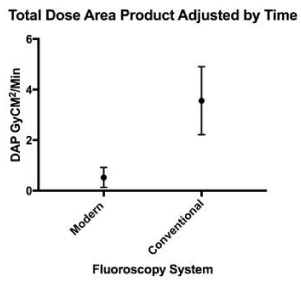
Of the 460 procedures performed, 350 were performed using the conventional system and 110 were performed using the modern system. Average ages in the two groups were 67.2 and 67.8 years in the modern and conventional groups, respectively. This study included majority males with 66.4% and 58.3% males in the modern and conventional groups, respectively. Additional baseline demographics and type of device placement are shown in table 1. More CRT devices were implanted utilizing the modern system than conventional system. Mean Dose Area Product (DAP) by device and for each group are shown in table 2. The DAP was higher with CRT device placement, compared to ICD or PPM placement using either system. A significantly lower DAP was found for all device placements with use of the modern system. When device placement was performed utilizing the more modern fluoroscopy system, DAP was reduced by more than two-thirds (CRT 28.65 ± 28.70 Gy·cm2 vs. 141.66 ± 121.24 Gy·cm2, ICD 8.09 ± 10.36 Gy·cm2 vs. 27.88 ± 30.29 Gy·cm2, PPM 4.97 ± 4.71 Gy·cm2 vs. 27.67 ± 30.94 Gy·cm2, in modern vs. conventional systems, respectively). These findings were unchanged when DAP was normalized to fluoroscopy pedal time, as shown in table 3. Figure 1 displays DAP adjusted by time between the two groups for all devices.
Discussion
This study confirms findings seen by others working with fluoroscopy in non-cardiac radiography [10,11], cardiac angiography [12-14] and cardiac electrophysiology procedures [8,9,15]. Newer imaging systems equipped with enhanced filtering and software systems can significantly decrease radiation exposure to both operator and patient. The decrease in radiation exposure between the modern imaging systems and conventional systems was comparable to prior studies with reductions of 40% to 70% in non-CRT device placement and nearly a 75% reduction during CRT implantation. Although we did not compare image quality between the two fluoroscopy systems in our study, others have clearly demonstrated that this new technology allows for a significant reduction in radiation exposure without sacrificing image quality [12,14].
This study was conducted at a large academic center with an electrophysiology training program. There is an obligation to minimize radiation exposure to protect both patients and providers as well as to make training as safe as possible. Academic and referral centers tend to have high complexity cases, and the presence of a trainee may extend the duration of radiation exposure in any particular case. Having modern equipment available is not only essential for training practitioners but also for ensuring their safety as they enter a lifelong career [16-17].
As procedure complexity and frequency increase, corresponding cumulative radiation exposure and consequences to providers increase. Various radiation protection equipment exist but are not always used [18] and proposed procedure limitations may have adherence issues [7]. This technology allows an alternative to decreasing provider exposure without significant drawbacks. Rapid and complete adoption of more modern imaging systems are logistically and financially difficult. Many institutions utilize a combination of both conventional and modern imaging systems. Cases requiring additional imaging time, such as CRT implantation, should be preferentially performed in laboratories with modern imaging technology. Ideally, all imaging systems should be upgraded to minimize exposure to ionizing radiation and maximize safety for patients and staff.
Our study had several limitations. These data represent the experience of a single center and this study was conducted in a retrospective, non-randomized manner limiting the generalizability of these findings. Additionally, the sample size of each type of device placement with each system was relatively small. Furthermore, the scope of this study was limited to radiation dose and does not evaluate objective image quality or whether image quality is affected by different radiation dosages.
In conclusion, radiation exposure was significantly lower during cardiac implantable electronic device implantation with use of a modern imaging system.
- Preston R. Radiation Biology: concepts for radiation protection. Health Phys. 2005; 88: 545-556. https://goo.gl/7nkcZC
- National Radiological Protection Board (NRPB) and Royal College of Radiologists (RCR). Patient dose reduction in diagnostic radiology: a report of the Royal College of Radiologists and the National Radiological Protection Board. Documents of the NRPB. Documents of the NRPB. 1990; 1: 3. https://goo.gl/r23pah
- Zhan C, Baine W, Sedrakyan A, Steiner C. Cardiac device implantation in the United States from 1997 through 2004: a population-based analysis. J Gen Intern Med. 2008; 23 Suppl 1: 13-19. https://goo.gl/aPPsxR
- Perisinakis K, Theocharopoulos N, Damilakis J, Manios E, Vardas P, Gourtsoyiannis N. Fluoroscopically guided implantation of modern cardiac resynchronization devices: radiation burden to the patient and associated risks. J Am Coll Cardiol. 2005; 46: 2335-2339. https://goo.gl/GpjwYE
- Shope TB. Radiation-induced skin injuries from fluoroscopy. Radiographics. 1996; 16: 1195-1199. https://goo.gl/v7NwdA
- Koenig TR, Mettler FA, Wagner LK. Skin injuries from fluoroscopically guided procedures: part 2, review of 73 cases and recommendations for minimizing dose delivered to patients. AJR Am J Roentgenol. 2001; 177: 13-20. https://goo.gl/kFnnwq
- Butter C, Schau T, Meyhoefer J, Neumann K, Minden HH, Engelhardt J. Radiation exposure of patient and physician during implantation and upgrade of cardiac resynchronization devices. Pacing Clin Electrophysiol. 2010; 33: 1003-1012. https://goo.gl/xFHTm3
- Dekker LR, van der Voort PH, Simmers TA, Verbeek XA, Bullens RW, Veer MV, Et al. New image processing and noise reduction technology allows reduction of radiation exposure in complex electrophysiologic interventions while maintaining optimal image quality: a randomized clinical trial. Heart Rhythm. 2013; 10: 1678-1682. https://goo.gl/NzV2Rs
- Hoffmann R, Langenbrink L, Reimann D, Kastrati M, Becker M, Piatkowski M, et al. Image noise reduction technology allows significant reduction of radiation dosage in cardiac device implantation procedures. Pacing Clin Electrophysiol. 2017; 40: 1374-1379. https://goo.gl/eoawiM
- Schernthaner R, Duran R, Chapiro J, Wang Z, Geschwind J, Lin M. A new angiographic imaging platform reduces radiation exposure for patients with liver cancer treated with transarterial chemoembolization. Eur Radiol. 2015; 25: 3255-3262. https://goo.gl/Mw6sP2
- Soderman M, Mauti M, Boon S, Omar A, Marteinsdottir M, Andersson T, et al. Radiation dose in neuroangiography using image noise reduction technology: a population study based on 614 patients. Neuroradiology. 2013; 55: 1365-1372. https://goo.gl/iuYws2
- Eloot L, Thierens H, Taeymans Y, Drieghe B, De Pooter J, Van Peteghem S, et al. Novel X-ray imaging technology enables significant patient dose reduction in interventional cardiology while maintaining diagnostic image quality. Catheter Cardiovasc Interv. 2015; 86: E205-E212. https://goo.gl/PZQERT
- Nakamura S, Kobayashi T, Funatsu A, Okada T, Mauti M, Waizumi Y, et al. Patient radiation dose reduction using an X-ray imaging noise reduction technology for cardiac angiography and intervention. Heart Vessels. 2016; 31: 655-663. https://goo.gl/pPn7hD
- Kastrati M, Langenbrink L, Piatkowski M, Michaelsen J, Reimann D, Hoffmann R. Reducing radiation dose in coronary angiography and angioplasty using image noise reduction technology. Am J Cardiol. 2016; 118: 353-356. https://goo.gl/FQPQYk
- Alkhorayef M, Sulieman A, Babikir E, Daar E, Alnaaimi M, Alduaij M, et al. Patient exposure during fluoroscopy-guided pacemaker implantation procedures. Appl Radiat Isot. 2018; 138:14-17. https://goo.gl/YvNuec
- van Dijk JD, Ottervanger JP, Delnoy PP, Lagerweij MC, Knollema S, Slump CH, et al. Impact of new X-ray technology on patient dose in pacemaker and implantable cardioverter defibrillator (ICD) implantations. J Interv Card Electrophysiol. 2017; 48: 105-110. https://goo.gl/bYbN1u
- Sharma M, Khalighi K. Reducing radiation exposure in an electrophysiology lab with introduction of newer fluoroscopic technology. Clin Pract. 2017; 7: 976. https://goo.gl/xXCCRW
- Marinskis G, Bongiorni MG, Dagres N, Lewalter T, Pison L, Blomstrom-Lundqvist C, Scientific Initiative Committee EHRA. X-ray exposure hazards for physicians performing ablation procedures and device implantation: results of the European Heart Rhythm Association survey. Europace. 2013; 15: 444-446. https://goo.gl/QSnjZp


Sign up for Article Alerts