Pituitary Carcinoma: a Case Report of a Rare Tumour Treated with Radiotherapy?
Aisling M Glynn, Guhan Rangaswamy*, Julianne O’Shea and Clare Faul
Department of Radiation Oncology, St. Luke’s Radiation Oncology Network, Dublin, Ireland
*Address for Correspondence: Guhan Rangaswamy, Department of Radiation Oncology, St. Luke’s Radiation Oncology Network, Rathgar, Dublin 6, Ireland, Tel: +353- 87 2-850-903; Fax: +353 -170-45590; ORCID: https://orcid.org/0000-0002-4703-3462; E-mail: guhan.rangaswamy@slh.ie
Submitted: 07 April 2019; Approved: 20 May 2019; Published: 25 May 2019
Citation this article: Glynn AM, Rangaswamy G, O’Shea J, Faul C. Pituitary Carcinoma: a Case Report of a Rare Tumour Treated with Radiotherapy. Int J Case Rep Short Rev. 2019;5(5): 024-027.
Copyright: © 2019 Glynn AM, et al. This is an open access article distributed under the Creative Commons Attribution License, which permits unrestricted use, distribution, and reproduction in any medium, provided the original work is properly cited
Keywords: Pituitary; Carcinoma; Radiotherapy; Macroadenoma; Metastases
Download Fulltext PDF
Background: Pituitary carcinomas are very rare, constituting only 0.1 or 0.2% of pituitary tumours and is only diagnosed when pituitary tumour non-contiguous with the sellar region is demonstrated. The prognosis is poor with a mean survival time of 2 to 2.4 years. We describe a case of pituitary carcinoma arising in a patient with a pituitary adenoma.
Methods: A sixty-eight-year-old male with a background history of recurrent pituitary adenoma presented in 2017 with ongoing headaches. He had a previous history of pituitary adenoma treated with surgery and RT on 2 separate occasions in 2002 and 2007. MRI showed an enhancing mass in the left suprasellar cistern Intra-operatively the tumour was stuck to the posterior brain stem. Histology showed cells with a high MIB-1 proliferation index, positive for chromogranin and in the context of an adenohypophyseal tumour which had extended beyond the sellar region to involve the 4th ventricle the features were felt to be that of a pituitary carcinoma. Staging scans showed no distant metastases. He was referred for consideration of re-radiation therapy.
Results: He went on to receive a five-fraction regime of stereotactic radiotherapy. A fractionated regime was used in view of the previous RT he had received and the previous dose to the optic chiasm. He tolerated treatment fairly well and completed the same with no major difficulties. However, he deteriorated shortly thereafter and passed away.
Conclusion: This case highlights the difficulty in treating pituitary carcinomas. No identifiable factors have been noted to correlate with increased survival. Radiation treatment is often used in its management but does not appear to change disease outcome. Several chemotherapy protocols have been used with disappointing results. Identifying those invasive pituitary tumours most likely to metastasize and treating them aggressively before they progress to pituitary carcinomas should be the focus of future studies.
Abbreviations
RT: Radiotherapy; MRI; Magnetic Resonance Imaging; CT: Computed Tomography; SRS: Stereotactic Radiosurgery; MDT: Multidisciplinary Team; CNS: Central Nervous System
Introduction
Whilst pituitary tumours are common, pituitary carcinomas are very rare. They arise from the pituitary gland which is an endocrine gland. It weighs about 0.5 grams and is an extension of the lower part of the hypothalamus at the base of the brain in a bony structure called the sella turcica and comprises of three lobes [1].The anterior pituitary (or adenohypophysis) regulates several physiological processes (growth, stress , reproduction and lactation) and the intermediate lobe secretes melanocyte-stimulating hormone. The posterior pituitary (or neurohypophysis) is connected to the hypothalamus by the median eminence via a small tube called the pituitary stalk (also called the infundibulum).Pituitary carcinomas constitute 0.1 or 0.2% of pituitary tumours [1]. They typically present in the third to fifth decade of life in patients with pre-existing pituitary adenomas [2]. Latency periods may vary from a few months in tumours that are treatment resistant despite surgical debulking and radiotherapy and progress to carcinomas to many years in tumours that respond to standard therapy and progress to carcinomas very slowly with a mean interval of 6.6 years [3]. Diagnosis of pituitary carcinomas is a challenge and are generally diagnosed when pituitary tumour non-contiguous with the sellar region is demonstrated. The hallmark of pituitary carcinoma is distant metastases [1]. They may arise as a consequence of previous radiotherapy in the treatment of a pituitary tumour, tumour seeding from previous surgery, malignant progression of a pituitary tumour or de novo carcinoma. The prognosis is poor with a mean survival time of 2 to 2.4 years [4].
We present a complex case of pituitary carcinoma arising on the longstanding background of a pituitary adenoma treated with radiotherapy at our institution.
Case Presentation
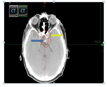
A sixty-eight-year-old male with a background history of recurrent pituitary adenoma presented in 2017 with ongoing headaches. He had a resection of a pituitary adenoma in 1997 and a further resection for a recurrence in 2000. He progressed again in 2002 and received radical external beam radiotherapy 45 Gray in 25 fractions. In 2007 he presented with deteriorating vison in his right eye. MRI scan showed a suprasellar mass causing compression of the optic chiasm. He went on to have hyper fractionated intensity modulated radiotherapy 50.4 Gray in 42 fractions treated twice daily (Figure 1).
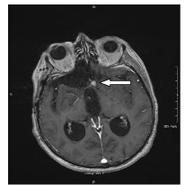
He was subsequently followed up with serial MRI scans. MRI done following his presentation in 2017 showed an enhancing mass in the left suprasellar cistern and the floor of 4th ventricle (Figure 2).
He went on to have a midline sub occipital craniotomy and near total excision of the mass. Intra-operatively the tumour was stuck to the posterior brain stem and histology showed cells with a high MIB-1 proliferation index, positive for chromogranin and in the context of an adenohypophyseal tumour which had extended beyond the sellar region to involve the 4th ventricle the features were felt to be that of a pituitary carcinoma. A staging CT TAP and MRI spine showed no distant metastases. Following a discussion at the National Neuro Oncology MDT meeting, he was referred for consideration of re-radiation therapy. Following a further discussion at the Stereotactic MDT meeting and a review of the previous radiotherapy plans from 2002 and 2007 the consensus was to proceed with re-radiation using fractionated Stereotactic Radiotherapy (SRS). He underwent a planning CT scan and this was fused with a planning MRI scan. A Gross Tumour Volume (GTV) was drawn on the fused slices using the planning MRI scan as a reference. A stereotactic RT plan was generated on iPlan. A five-fraction treatment regime was used instead of a single fraction in view of the radiotherapy he had already received and the previous dose to the optic chiasm. He was prescribed 25 Gray in 5 fractions stereotactic radiotherapy to the 80% isodose line delivered over 5 days. He tolerated treatment fairly well and completed the same with no major difficulties. Given that he had recurrent disease the intent of treatment was to achieve as much local control as was feasible Despite this the patient deteriorated clinically as a result of his underlying disease and passed away six weeks following the completion of his treatment.
Discussion
This case highlights the difficulty in diagnosing and treating pituitary carcinomas. Whilst they commonly present as early recurrence after pituitary surgery, followed by repeated surgeries for local re-growth and tumour extension, there are no factors that predict their occurrence reliably. The majority arise from a macroadenoma that show suprasellar extension [5]. The hallmark of pituitary carcinoma is distant metastases. 47% of metastases occurs systemically to sites like the liver, kidney eyes, heart, lung, neck nodes, pancreas, ovary and bone [6] and 40% metastasize to the CNS sites like the cerebrum, cerebellum, spinal cord and leptomeninges. The most common pituitary carcinomas are adrenocorticotrophic hormone secreting carcinomas, followed by prolactin and growth hormone secreting carcinomas [7]. The treatment of pituitary carcinomas includes surgery usually via the transsphenoidal route, radiotherapy, medical treatment including dopamine antagonists and somatostatin analogues [1,8] and chemotherapy protocols that have been used with disappointing results [5,9].
External beam radiotherapy has been used for prevention of regrowth of tumour in large, incompletely excised tumours and for the local control of expanding tumours and metastatic deposits [8]. Stereotactic Radiosurgery (SRS) involves precise delivery of high-dose radiation typically at a single visit, which offers good efficacy and patient convenience and is preferred in patients who have already had conventional radiotherapy and have developed a recurrence or metastases [8]. However, the efficacy of the different forms of radiotherapy like fractionated RT, SRS, Cyberknife and Proton beam therapy is difficult to estimate given the rarity of these tumours, differences in dose and the technique used in treatment delivery [10].
Conclusion
Our case highlights the difficulty and challenges faced in the management of pituitary carcinomas. As demonstrated by our case pituitary carcinomas are locally invasive and characterised by multiple recurrences with limited therapeutic options particularly in the recurrent setting. Radiotherapy can sometimes lead to disease stabilisation. However, the management of pituitary carcinomas is mainly palliative and may not significantly prolong survival to any major extent [11]. There are several published reports of long-term survivors of pituitary carcinoma particularly in patients who received aggressive treatment [12]. Given the rarity of these tumours randomized trials looking at establishing an effective treatment strategy is not possible. The aim of future studies should be to try and identify those invasive pituitary tumours most likely to metastasize and treating them aggressively before they progress to pituitary carcinomas [8,13].
- Ragel BT, Couldwell WT. Pituitary Carcinoma: a review of the literature, Neurosurg Focus. 2004; 15: 16. https://bit.ly/2HtDyMB
- Kontogeorgos G. Classification and pathology of pituitary tumors. Endocrine. 2005; 28: 27-35. https://bit.ly/2JvTekJ
- Roncaroli F, Scheithauer BW, Horvath E, Erickson D, Tam CK, Lloyd RV, et al. Silent subtype 3 carcinoma of the pituitary: a case report. Neuropathol Appl Neurobiol. 2010; 36: 90-94. https://bit.ly/2YLeRkR
- Pernicone PJ, Scheithauer BW, Sebo TJ, Kovacs KT, Horvath E, Young WF Jr, et al. Pituitary Carcinoma: a clinicopathologic study of 15 cases. Cancer. 1997; 79: 804-812. https://bit.ly/2HGLFnX
- Lopes MB, Scheithauer BW, Schiff D. Pituitary Carcinoma: diagnosis and treatment. Endocrine. 2005; 28: 115-121. https://bit.ly/2QcFuMq
- Scheithauer BW, Fereidooni F, Horvath E, Kovacs K, Robbins P, Tews D et al. Pituitary Carcinoma: an ultrastructural study of eleven cases. Ultrastruct Pathol. 2001; 25: 227-242. https://bit.ly/2M23kfe
- Kaltsas GA, Grossman AB. Malignant pituitary tumours. Pituitary. 1998; 1: 69-81
- Heaney AP. Pituitary Carcinoma: difficult diagnosis and treatment, J Clin Endocrinol Metab. 2011; 96: 3649-3660. https://bit.ly/2HsWYRP
- Kaltsas GA, Mukherjee JJ, Plowman PN, Monson JP, Grossman AB, Besser GM. The role of cytotoxic chemotherapy in the management of aggressive and malignant pituitary tumours. JClin Endocrinol Metab. 1998: 4233-4238. https://bit.ly/2VJAzDW
- Rowland NC, Aghi MK. Radiation treatment strategies for acromegaly. Neurosurg Focus. 2010; 29: 12. https://bit.ly/2HtA38V
- Sironi M, Cenacchi G, Cozzi L, Tonnarelli G, Iacobellis M, Trere et al. Progression on metastatic neuroendocrine carcinoma from a recurrent prolactinoma: a case report. J Clin Pathol. 2002; 55: 148-151. https://bit.ly/2EmnNFA
- Landman RE, Horwith M, Peterson RE, Khandji AG, Wardlaw SL. Long-term survival with ACTH-secreting carcinoma of the pituitary: a case report and review of the literature. J Clin Endocrinol Metab. 2002; 87: 3084-3089. https://bit.ly/2JvTal5
- Gregory A. Kaltsas, Panagiotis Nomikos, George Kontogeorgos, Michael Buchfelder, Ashley B. Grossman. Clinical Review: diagnosis and management of pituitary carcinomas. The Journal of Clinical Endocrinology & Metabolism. 2005; 90: 3089-3099. https://bit.ly/30AOsb2


Sign up for Article Alerts