Pancreatic Panniculitis: a Lesser-Known Cutaneous Complication of Acute Pancreatitis?
Rohit Gupta1*, Nadia Shirazi2 and Rashmi Jindal3
1Department of Gastroenterology, All India Institute of Medical Sciences, Rishikesh
2Department of Pathology, Himalayan Institute of Medical Sciences, Swami Rama Himalayan University, Jolly Grant, Dehradun
3Department of Dermatology, Himalayan Institute of Medical Sciences, Swami Rama Himalayan University, Jolly Grant, Dehradun
*Address for Correspondence: Rohit Gupta, Department of Gastroenterology, All India Institute of Medical Sciences, Rishikesh, India, Tel: + 917-579-067-715; E-mail: docgupta1976@gmail.com
Submitted: 20 April 2019; Approved: 30 May 2019; Published: 03 June 2019
Citation this article: Gupta R, Shirazi N, Jindal R. Pancreatic Panniculitis: a Lesser-Known Cutaneous Complication of Acute Pancreatitis. Int J Case Rep Short Rev. 2019;5(6): 028-030.
Copyright: © 2019 Gupta R, et al. This is an open access article distributed under the Creative Commons Attribution License, which permits unrestricted use, distribution, and reproduction in any medium, provided the original work is properly cited
Keywords: Pancreatitis; Alcoholic; Panniculitis; Fat necrosis
Download Fulltext PDF
Pancreatic panniculitis is a rare complication of pancreatic disease seen in 0.3-3% of all patients, most commonly those with acute or chronic pancreatitis. It presents with painful, tender erythematous nodules, which may undergo spontaneous ulceration. Seldom they may be associated with discharge of oily, brown, viscous material due to underlying liquefactive necrosis of adipocytes. Treatment is directed at management of underlying disease. We present a case of pancreatic panniculitis in a middle aged alcoholic male.
Abbreviations
PP : Pancreatic panniculitis
Introduction
Pancreatic Panniculitis (PP) is a rare entity that was first described by Chiari in 1883, and it is most frequently associated with pancreatic diseases [1]. PP is a rare complication of pancreatic disease appearing in approximately 2% to 3% of all patients, most commonly in acute or chronic pancreatitis, but also in patients with carcinoma of the pancreas, more frequently with acinar cell carcinoma [2]. PP typically presents with tender, edematous and erythematous to red brown subcutaneous nodules that spontaneously ulcerate and exude an oily brown substance that results from liquefaction necrosis of adipocytes. The lesions most commonly develop on the lower legs; other sites include thighs, buttocks, arms, abdomen, and chest and scalp [3]. Its pathogenesis is still not well understood but the release of pancreatic enzymes in the setting of pancreatic injury may play an important role, leading to fat necrosis in subcutaneous tissue and elsewhere [4].
We present a case of 60 years old male patient who presented with acute attack of pancreatitis with development of tender skin nodules and was diagnosed as Panniculitis.
Case Report
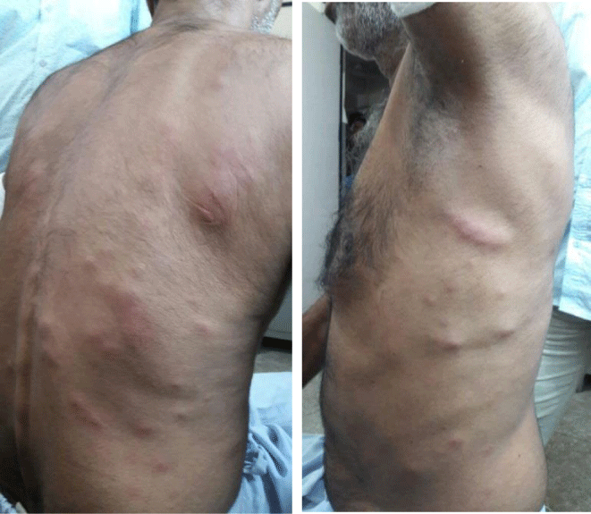
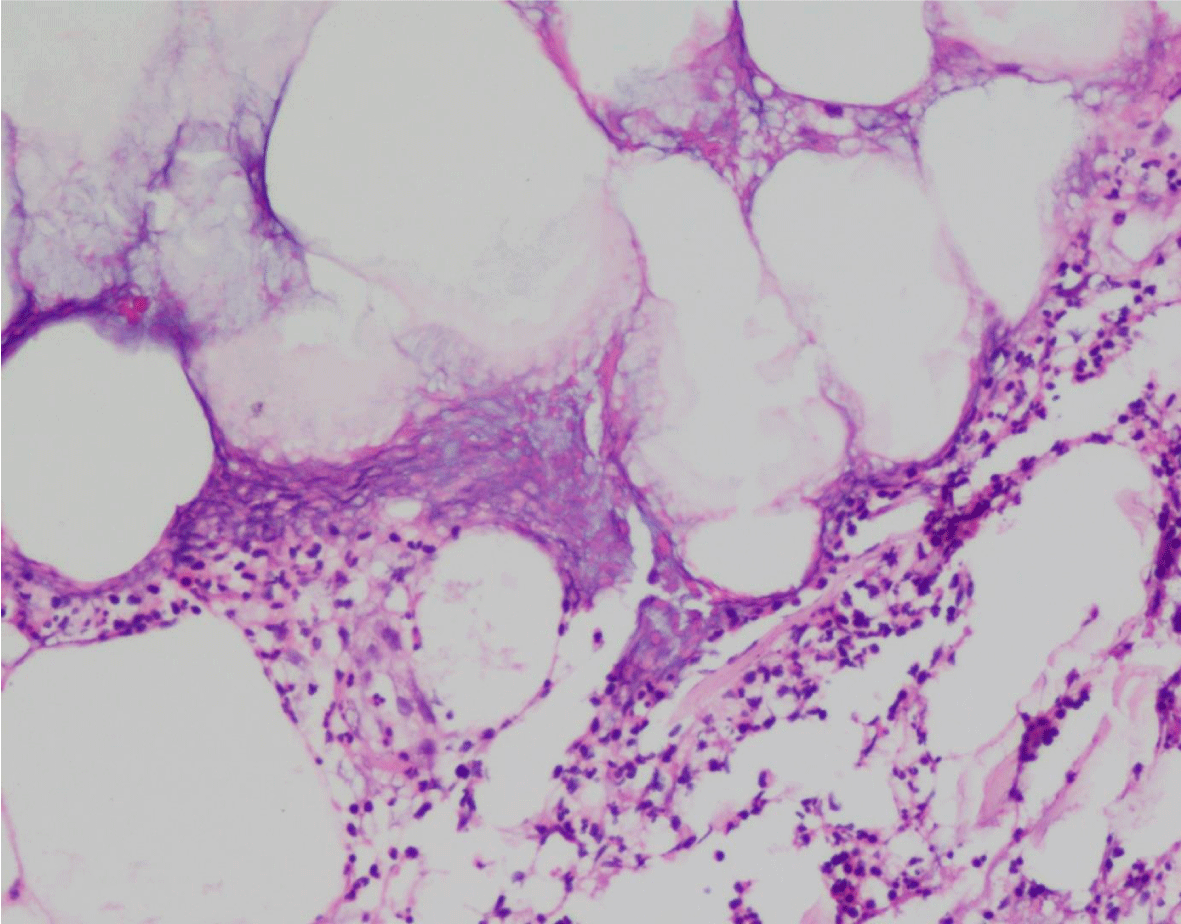
A 60 years old male patient with history of alcohol intake (250ml/ d) presented to Gastroenterology outpatient department with complaints of severe pain in abdomen associated with vomiting and abdominal distension. Patient also gave history of development of multiple painful subcutaneous nodules on his abdomen and back and upper limb, which he developed suddenly in 2 weeks with the onset of his abdominal complaints. There was no past history of joint pain, drug intake or weight loss. On examination, patient’s vitals were stable, abdomen examination showed diffusely tender abdomen with no obvious palpable lump. Dermatological examination showed presence of multiple tender erythematous nodules of 1-2cm in size involving trunk and bilateral upper limbs (figure 1). A clinical diagnosis of acute pancreatitis with pancreatic panniculitis, or atypical Mycobacterial infection was made. His laboratory investigations were ordered and skin biopsy was performed. Total Leukocyte Count was 13.8 x 10⁶ cells/cu mm, lipase was mildly raised however, serum amylase was markedly elevated (9124 U/L). Blood culture was sterile. Computed Tomography (CT scan) of abdomen showed features of acute pancreatitis however no gall stones, liver cirrhosis or pancreatic tumor was identified. Patient was non-reactive for HIV or HBs Ag. Skin biopsy showed an atrophic epidermis with mild inflammation in the dermis. Subcutaneous fat showed lobular and septal panniculitis admixed with fibrinoid necrosis and occasional ‘ghost cells’ (figure 2). Ziehl- Nelson stain was negative for Acid Fast Bacilli. A diagnosis consistent with pancreatic panniculitis was made on histopathology. The patient received primarily supportive medical treatment with intravenous administration of fluids and analgesics. The nodules resolved spontaneously after 2 weeks and his amylase and lipase levels returned to normal.
Discussion
Pancreatic panniculitis is a rare complication in the setting of pancreatic disease, in which fat necrosis takes place in the subcutaneous tissue and elsewhere. The exact patho- physiology is becoming clear but not entirely understood. The release of pancreatic enzymes, such as lipase, trypsin, amylase, phosphorilase and trypsin, may be involved in increasing the micro vascular permeability allowing hydrolysis of neutral fat. The resultant glycerol and free fatty acids, leads to fat necrosis and a brisk inflammatory response. The pathogenic role of pancreatic lipase is supported by the finding of the enzyme in the areas of subcutaneous necrosis, and also anti-lipase monoclonal antibodies within the necrotic tissue. Pancreatic disorders are much more common when compared to pancreatic panniculitis which might indicate that there are still unrecognized factors involved in the development of Pancreatic Panniculitis. Also there are some cases in the literature that have been described with normal levels of pancreatic enzymes further supporting the likely involvement of other unidentified factors in the causality of this entity. An immune complex mechanism has been described with pancreatic panniculitis.
The dermatologic manifestations are independent of the severity of pancreatic pathology and precede the diagnosis of pancreatic pathology about 40% of the times s. The lesions are generally located in the lower extremities as tender, erythematous nodules that are red-brown in color, which may ulcerate with drainage of oily fluid. The clinical features of pancreatic panniculitis are similar despite the variety of pancreatic disorders it is associated with. It is important to differentiate PP from other forms of panniculitis such as Erythema nodosum, erythema induratum, lupus panniculitis, infectious, traumatic or alpha 1- antitrypsin deficiency panniculitis.
PP is usually confirmed with a subcutaneous biopsy. The main histopathologic feature is a mostly lobular panniculitis without vasculitis. But, in the very early stage, a septal pattern has been described, which results from enzymatic damage of the endothelial septa, allowing pancreatic enzymes to cross from blood to fat lobules resulting in coagulative necrosis of the adipocytes, which leads to pathognomonic “ghost cells” (enucleated necrotic cells that have a thick wall with a fine basophilic granular material within their cytoplasm from dystrophic calcification).
Specific subgroups have known to have a higher association (alcoholic men, patients with acinar neoplasms), and PP may be the harbinger of pancreatic pathology by several days or even months [2, 5].
The treatment of PP is mainly directed towards the underlying pancreatic pathology, following which a complete to near complete resolution of symptoms is experienced as illustrated in our report. Corticosteroids may provide some symptomatic relief however NSAID’s or immune suppressors are usually not effective for the treatment of skin lesions.
Conclusions
Pancreatic panniculitis is a not a very common complication of acute pancreatitis and high index of suspicion is required for the diagnosis. There is no specific treatment and usually settles with resolution of pancreatitis.
- Chiari H. Uber die Sogenannte Fettnekrose. Prager Med Wochenschr. 1883; 8: 285-286.
- Poelman SM, Nguyen K. Pancreatic panniculitis associated with acinar cell pancreatic carcinoma. J Cutan Med Surg. 2008; 12: 38-42. https://bit.ly/2Knhjdn
- Bagazgoitia L, Alonso T, Rios Buceta L, Ruedas A, Carrillo R, Munoz Zato E. Pancreatic panniculitis: an atypical clinical presentation. Eur J Dermatol. 2009; 19: 191-192. https://bit.ly/2IfuNoL
- García-Romero D, Vanaclocha F. Pancreatic panniculitis. Dermatol Clin. 2008; 26: 465-470.
- Zundler S, Erber R, Agaimy A, Hartmann A, Kiesewetter F, Strobel D, et al. Pancreatic panniculitis in a patient with pancreatic-type acinar cell carcinoma of the liver--case report and review of literature. BMC Cancer. 2016; 20: 16: 130. https://bit.ly/2WAPZyA


Sign up for Article Alerts