Myofascial Release Technique and Ultrasound Guided Dry Needling in Management of Complex Regional Pain Syndrome Type 1: A Case Report?
Dipti Geete1*, KV Mate2, Chhaya Verma3 and Esther Katarnawre4
1Physiotherapy School and Centre, Seth G. S. Medical College, King Edward Memorial Hospital, Parel, Mumbai, India
2Center for Outcomes Research and Evaluation, McGill University Health Center Research Institute, Montreal, Canada
3Nair Hospital & T. N. Medical College, Mumbai Central, Mumbai, India
4Lokmanya Tilak Municipal Medical College and General Hospital
*Address for Correspondence: Dipti B. Geete, Physiotherapy School and Centre, Seth G. S. Medical College, King Edward Memorial Hospital, Parel, Mumbai, India.Tel: +91- 98-69-70-14-64; E-mail: dipti_geete@yahoo.com
Submitted: 30 May 2019; Approved: 20 June 2019; Published: 24 June 2019
Citation this article: Geete D, Mate KV,Verma C, Katarnawre E. Myofascial Release Technique and Ultrasound Guided Dry Needling in Management of Complex Regional Pain Syndrome Type 1: A Case Report. Int J Case Rep Short Rev. 2019;5(6): 031-036.
Copyright: © 2019 Geete DB, et al. This is an open access article distributed under the Creative Commons Attribution License, which permits unrestricted use, distribution, and reproduction in any medium, provided the original work is properly cited
Keywords: Complex regional pain syndrome; Myofascial release; Intramuscular stimulation; Physiotherapy
Download Fulltext PDF
Complex Regional Pain Syndrome (CRPS) results in multitude of impairments that limits functioning and impacts a person’s quality of life. Various multi- and trans-disciplinary treatment options have been reported in the literature to maximize function and return to pre-injury level. The healthcare pathways in management of people with CRPS require large facilities and efficient organisation of professionals which are currently missing in low-resource settings in urban India. The objective of this case report is to support a potential combination of myofascial release techniques and intramuscular stimulation using dry needling to improve outcomes in an individual with CRPS in an urban tertiary care hospital. A 48-year-old man diagnosed with CRPS-I following traumatic radial head dislocation was treated with a structured plan over a period of 9 months with a combined therapy that included Myofascial Release (MFR) techniques, Ultrasound guided Dry Needling (USGDN) and conventional physiotherapy treatment. The combined intervention showed decrease in pain, improved range of motion and better scores on Disabilities of the Arm, Shoulder and Hand (DASH) scale. Combination of MFR and USGDN may potentially be used as a treatment strategy in management of people with CRPS-1, a direction for future research in this area and could be further investigated.
Introduction
Complex Regional Pain Syndrome (CRPS) is a painful condition characterised by regional pain, allodynia, temperature, sudomotor and vasomotor changes following a trauma [1]. CRPS is classically divided into two types, Type 1 called as CRPS-I (formerly known as Reflex Sympathetic Dystrophy) and Type 2 called as CRPS-II (also known as Causalgia) [2,3]. Literature reports an association of former with no nerve injury and later associated with nerve injury. A systematic review on effectiveness of physical therapy interventions in management of CRPS suggests that there is lack of evidence on effectiveness of physical therapy interventions. The review recommended that therapy should be directed to ameliorate symptoms and improve function delivered through an interdisciplinary team [4].
The presented case report is aimed to explore a possible multidisciplinary approach in management of an individual diagnosed with CRPS in low-resource urban tertiary care hospital. Health care service delivery centres in a Metropolitan Mumbai city are busy with a large number of patients in need for rehabilitation interventions. This results in smaller time spend between patient-therapist interaction. In order to maximize this time, in absence of clinical guidelines and with limited resources, a multidisciplinary approach to management of CPRS seems most realistic.
A review by Lee JW, et al. (2018) on the Diagnosis and Management of CRPS-I has given few important insights on CRPS. The author concludes that diagnosis of CRPS is made primarily on a clinical basis, and no specific test is known to confirm or exclude CRPS diagnosis. Numerous therapeutic methods have been introduced, but none have shown definitive results. When symptoms persist, patients experience permanent impairment and disability. Several clinical diagnostic criteria have been reported in the literature to classify different types of CRPS. Among these different measures used, the Budapest diagnostic criteria are widely accepted and frequently cited in the literature. Therefore, this review emphasized the need for early diagnosis and treatment in people with CRPS-1 so as to prevent permanent impairments and resulting disability.
The overall goal in management of CRPS is the return of affected extremity to preexisting functional status. There is no evidence on any preventive steps that could be taken so as to avoid CRPS following surgery, but prolonged immobilization has been shown to be associated with higher incidence of CRPS [5]. This emphasis the need for early post-surgical rehabilitation. During initial stages of regaining movement, pain is the most limiting factor to achieve any gains. A multidisciplinary approach seems to provide a combination pharmacotherapy to provide adequate pain control that will allow gain in movements.
There are several movement or mobilization techniques reported in the literature that are targeted to reduce pain while permitting large gains in range of movement. Myofascial Release (MFR) is one such technique to maintain the flexibility of muscle and fascial planes. Myofascial pain syndrome have been successfully treated using intramuscular stimulation using dry needling [6-8].
Yeuh-Ling-Hsieh et al., used dry needling for Myofascial Trigger Point (MTrP) in rabbit skeletal muscles and showed that use of dry needle modulates a variety of biochemical associated with pain, inflammation and hypoxia [9]. This study investigated the activities of β endorphin, substance P, Tumor Necrosis Factor- α (TNF- α), Cyclooxygenase-2 (COX-2), Hypoxia-Inducible Factor-1 α (HIF-1α), Vascular Endothelial Growth Factor (VEGF), and inducible isoform of nitric oxide synthases (iNOS) after different dosages of DN at the MTrP of a skeletal muscle in rabbit. The study showed that treatment with one dosage increased β endorphin levels in Biceps Femoris (BF) and serum and reduced substance P in the BF & Dorsal Root Ganglion (DRG). Treatment with five dosage on the other hand reversed the effects and increased levels of TNF- α, COX-2, HIF-1α, iNOS and VGEF production in the BF. The study concluded that DN at the MTrP modulates various biochemicals associated with pain, inflammation, and hypoxia in a dose-dependent manner [9].
Lobo CC, et al. conducted a study to test the efficacy potential of Deep Dry Needling (DDN) on latent Myofascial Trigger points The study showed that a single PT intervention combined with DDN on a single latent MTrP, in conjunction with one active MTrP, in the infraspinatus muscles may increase the Pressure Pain Thresholds (PPT) of the extensor carpi radialis brevis muscle area immediately following and one week after the intervention in older adults with nonspecific shoulder pain. This study may contribute to the consideration of latent MTrPs DDN to reduce the mechanosensitivity of the distal musculature of the upper limb [10].
Shah JP and colleagues showed that Myofascial Pain Syndrome (MPS) could be acute or chronic and involved both muscle and fascia. Investigators demonstrated use of dry needling to provide symptom relief and change in the status of the trigger point, although the mechanism by which this works has not yet been demonstrated [11]. The authors reported that the pathogenesis and pathophysiology of MTrP and their role in MPS is unclear. Moreover, earlier theories on pathogenesis of MTrPs and MPS including muscle overuse and mechanical limitations have neither been proven nor disproven in the literature. Current therapies such as USGDN and MFR, considered to have potential to minimise the impairments resulting from CRPS and improve functional outcome, were added to the management of this patient with CRPS-I in addition to conventional physiotherapy.
Materials and Methods
Based on the findings in the literature, this case report applied two of these techniques, MFR and USGDN in a 48 year old right dominant male who was referred to outpatient physical therapy services. He worked as a plumber and job analysis revealed use of unilateral and bilateral hand activities that required both power and precision action of upper extremity. He reported a fall on his back with an outstretched hand from approximately 10-15 feet. Emergency Medical Services (EMS) visit showed right posterior dislocation of elbow with fracture of radial head and a grade 1 anterior wedge compression fracture of 12th thoracic vertebral body with no neurological deficit. He was managed conservatively with an above-elbow plaster cast for six weeks and recommended bed rest for spinal fracture. On subsequent follow-up, surgical excision of radial head was performed as he displayed type 4 Mason fracture followed with additional 3 weeks of immobilisation in a plaster cast with forearm in mid-prone and elbow at 90° of flexion. At the end of 10 weeks of conservative management and post-immobilisation for spinal fracture, the patient was referred to physical therapy services.
The physical examination findings shown below pertains to right upper extremity
Upper extremity was held in protective posture with shoulder in elevation, elbow in mid-flexion and mid-prone position and wrist was in flexion.
Pain score of 4 out of 10 on Visual Analog Scale [12]
Swelling over the dorsum of hand
Shiny and dark shade of skin on dorsal aspect of hand
Stiffness (Range of Motion) assessed By Goniometer Scale [13]
Patient reported nociceptive sensation in response to touch (allodynia)
Atrophy of supraspinatus, infraspinatus, deltoid, biceps, triceps and forearm muscles of right upper limb.
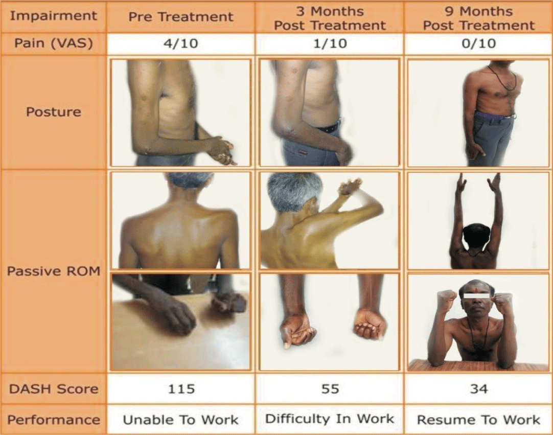
Trigger points and taut bands were present in upper trapezius, rhomboids, teres major, supinator, pronator and brachioradialis (Figure 1).
The clinical findings were suggestive of CRPS-I using Budapest diagnostic Criteria [14,15].Patient management plan involved a multidisciplinary team that involved a pain physician and a physiotherapist. Physiotherapy intervention was directed towards improving functional use of right upper extremity. SMART (Specific, Measurable, Attainable, Relevant and Timely) goals were set with a home exercise plan. Supervised exercise program were provided for three times per week for six weeks and included active exercises, strengthening exercises, myofascial release techniques and desensitization techniques. The patient was followed for nine months during which exercises were progressed based on improvement till full functional return was achieved.
Pain management included injection of Bupivacaine, Kenacort and 10 milligrams of oral Tryptomer. The patient also received two 30-minute sessions per week of ultrasound-guided dry needling to upper trapezius, supraspinatus, infraspinatus, teres major, and biceps, subscapularis, triceps, supinator and brachioradialis. The needle size was determined on the bulk of muscles and both flexor and extensor group of muscles were targeted in both the session. For restricted range of motion at shoulder joint, the patient additionally received ultrasound guided steroid injection to triceps and brachioradialis. Each session of dry needling was followed by the supervised exercise program. The patient was requested to maintain a log of exercises in diary carried out as home program.
The outcome measures recorded at three time points during the therapy are shown in table 1
Physiotherapy management
Posture correction in front of mirror.
Manual therapy: a) Myofascial release was done on following muscles using both direct & indirect techniques: Latissimus Dorsi, Levator scapulae, Teres Minor, Upper trapezius, Serratus anterior, Subscapularis muscles Biceps Brachii.
Brachioradialis, supinators, pronators, and flexor group of muscles.
b) Massage: Pick-up, Rolling & Wringing techniques applied to the Upper trapezius teres minor muscles.
c) Muscle energy techniques: Pectoralis Minor Muscle to correct anterior tipping of shoulder (10)
- Contract-Relax technique given to the shoulder internal rotators.
d) Joint mobilizations
Scapula: Upward rotation & depression was given to correct elevation downward rotation of scapula.
Shoulder joint: Maitland mobilization
-Posterior Glide-to improve Flexion & Internal rotation.
-Inferior Glide-to improve Abduction.
- Elbow- Movement with mobilization given to improve flexion range.
Wrist & Hand: - Dorsal glide to improve flexion, ventral glide to Improve extension and flexion at proximal and distal inter-phalangeal joints.
The regained mobility was maintained by giving full range of motion exercises
Therapeutic exercisesa) Shoulder gleno-humeral complex
Stretching Exercises: Levator scapulae, upper trapezius, posterior capsule, shoulder internal and external rotators
Posterior tipping exercise to correct anterior tipping of scapula
Table-top exercise to maintain shoulder range of motion
Shoulder mobility exercises with wand
Rotator cuff strengthening exercises
b) Elbow joint
Active exercise to improve & maintain range of motion
Strengthening Exercises to bicep & triceps muscles
c) Radio-Ulnar joint
Pronator-Supinator bar exercise
Auto-stretching exercise
d) Wrist and hand
Stretching exercises to wrist long flexors muscles
Active exercise to improve and maintain range of motion
Paper crumpling exercise
Soft ball squeezing, peg-board exercises
Different types of pinch and grip exercises
e) Brisk walking for 30 to 40 minutes
f) Relaxation for 10 minutes
Results
The combined effect of MFR and USGDN was used in management of this patient with CRPS. This combined intervention showed improvement in the range of motion, eliminating pain and decreased DASH score (improved function).
Discussion
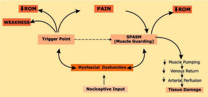
There is a paucity in the literature of USGDN techniques and Myofascial Release techniques (MFR) in the management of CRPS 1. The possible mechanism that could be at play is explained below [16,17]. The abnormal sympathetic nervous system activity in CRPS is suggested as the cause of the structural changes like ischemia and atrophy of the affected muscles [18]. Studies have shown that changes in the affected muscles is a direct consequence of the CRPS (primary phenomenon) rather than as a consequence of pain associated disuse [19]. A hypothetical model for this mechanism of effect is displayed in (Figure 2).
Pain additionally has an effect on muscle spasm causing muscle guarding and protective positioning of the affected part. Long term shortening of muscle results in reduced arterial perfusion and oxygen delivery to the tissue (Allen RJ., Koshi. LR.). Consequently, this affects the joint play causing connective tissue shortening and adhesions (Allen RJ., Koshi. LR.)
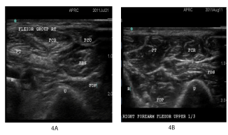
The therapeutic intervention i.e. MFR and intra muscular stimulation using dry needling directly impacts the muscles. Dry needling in treatment of CRPS here stimulates the myofascial trigger points which relieves pain and relaxes the shortened muscle. Literature reports that MFR realigns fascia, removes fascial restriction, and release bonds between the integuments, muscles and bones [20]. Functional, biomechanically efficient movement depends on intact and properly distributed fascia. When the structure has been returned to a balanced state, it is realigned with gravity. The body’s inherent ability to self-correct returns, thus restoring optimum function and performance with least amount of energy expenditure. MFR in combination with IMS helps to increase the extensibility and decrease the dominance of the muscle causing dysfunctional movement which ultimately leads to functional restoration. Hence it could be a possible management strategy in the management of CRPS 1 [21]. (Figure 3) (Figure 4 A & B).
Acknowledgement
The authors would like to thank Dr. Lakshmi V, Dr. Pai R. from Ashirwad Institute for Pain Management and Research for their contribution and support as a part of multidisciplinary team involved in therapy of this patient. And like to thank Chetali Khadye is Physiotherapist.
- Merskey HE. Classification of chronic pain. Descriptions of chronic pain syndromes and definitions of pain terms. Prepared by the international association for the study of pain, subcommittee on taxonomy. Pain Suppl. 1986; 3: 1-226. https://urlzs.com/sFeKi
- Stanton Hicks M, Janig W, Hassenbusch S, Haddox JD, Boas R, Wilson P. et al. Reflex sympathetic dystrophy: changing concepts and taxonomy. Pain. 1995; 63: 127-133. https://bit.ly/2N0hJt1
- Harden RN. Interdisciplinary management/functional restoration. Journal of Neuropathic Pain & Symptom Palliation. 2006; 2: 57-68. https://urlzs.com/vhRMf
- Dommerholt J. Complex regional pain syndrome-2: physical therapy management. Journal of bodywork and movement therapies. 2004; 8: 241-248. https://urlzs.com/A3DfL
- Lee JW, Lee SK, Choy WS. Complex regional pain syndrome type 1: diagnosis and management. J Hand Surg Asian Pac Vol. 2018; 23: 1-10. https://urlzs.com/Ak91V
- Kalichman L, S. Vulfsons, Dry needling in the management of musculoskeletal pain. J Am Board Fam Med. 2010; 23: 640-646. https://urlzs.com/aPH4C
- Kietrys DM, Palombaro KM, Azzaretto E, Hubler R, Schaller B, Schlussel JM, et al, Effectiveness of dry needling for upper-quarter myofascial pain: a systematic review and meta-analysis. J Orthop Sports Phys Ther.2013; 43: 620-634. https://urlzs.com/LUAaF
- Gunn CC. Neuropathic myofascial pain syndromes. Management of Pain. 3rd ed. Philadelphia: Lippincott Williams & Wilkins; 2001: 522-529.
- Hsieh YL, Yang SA, Yang CC, Chou LW. Dry Needling at Myofascial Trigger Spots of Rabbit Skeletal Muscles Modulates the Biochemicals Associated with Pain, Inflammation, and Hypoxia. Evid Based Complement Alternat Med. 2012; 2012: 1-12. https://urlzs.com/NFDEk
- Calvo Lobo C, Pacheco da Costa S, Hita Herranz E. Efficacy of deep dry needling on latent myofascial trigger points in older adults with nonspecific shoulder pain. J Geriatr Phys Ther. 2017; 40: 63-73. https://urlzs.com/GHU1i
- Shah J, Thaker N, Heimur J, Aredo J, Sikdar S, Gerber L. Myofascial trigger points then and now: a historical and scientific perspective. PM R. 2015; 7:746-761. https://urlzs.com/SnP43
- McCormack HM,Horne DJ, Sheather S. Clinical applications of visual analogue scales: a critical review. Psychol Med. 1988; 18: 1007-1019. https://urlzs.com/Pm4M1
- Van Rijn S, Zwerus E, Koenraadt K, Jacobs W, van den Bekerom M, Eygendaal D. The reliability and validity of goniometric elbow measurements in adults: A systematic review of the literature. Shoulder Elbow. 2018; 10: 274-284. https://urlzs.com/ndFhH
- Harden RN, Bruehl S, Perez RS, Birklein F, Marinus J, Maihofner C, et al. Validation of proposed diagnostic criteria (the “Budapest Criteria”) for complex regional pain syndrome. Pain. 2010; 150: 268-274. https://urlzs.com/b1TXb
- Reinders MF, Geertzen JHDijkstra PU, Complex regional pain syndrome type I: use of the International Association for the Study of Pain diagnostic criteria defined in 1994. Clin J Pain. 2002; 18: 207-215. https://urlzs.com/b1TXb
- Borstad JD, Ludewig PM. The effect of long versus short pectoralis minor resting length on scapular kinematics in healthy individuals. J Orthop Sports Phys Ther. 2005.35: 227-38. https://urlzs.com/BcdQb
- Edwards J, Knowles N. Superficial dry needling and active stretching in the treatment of myofascial pain - a randomised controlled trial. Acupunct Med. 2003; 21: 80-86. https://urlzs.com/L3tk3
- Nishida Y, Saito Y, Yokota T, Kanda T, Mizusawa H, et al. Skeletal muscle MRI in complex regional pain syndrome. Intern Med. 2009; 48: 209-212. https://urlzs.com/bFRvM
- Vas LC, Pai R, Radhakrishnan M. Radhakrishnan M, Ultrasound appearance of forearm muscles in 18 patients with complex regional pain syndrome 1 of the upper extremity. Pain Practice. 2013; 13: 76-88. https://urlzs.com/33UPb
- Carol M.Davis, Rose G. Complementary therapies in rehabilitation: Evidence for efficacy in therapy, prevention and wellness. 2nd ed. Thorofare, NJ: 2004.
- Donatali R, Wooden M. Orthopaedic physical therapy. 3rd ed. Elsevier health sciences; 2009.


Sign up for Article Alerts