Perivascular Epithelioid Cell Tumor (PECom) of Vagina: A Rare Occurrence?
Sheikh Zahoor1, Shah Naveed2*, Abdul Wahid Mir1, Azhar Jan Batoo1, Ifrah3, Tabish Maqbool3 and Jasif3
1Associate Professor Department of Surgical Oncology, SKIMS, India
2Assistsant Professor Department of Surgical Oncology, SKIMS, India
3Senior Resident Department of Surgical Oncology, SKIMS, India
*Address for Correspondence: Shah Naveed, Department of Surgical Oncology, SKIMS, India, E-mail: kingshahnaveed@yahoo.co.in
Submitted: 04 March 2020; Approved: 24 June 2020; Published: 26 June 2020
Citation this article: Zahoor S, Naveed S, Mir AW, Batoo AJ, Ifrah, et al. Perivascular Epithelioid Cell Tumor (PECom) of Vagina: A Rare Occurrence. Int J Case Rep Short Rev. 2020;6(6): 021-022.
Copyright: © 2020 Zahoor S, et al. This is an open access article distributed under the Creative Commons Attribution License, which permits unrestricted use, distribution, and reproduction in any medium, provided the original work is properly cited
Download Fulltext PDF
Introduction
Perivascular epithelioid cell tumor (PEComa) is a rare subtype of mesenchymal origin tumors, and is composed of perivascular epithelioid cells with specific histologic and immunohistochemical features [1]. PEComas can occur at any anatomic site and include angiomyolipomas, lymphangioleiomyomatosis, clear cell “sugar” tumors of the lung, and PEComa not otherwise specified [2,3]. PEComa of the female gynecological tract is a rare entity. When considering PEComas of the female genital tract, the uterus is the most common location. Involvement of the ovary in the context of a primary uterine PEComa, in the absence of systemic disease associated with tuberous sclerosis, however, has only been reported in 1 previous case [4]. One case of primary PEcoma of vagina was reported from china [5]. Second case was reported in a eight year old girl [6].
Case Report
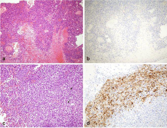
Our patient was a 36-year-old woman who presented with vaginal mass. Her past medical history was unremarkable. Initial findings were a 2/2 cm of vaginal mass on pelvic examination. A punch biopsy of vaginal mass was performed. Histologic and immunohistochemical findings (positive for SMA, but negative for HMB45) were malignant tumor, suggestive of leiomyosarcoma. Pelvic Magnetic Resonance Imaging (MRI) revealed a 2.2 × 2 cm sized heterogeneous enhancing mass in left lateral vaginal wall. Subsequent chest Computerized Tomography (CT) was normal.. Consequently, the patient underwent wide local excision. Histologic examination revealed that the tumor cells were predominantly composed of pleomorphic spindle and epithelioid cells in fascicles with interspersed lymphocytic aggregates. The tumor cells showed many bizzare and multinucleated forms, however mitotic activity was negligible. Immunohistochemistry showed tumor cells expressed MiTF(focal), TFE 3, SMA(focal) and desmin and were immuno negative for CD 21, CD 23, CD 35 , CD 68 , HMB 45 and CD 10. The final pathologic diagnosis was of perivascular epithelioid cell tumor (PEComa) and was benign.
Discussion
Perivascular epithelioid cell tumor (PEComa) is recently described entity in the gynecological tract. It occurs most commonly in the retroperitoneum, abdominopelvic region, and uterus.
Since PEComas are nearly always immunoreactive for both melanocytic (HMB-45, melan-A, MiTF) and smooth muscle (actin, desmin, caldesmon) markers, characteristic histologic and immunohistochemical findings provide the most accurate means of diagnosis [7]. Although PEComas are often benign, there have been reported cases of malignant tumors. To distinguish between benign and malignant uterine PEComas, the Folpe criteria are often used [8]. According to the Folpe criteria, a tumor is considered malignant if it contains at least 2 worrisome features, defined as size of at least 5 cm, high nuclear grade and cellularity, a mitotic rate of at least 1 per 50 HPF and necrosis or vascular invasion. In 2015, a modification of the Folpe criteria was proposed, defining malignant tumors as those containing any necrosis or at least 1 worrisome feature, defined as an invasive edge, size of at least 5 cm, a mitotic rate of at least 2 to 3 per 50 HPF, and lymphovascular invasion [9].
Conclusion
Distinguishing among mesenchymal neoplasms, including PEComas, endometrial stromal sarcomas, and leiomyosarcomas, can be difficult. Careful analysis of morphologic and immunohistochemical features is of the utmost importance.
- Musella A, De Felice F, Kyriacou AK, Barletta F, Di Matteo FM, Marchetti C, et al. Perivascular epithelioid cell neoplasm (PEComa) of the uterus: A systematic review. Int J Surg. 2015; 19: 1-5. Doi: 10.1016/j.ijsu.2015.05.002
- Bonetti F, Pea M, Martigoni G, C Doglioni, Zamboni G, Capelli P, et al. Clear cell (“sugar)” tumor of the lung is a lesion strictly related to angiomyolipoma - the concept of a family of lesions characterized by the presence of the perivascular eptihelioid cells (PEC). Pathology. 1994; 26: 230-236. Doi: 10.1080/00313029400169561
- Zamboni G, Pea M, Martignoni G, Zancanaro C, Faccioli G, Gilioliet E, et al. Clear cell “sugar” tumor of the pancreas. A novel member of the family of lesions characterized by the presence of perivascular epithelioid cells. Am J Surg Pathol. 1996; 20: 722-730. Doi: 10.1097/00000478-199606000-00010
- Megan Fitzpatrick, Tanya Pulver, Molly Klein, Paari Murugan, Mahmoud Khalifa, Khalid Amin. Perivascular epithelioid cell tumor of the uterus with ovarian involvement: A Case Report and Review of the Literature. Am J Case Rep. 2016; 17: 309-314. Doi: 10.12659/ajcr.896401
-
Ye HY, Chen JG, Luo DL, Jiang ZM, Chen ZH. Perivascular epithelioid cell tumor (PEComa) of gynecologic origin: A clinicopathological study of three cases. Eur J Gynaecol Oncol. 2012; 33: 105-108. https://bit.ly/3hS1dXc -
Lin Yin Ong, Wei Sek Hwang, Adelina Wong, Mei Yoke Chan, Chan Hon Chui. Perivascular epithelioid cell tumour of the vagina in an 8 year old girl. Journal of paediatric surgery. 2007; 42: 564-566. https://bit.ly/2V8yHXO - Folpe AL, Mentzel T, Lehr HA, Fisher C, Balzer BL, Weiss SW. Perivascular epithelioid cell neoplasms of soft tissue and gynecologic origin: A clinicopathologic study of 26 cases and review of the literature. Am J Surg Pathol. 2005; 29: 1558-1575. Doi: 10.1097/01.pas.0000173232.22117.37
- Folpe AL, Mentzel T, Lehr HA, Cyril Fisher, Bonnie L Balzer, Sharon W Weiss. Perivascular epithelioid cell neoplasms of soft tissue and gynecological origin: A clinicopathologic study of 26 cases and review of the literature. Am J Surg Pathol. 2005; 29: 1558-1578. Doi: 10.1097/01.pas.0000173232.22117.37 Conlon N, Soslow RA, Murali R. Perivascular epithelioid tumours (PEComas) of the gynaecological tract. J Clin Pathol. 2015; 68: 418-26. Doi: 10.1136/jclinpath-2015-202945


Sign up for Article Alerts