Palatal Low Grade Myofibroblastic Sarcoma: Report of a Rare Case?
Shuchita Singh1*, Lavleen Singh2, Arvind Kumar Kairo1, Asit Ranjan Mridha2 and Alok Thakar1
1Department of Otorhinolaryngology, All India Institute of Medical Sciences, New Delhi, India
2Department of Pathology, All India Institute of Medical Sciences, New Delhi, India
*Address for Correspondence: Shuchita Singh, Department of Otorhinolaryngology, All India Institute of Medical Sciences, New Delhi, India, E-mail: drshuch@yahoo.com
Submitted: 20 June 2020; Approved: 22 July 2020; Published: 23 July 2020
Citation this article: Singh S, Singh L, Kairo AK, Mridha AR, Thakar A. Palatal Low Grade Myofibroblastic Sarcoma: Report of a Rare Case. Int J Case Rep Short Rev. 2020;6(7): 025-028.
Copyright: © 2020 Singh S, et al. This is an open access article distributed under the Creative Commons Attribution License, which permits unrestricted use, distribution, and reproduction in any medium, provided the original work is properly cited
Keywords: Myofibroblastic sarcoma; Surgery; Histo-pathology; Spindle cell tumor; Locally invasive
Download Fulltext PDF
A low-grade myofibroblastic sarcoma is a rare malignant tumor of myofibroblastic differentiation, first recognized as a separate entity in 2002.
Case report: Here we are describing a case of palatal low grade myofibroblastic sarcoma in a middle-aged man presenting to us as a non healing palatal ulcer. The patient underwent surgical excision with adequate margins all around which on histology showed a spindle cell tumor.
Conclusion: The case is being presented not only for its rarity, but also to stress upon the apparent benign presentation and indolent course which could evade an early diagnosis. Due to inconclusive clinical picture and radiology, a detailed histo-pathological examination is essential for accurate diagnosis. Surgical excision with adequate clearance is the treatment of choice.
Introduction
Myofibroblasts are mesenchymal cells simulating fibroblasts and smooth-muscle cells, morphologically and functionally. This cell was first described by Gabbiani et al, in 1971 [1]. The malignant potential of these cells is still debatable. The neoplasms arising from these cells have been named as myofibrosarcoma or myofibroblastic sarcoma by various authors [2]. A Low-Grade Myofibroblastic Sarcoma (LGMS) is a rare malignant tumor of myofibroblastic differentiation [3]. Here we are presenting a case of palatal LGMS in a middle aged male patient. The index case is being presented for its rarity, apparently benign presentation and indolent course which could hamper the early diagnosis of the case thus delaying prompt treatment.
Case Report
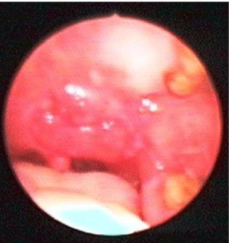
A 48-year-old male patient presented to the Department of Otorhinolaryngology, All India Institute of Medical Sciences, New Delhi, with the chief complaint of a firm 3x4 cm ulcerated lesion over left side of the palate, at the junction of soft and hard palate reaching midline (Figure 1) for last 6 months. It was not associated with pain, bleeding, loosening of tooth, nasal regurgitation, change in voice, breathing difficulty or trismus. Patient had been a chronic smoker for last 20 years, smoking 20-25 cigarettes/day. He had hypertension and diabetes mellitus which were controlled on oral medications. Contrast enhanced CT of head and neck showed a superficial mucosal lesion over left posterior part of palate, predominantly soft palate reaching up to midline, with no associated lymphadenopathy or underlying bone erosion indicating a benign pathology. Incisional biopsy from the lesion showed a malignant spindle cell tumor in the sub-epithelium.
Based on the clinical and histopathological findings, the patient was taken up for surgery in the form of wide local excision. A 3x4 cm tumor was present over the left side of soft palate extending from superior pole of left tonsil laterally to uvula medially. Wide local excision was done with adequate margin all around and reconstructed with palatal mucosal rotation flap based on right sided greater palatine artery.
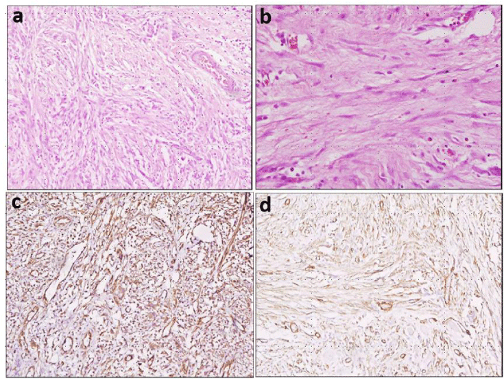
The gross examination of the specimen showed a 3x3x1 cm tumor with ulceration of the overlying mucosa. The histopathological examination (Figure 2) revealed an infiltrative spindle cell tumor in the sub-epithelium with erosion of the overlying epithelium. The individual cells showed eosinophilic cytoplasm, oval nuclei with mild nuclear pleomorphism and occasional mitosis (1-2/10 hpf) (Figure 2 a,b) . No areas of necrosis were identified. The tumor cells were immunopositive for vimentin (Figure 2c) and Smooth Muscle Actin (SMA) (Figure 2d) , while negative for S100, Cytokeratin (CK), ALK, desmin, myogenin, CD 34 and p40. The differential diagnoses considered were low grade myofibroblastic sarcoma, Nodular Fasciitis (NF), sarcomatoid carcinoma, neurofibrosarcoma and Inflammatory Myofibroblastic Tumor (IMT). Since the tumor was infiltrative and lacked the loose feathery appearance, nodular fasciitis was ruled out. On account of immunonegativity for pancytokeratin, p40, S100 and ALK, the other differential diagnoses except LGMS were also excluded (Table 1). Thus the final histopathological diagnosis of low grade myofibroblastic sarcoma was proffered. The patient was sequentially followed up at monthly intervals and was free of tumor till the last follow up (20 months).
Discussion
Myofibroblast is an essential constituent of granulation tissue and tumor stroma, however, uninhibited proliferation of these cells may result in tumorigenesis. LGMS was a controversial entity till 2002, when it was first classified as a distinct neoplasm in the World Health Organization (WHO) classification of soft tissue tumors [3]. WHO defined it as a distinct atypical myofibroblastic tumor often with fibromatosis-like features and a strong predilection for the head and neck. Intermediate and high grade myofibroblastic sarcomas have also been documented in the scientific literature [4]. High-grade myofibroblastic sarcomas, also known as pleomorphic sarcomas are composed of atypical spindle, polygonal and giant cells with myofibroblastic differentiation and high mitotic rates [5].
LGMS generally occurs in adulthood with a mean age of 40.7 years and a male to female ratio of 4:3 [6]. The most frequently affected sites are oral cavity (especially the tongue), followed by limbs, trunk, or abdominal and pelvic cavities [3,7]. Although head and neck is the commonest site for LGMS, less than 40 cases of this region have been reported till date [5]. To the best of our knowledge, the index case is the fourth case occurring in the palate.
On microscopy LGMS is composed of interlacing fascicles of slender spindle cells with scant to moderate amount of eosinophilic to amphophilic cytoplasm and fusiform nuclei. There is mild nuclear pleomorphism and a low mitotic rate. The tumor cells are immunopositive for vimentin, smooth muscle actin, muscle-specific actin, calponin and fibronectin, rarely immunopositive for desmin, and immunonegative for laminin, type IV collagen, S-100, EMA, cytokeratin or CD34 [8]. The main differential diagnoses of LGMS include Nodular Fasciitis (NF) while others are Leiomyosarcoma (LMS), Inflammatory Myofibroblastic Tumor (IMT) and fibromatosis (Table 1).
The recurrence rate is highest in Sino-nasal tumours, around 38.2%, followed by jawbone, and deep tissue space, and it increases from 21.4% to 46.2% as the tumor size gets above 3 cm [8]. Early detection is difficult owing to the indolent course of the disease, apparent asymptomatic nature of the tumor, more so when they are deep-seated. LGMS is known to cause local recurrences more often than distant metastasis [9].
Surgery still remains the mainstay treatment modality and the role of chemo-radiation is debatable [9]. Following 6 months post-surgery, our patient is still free of disease.
To conclude, LGMS is a rare tumor with a limited malignant potential. Since the clinical picture is equivocal and radiological evaluation is frequently on-specific, a detailed histo-pathological examination is sine-qua-non to reach an accurate diagnosis. Though due to paucity of cases the exact treatment protocol for LGMS has not been established, but surgical excision with adequate clearance has resulted in desirable treatment response. Larger case series and more case reports would be pivotal in determining the exact biological behaviour, pathogenesis, prognostic indicators and ideal treatment modality for this enigmatic entity.
- Gabbiani G, Ryan GB, Majno G. Presence of modified fibroblasts in granulation tissue and their possible role in wound contraction. Esperientia. 1971: 27: 549-550. https://bit.ly/3juYGTB
- Mentzel T, Fletcher JA. Low grade myofibroblastic sarcoma. In: Fletcher CD, Unni KK, Mertens F, ed. Pathology and genetics of tumours of soft tissue and bone. Lyon: IARC Press. 2002: 94-95.
- Montgomery E, Goldblum JR, Fisher C. Myofibrosarcoma: A clinicopathologic study. Am J Surg Pathol. 2001; 25: 219-228. DOI: 10.1097/00000478-200112000-00005
- Bisceglia M, Tricarico N, Minenna Pet al. Myofibrosarcoma of the upper jawbones: A clinicopathologic and ultrastructural study of two cases. Ultrastruct Pathol. 2001; 25: 385-397. DOI: 10.1080/019131201317101261
- Fisher C. Myofibroblastic malignancies.Adv Anat Pathol. 2004; 11: 190-201. DOI: 10.1097/01.pap.0000131773.16130.aa
- Yamada T, Yoshimura T, Kitamura N, Sasabe E, Ohno S, Yamamoto T. Low-grade myofibroblastic sarcoma of the palate. Int J Oral Sci. 2012; 4: 170-173. DOI: 10.1038/ijos.2012.49
- FujiwaraM, Yuba Y, Wada A, Izumi Imaoka, Noriaki Shintaku, Setsuko Miyanishi. Myofibrosarcoma of the nasal bone.AmJOtolaryngol. 2005; 26: 265-267. DOI: 10.1016/j.amjoto.2004.11.017
- Demarosi F, Bay A, Moneghini L et al. Low-grade myofibroblastic sarcoma of the oral cavity. Oral Surg Oral Med Oral Pathol Oral Radiol Endod. 2009; 108: 248-254. DOI: https://doi.org/10.1016/j.tripleo.2009.03.031
- Niedzielska I, Janic T, Mrowiec B. Low-grade myofibroblastic sarcoma of the mandible: A case report. J Med Case Reports. 2009; 10: 8458. DOI: 10.4076/1752-1947-3-8458


Sign up for Article Alerts