Assessment of Serum Interleukin 23, 17 Level and its Relation to Disease Control in Psoriasis, Atopic Dermatitis and Lichen Planus: A Serological Study
Ahmed Aladl1*, Al-Sadat Mosbeh1 and Sameh Salama2
1Department of Dermatology, Faculty of Medicine, Al-Azhar University, Cairo, Egypt2Department of Clinical Pathology, Faculty of Medicine, Al-Azhar University, Cairo, Egypt
*Address for Correspondence: Ahmed Aladl, Department of Dermatology, Faculty of Medicine, Al-Azhar University, Cairo, Egypt, ORCID ID: orcid.org/0000-0002-8328-4947; E-mail: draladl80@gmail.com
Submitted: 21 June 2020; Approved: 30 June 2020; Published: 02 July 2020
Citation this article: Aladl A, Mosbeh AS, Salama S. Assessment of Serum Interleukin 23, 17 Level and its Relation to Disease Control in Psoriasis, Atopic Dermatitis and Lichen Planus: A Serological Study. Sci J Clin Res Dermatol. 2020;5(1): 003-011.
Copyright: © 2020 Aladl A, et al. This is an open access article distributed under the Creative Commons Attribution License, which permits unrestricted use, distribution, and reproduction in any medium, provided the original work is properly cited
Download Fulltext PDF
Psoriasis, Lichen Planus (LP), and atopic dermatitis are common chronic inflammatory skin diseases. The exact pathogenesis of these diseases until now is not fully determined. They may result from complex interactions of innate and adaptive immune responses based on a strong genetic predisposition and triggered by environmental factors.
Although it is becoming evident that many Th1 diseases including psoriasis have a strong IL17 signal, the importance of the Th17 cells in AD and lichen planus is still unclear, leading us to investigate its role in these diseases, as compared with psoriasis. This study aimed at the detection of serum interleukin 23 and interleukin 17 in psoriasis, lichen planus and atopic dermatitis, and whether these interleukins could be candidate biomarkers of the diseases activity.
A total of sixty adult patients were included in this study; they were divided into 3 groups, 20 patients each, of psoriasis, atopic dermatitis and lichen planus (13 patients with combined cutaneous and oral lp and 7 patients with cutaneous lp only), 8 of lichen planus patients were HCV positive. In addition to twenty healthy subjects taken as control. The activity of the diseases was estimated at the time of examination using specific activity index for each disease. The serum levels of IL23 and IL17A had been estimated by ELISA. It has been found that these diseases had significantly higher serum levels of IL17A and IL23 than the control group. The serum levels of both interleukins were higher in psoriasis compared with lichen planus and atopic dermatitis, but the difference between psoriasis and lichen planus was not statistically significant. There was no correlation between the serum levels of IL23, IL17 and the clinical severity of the three diseases. On the other hand, a significant positive correlation between IL23 and IL17was found in the three groups of patients.
In conclusion, our findings demonstrated that IL23 and IL17 may play a role in the pathogenesis of the three diseases but not the maestro in controlling these diseases and cannot be used as a marker of their severity. On the other hand, the significant positive correlation between IL23 and IL17in the three diseases confirm that IL23 is important for IL17 production and suggest that the factors affect IL23 will be reflected on IL17 levels.
Introduction
Inflammatory skin diseases involve a very wide range of skin diseases as psoriasis, atopic dermatitis and lichen planus. These diseases are multifactorial, with complex interactions of immune responses based on a strong genetic predisposition and triggered by environmental factors [1].
Interleukin (IL)-23 is a heterodimeric cytokine closely related to IL-12. IL-23 is produced by sentinel dendritic cells and macrophages within a few hours after exposure to lipopolysaccharide and other microbial products [2]. If the IL-23/IL-17 immune pathway becomes dysregulated, there is a danger of breaking tolerance to‘self’ tissues antigens, leading to severe autoimmune diseases and chronic inflammation [3].
Psoriasis is currently accepted as a T-helper (Th) 1⁄Th17 inflammatory disease, and it is therefore associated with an increase of Th1 and Th17 cytokines. These cytokines induce the pathological features of psoriasis as epidermal hyperplasia, hypogranulosis, acanthosis and dermal inflammation [4]. Moreover, some of the Th17 pathway-related genes, IL-23A subunit, IL-23R, IL23B subunit, have been identified as psoriasis susceptibility genes. Responses to tumor necrosis factor (TNF) a-blocking therapy and narrow-band ultraviolet B light therapy are correlated with the suppression of Th17 pathway [5].
Lichen planus is a mucocutaneous inflammatory disease characterized by a T-cell mediated immune response against epithelial cells, causing epithelial cell damage and subepithelial band-like infiltration of T lymphocytes. The mechanisms involved in this disease remain unclear [6].
Th2 cells are important contributors to mucosal host defense, and IL-22 is central to host protection against bacterial infections at barrier sites [7]. Recent studies revealed that IL-23 is required for IL-22 production, and IL- 23 is also regarded as a pivotal cytokine for the pathogenesis of inflammatory and autoimmune diseases so IL-23 may also play roles in the development and maintenance of cutaneous and oral LP [8].
Aim of the Work
The aim of the present study was to detect the role of Interleukin 23 and interleukin17 in the pathogenesis of psoriasis, atopic dermatitis and lichen planus.
Subjects and Methods
Ethical considerations
All participants signed a written informed consent and filled a written survey including demographic and clinical data.
Study setting
The study was carried out in the Department of Dermatology and Venereology, Al-Hussain University Hospital during the period from January 2018 to December 2019.The ELISA test was done at the Clinical Pathology Department in the same Hospital.
Study type
Case control study.
Sampling
Sample type: Systematic random sample
Setting: Out-patient clinic of Dermatology & Venereology Department, Al-Hussain University Hospital during the period from January 2018 to December 2019.
Sample selection: The study included 60 Patients. They were assigned to three groups.
The first group included 20 patients with psoriasis vulgaris, the severity of which was measured by using Psoriasis Area and Severity Index (PASI) according to the method described by Robinson (2012) (9) (Figure 1).
PASI calculation
A representative area of psoriasis is selected for each body region. The intensity of redness, thickness and scaling of the psoriasis is assessed as none (0), mild (1), moderate (2), severe (3) or very severe (4).
Calculation for intensity
The three intensity scores are added up for each of the four body regions to give subtotals A1, A2, A3, A4.
Each subtotal is multiplied by the body surface area represented by that region.
• A1 x 0.1 gives B1
• A2 x 0.2 gives B2
• A3 x 0.3 gives B3
• A4 x 0.4 gives B4
Area
The percentage area affected by psoriasis is evaluated in the four regions of the body. In each region, the area is expressed as nil (0), 1-9% (1), 13-29% (2), 30-49% (3), 50-69% (4), 70-89% (5) or 90-100% (6).
• Head and neck
• Upper limbs
• Trunk
• Lower limbs
Calculations for area
Each of the body area scores is multiplied by the area affected.
• B1 x (0 to 6)= C1
• B2 x (0 to 6)= C2
• B3 x (0 to 6)= C3
• B4 x (0 to 6)= C4
Total score
The PASI score is C1 + C2 + C3 + C4.
The second group included 20 patients with atopic dermatitis, the severity was evaluated by the SCORING AD index (SCORAD). The SCORAD index consists of the interpretation of the extent of the disorder, the intensity, that is, composed of six items (erythema, edema/papules, effect of scratching, oozing/crust formation, lichenification and dryness) and two subjective symptoms (itch and sleeplessness). The maximum score is 103 points Marjolein, et al. [10].
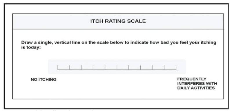
The third group included 20 patients with lichen planus, the severity of the disease was evaluated by visual analogue scale (VAS). VAS is usually a horizontal line, in which the left end marked (0) as no pruritis or pain and the right end (10) marked as worst imaginable pruritis or pain, anchored by word descriptors at each end, as illustrated in figure 1. The patients mark on the line the point that they feel represents their perception of their current state
All the patients in each group were diagnosed clinically and their disease severity was measured by the same physician. None of the patients received any treatment for at least 1 month prior to inclusion in the study. Different clinical variables, including age, sex, disease duration, type and severity were evaluated for all patients before enrollment into the study.
Exclusion criteria
Patients presenting with other skin diseases, diabetes mellitus, inflammatory, autoimmune, cardiovascular or kidney diseases were excluded from the study.
Twenty age and sex matched healthy controls were recruited from health care personnel, medical students and patients present at the outpatient clinic.
Methods
Sample collection
Three ml venous blood samples were collected in sterile plane tube and were allowed to stand for 30 minutes at room temperature then centrifuged at 300 g for 5 minutes. Sera immediately separated and stored at -20 C until the time of analysis.
Cytokine detection
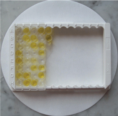
IL17A detection: The Koma biotech inc. Human IL-17A ELISA (K0331207) kit was used according to the manufacturer’s instructions as follow:
'1- Add 200ul of washing solution to each well. Aspirate the wells to remove liquid and wash the plate 3 times using 300ul of washing solution per well. After the last wash, invert plate to remove residual solution and blot on paper towel.
2- Add 100ul of sample to each well in duplicate. Cover with the plate sealer. Incubate at room temperature for at least 2 hours.
3- Aspirate the wells to remove liquid and wash the plate 4 times like as step1.
4- Add 100ul of the diluted detection antibody per well. Cover with the plate sealer. Incubate at room temperature for 2 hours.
5- Aspirate and wash plate 4 times like as step 1.
6- Add 100ul of the diluted Color Development Enzyme (1:20 dilute) per well. Cover with the plate sealer. Incubate 30 minutes at room temperature.
7- Aspirate and wash plate 4 times like as step 1.
8- Add 100ul of color development solution to each well. Incubate at room temperature for a proper color development (12-22 minutes). To stop the color reaction, Add 100ul of the stop solution to each well.
9- Using a microtiter plate reader, read the plate at 450 nm wavelength.
IL23 detection: The eBioscience Human IL-23 ELISA (BMS2023/3 and BMS2023/3TEN) kit was used according to the manufacturer’s instructions as follow:
a. Wash the microwell strips twice with approximately 400 μl Wash Buffer per well with thorough aspiration of microwell contents between washes. Allow the Wash Buffer to sit in the wells for about 10 – 15 seconds before aspiration. After the last wash step, empty wells and tap microwell strips on absorbent pad or paper towel to remove excess Wash Buffer. Use the microwell strips immediately after washing.
b. Add 100 μl of Sample Diluent in duplicate to all standard wells. (concentration of standard 1, S1 = 2000 pg/ml). Continue this procedure 6 times, creating two rows of human IL-23 standard dilutions ranging from 2000.0 to 15.6 pg/ml.
Discard 100 μl of the contents from the last microwells used.
c. Add 100 μl of Sample Diluent in duplicate to the blank wells.
d. Add 50 μl of Sample Diluent to the sample wells.
e. Add 50 μl of each sample in duplicate to the sample wells.
f. Cover with an adhesive film and incubate at room temperature (18° to 25°C) for 2 hours.
g. Prepare Biotin-Conjugate.
h. Remove adhesive film and empty wells. Wash microwell strips 5 times according to point b.
i. Add 100 μl of Biotin-Conjugate to all wells.
j. Cover with an adhesive film and incubate at room temperature (18° to 25°C) for 1 hour.
k. Prepare Avidin-HRP.
l. Remove adhesive film and empty wells. Wash microwell strips 5 times according to point b. of the test protocol. Proceed immediately to the next step.
m. Add 100 μl of diluted Avidin-HRP to all wells.
n. Cover with an adhesive film and incubate at room temperature (18° to 25°C) for 30 minutes.
o. Remove adhesive film and empty wells. Wash microwell strips 5 times according to point b.
p. Pipette 100 μl of TMB Substrate Solution to all wells.
q. Incubate the microwell strips at room temperature (18° to 25°C) for about 15 minutes. Avoid direct exposure to intense light.
The color development on the plate should be monitored and the substrate reaction stopped before positive wells are no longer properly recordable.
r. Stop the enzyme reaction by quickly pipetting 100 μl of Stop Solution into each well. It is important that the Stop Solution is spread quickly and uniformly throughout the microwells to completely inactivate the enzyme. Results must be read immediately after the Stop Solution is added or within one hour if the microwell strips are stored at 2 - 8°C in the dark.
s. Read absorbance of each microwell on a spectro-photometer using 450 nm as the primary wavelength (optionally 620 nm as the reference wavelength; 610 nm to 650 nm is acceptable). Blank the plate reader according to the manufacturer’s instructions by using the blank wells. Determine the absorbance of both the samples and the standards.
Statistical analysis
Data were checked, entered and analyzed by using SPSS (version 19). Data were represented as mean ± SD for quantitative variables. Number and percentage were used for categorical variables. Chi–square (X2) or Fisher exact test were used when appropriate. P<0.05 was considered statistically significant. T-test was used to compare means. Kruskal Wallis test was used for the nonparametric variables.
Results
Characteristics of the studied cases
This study included 60 patients; they were assigned to three groups (Table 1 & 2).
The first group included 20 psoriatic patients (14 males and 6 females), their ages ranged from 21-63 years with a mean of 43.4 and SD 10.2. Their PASI score ranged from 3.40 to 40 with an average of 14 ± 10.9. Their disease duration ranged from 3-30 years. They were classified in to sever (9 patients, PASI more than 20), moderate (7 patients PASI less than 20) and mild (4 patients, PASI less than 10) (Table 2).
The second group included 20 patients (13 male and 7 female) with atopic dermatitis. Their ages ranged from 17-58 years with a mean of 39.4 and SD of 10.3. Their disease duration ranged from 2-13 years. The disease severity was evaluated by the SCORING AD index (SCORAD). Their SCORAD ranged from 20 to 61with an average of 36.5 ± 12.9. They were classified in to sever (6 patients, SCORAD more than 40), moderate (11 patients, SCORAD ranged from 15-40) and mild (3 patients, SCORAD less than 15).
The third group, Included 20 patients (8 male and 12 female) with lichen planus (13 patients, 9 females and 4 males, with combined cutaneous and oral lp) and (7 patients, 3 females and 4 males, with cutaneous lp only) their ages ranged from 17-68 years with a mean of 40.1 and SD of 14.3. Their disease duration ranged from 1 month to 17 years. Eight patients was HCV positive. The disease severity was evaluated by Visual Analogue Scales (VAS). Their VAS ranged from 0 to 9 with an average of 6.1 ± 1.9. They were classified in to sever (6 patients, VAS more7), moderate (10patients, VAS 4-6) and mild (4 patients, VAS less than 4).
The Control group Included 20 healthy controls (12 male and 8 female). Their ages ranged from 18-70 years with a mean of 41.4 and SD of 6.4.
There were no statistically significant differences between the studied groups as regarding age and sex.
Serum IL23 and IL17 levels among the studied groups
The serum levels of IL23 and IL17A of the three diseases were significantly higher than the control group. The mean serum level of IL23 of the control group was (19 pg/ml), while it was (41.2 pg/ml) in psoriasis, (27 pg/ml) in AD and (37.5pg/ml) in lichen planus. The mean serum level of IL17A was (7.4 +5.3 pg/ml) in control group, (24.8 + 4.1 pg/ml) in psoriasis, (20.5 + 3.8 pg/ml) in AD and (23.3 + 2.17 pg/ml) in lichen planus (Table 3).
When comparing the serum levels of IL23 and IL17 in the three diseases , their levels were significantly higher in psoriasis and lichen planus than atopic dermatitis, while the difference of the serum levels of IL23 and IL17 between psoriasis and lichen planus was not statistically significant (Table 4). As regard lichen planus patients, the serum levels of IL23 and IL-17 were significantly higher in patients with both cutaneous and oral LP compared with patients of cutaneous LP only.
There was no statistically significant difference in serum IL-17 and IL23 levels as regards gender, age and disease duration (Table 5,6).
An analysis of the correlations between the serum levels of IL23 and IL17 was conducted, a significant positive correlation between the IL23 and IL17 values was found in the three groups of patients (p < 0.05)
Correlation between serum interleukins levels and disease severity of the studied patients
No significant correlation between the serum levels of IL23, IL17 and the severity of either psoriasis, atopic dermatitis or lichen planus assessed by PASI, SCORAD and VAS respectively (Table 7,8 & 9).
Comparison between serum interleukin levels of patients with cutaneous LP only and those with both cutaneous and oral LP.
The serum levels of IL23 and IL-17 were significantly higher in patients with both cutaneous and oral LP compared with patients of cutaneous LP only (Table 10).
Discussion
Psoriasis, lichen planus and atopic dermatitis are chronic relapsing inflammatory skin diseases. They may lead to marked disability affecting the physical and emotional wellbeing of patients. The combined cost of long-term therapy and social burden of this disease have a major impact not only on the patient, but also on health care systems. Recent studies indicate that severe, long-standing psoriasis is associated with co-morbidities, as well as a shorter lifespan due to cardiovascular mortality [11,13].
These diseases are multifactorial, with complex interactions of innate and adaptive immune responses based on a strong genetic predisposition and triggered by environmental factors. The exact pathogenesis of these diseases until now is not fully determined [14].
The interleukin-17 (IL17) cytokines are emerging as key players in immune responses. The first member to be identified, IL17A. The other members, IL 17B to IL17F were subsequently identified based on their homology to IL17A. Importantly, IL17A dysregulation favors chronic inflammation, autoimmune disease and tumor development [3].
IL17A upregulates the expression of multiple neutrophil chemo-attracting chemokines on keratinocytes, including CXCL1, CXCL3, CXCL5, CXCL6 and CXCL8 and also induces expression of antimicrobial peptides on keratinocytes, as Human Beta Defensin 2, S100A7, S100A8 and S100A9 [15]. IL17A acts on a variety of other cell types besides keratinocytes, including myeloid dendritic cells (mDCs), macrophages, fibroblasts and endothelial cells, leading to production of different types of cytokines and chemokines [16].
In the current study, we evaluated IL17A and IL23 serum levels in psoriasis, cutaneous lichen planus and atopic dermatitis using ELISA technique. The results revealed that these diseases had significantly higher serum levels of IL17A and IL23 than the control group. The serum levels of both interleukins were higher in psoriasis compared with lichen planus and atopic dermatitis, however the difference between psoriasis and lichen planus was not statistically significant.
There was no statistically significant difference in serum IL17 and IL23 levels as regards gender, age and disease duration. In addition, an analysis of the correlations between the serum levels of IL23 and IL17 was conducted, a significant positive correlation between IL23 and IL17 was found in the three groups of patients (p < 0.05) in accordance with Stoma, et al. [17] who reported positive correlation of the serum levels of IL23 with IL17 in psoriatic patients.
As regard psoriasis, several recent studies have identified high levels of IL17 and IL23 expression in skin lesions and in serum, that positively correlated with disease severity measured by PASI score [18]. In the current study, the serum levels of IL23 and IL17 in psoriasis were higher than the control group, however, there was no significant correlation between the serum levels of IL23or IL17 and psoriasis severity measured by PASI score.
On the other hand, in previous study they could not detect IL23 or IL17 in the serum of psoriatic patients, while reported higher IL22serum levels than the controls that positively correlated with PASI score. So, it was suggested that IL23 and IL17 might be involved in the very early phase of psoriasis development or be present only in the lesioned skin.
Similarly, Stoma et al. reported absence of significant differences in the serum IL17 and IL23 levels between the psoriatic patients and control group, however, a significantly higher level of IL22 was observed in psoriatic patients compared with the healthy controls that positively correlated to psoriasis severity measured by PASI. According to these findings Stoma, et al. [17] suggested that TH17 might be more important in the very early phase of psoriasis development while Th22 role is more significant in the other phases of psoriasis. This may raise the question about the exact role of IL-17 and IL-23 in the pathogenesis of psoriasis.
The diagnosis of AD remains clinical, because there is currently no reliable biomarker that can distinguish the disease from other entities. Discovery of new T-lymphocyte subsets and novel cytokines and chemokines have generated potential biomarkers. These include serum levels of macrophage-derived chemoattractant (MDC), interleukins (IL)-12, -16,-18,-22 and -31. But to date, none have shown reliable sensitivity or specificity for AD to support general clinical use for diagnosis or monitoring [20].
This study revealed higher serum levels of both IL23 and IL17 in atopic patients compared with the control group in accordance with Yassky et al. [21] who reported higher numbers of TH17 cells in the peripheral blood and in skin lesions of patients with AD compared with healthy control.
On the other hand, IL23 and IL17 serum levels in AD were lower than in psoriasis. This was in agreement with Eyerich et al. [22] who reported lower levels of IL-17 in patients with AD compared with that seen in patients with psoriasis and explained their results by the fact that the cytokines produced by TH2 cells (IL-4 and IL-13) inhibit IL-17 production from T cells, which further reduces TH17 cell activity.
Another evidence that supports the more important role of IL-23 in the pathogenesis of psoriasis as compared with AD was shown by Boguniewicz, et al. [23] who detected higher levels of IL23 in the skin lesions of psoriasis than those detected in AD lesions.
The correlation between the serum levels of IL23, IL17 and AD severity measured by SCORAD index was insignificant. This was not in agreement with the results reported by Cuparri, et al. [24] who have found significant positive correlation between the serum IL-17 levels and the disease severity. This may be explained by the different age group in their study (children) versus adults in our study.
Some studies established Th22 cells as an important component of mucosal antimicrobial host defence, IL-22 is central to host protection against bacterial infections at barrier sites, and IL-22 production was strictly IL-23 dependent. The increased levels of IL-22 and IL-23 in oral LP might be due to action of the oral microorganisms and tissue antigens of uncertain origin that initiate and maintain the inflammatory response [25,26].
Other mechanisms have been implicated in the pathogenesis of LP including mast cell degranulation and matrix metalloproteinase (MMP) activation. IL-17 has been shown to up regulate or synergize with local inflammatory mediators and promote extra cellular matrix injury through stimulation of production of MMP [6].
There are few numbers of studies that evaluate the serum level of IL23 and IL17 in cutaneous lichen planus. The results of the current study demonstrated higher levels of IL23 and IL17 in LP than the control group in accordance with previous studies [26,27], Chen, et al. [26]. The difference of the serum levels of IL23 and IL17 between psoriasis and lichen planus was not statistically significant but IL23 and IL17 were significantly higher in LP than AD. All these finding support the possible role of IL23 and IL17 in the pathogenesis of LP.
More focus on LP revealed that the patients who suffered from both cutaneous and oral LP had higher serum levels of IL23 and IL-17 than those presenting with cutaneous LP only. This was in accordance with the results reported by Chen, et al. [26] who have found that IL23 expression level in the oral LP lesions was significantly higher than cutaneous LP.
No significant correlation was found between the serum levels of IL23 and IL17 and the severity of lichen planus as measured by VAS. Also, there was no significant difference of the serum levels of IL23 and IL17 between HCV positive and HCV negative patients. This finding was also observed by Shaker and Hassan (2012) who have reported absence of significant difference of the serum levels of IL17 between HCV positive and HCV negative patients [27,28].
We found that IL23 and IL17 may play a role in the pathogenesis of the three diseases but not the maestro in controlling these diseases and cannot be used as a marker of their severity. On the other hand, the significant positive correlation between IL23 and IL17in the three diseases confirm that IL23 is important for IL17 production and suggest that the factors affect IL23 will be reflected on IL17 levels.
Conclusion
In conclusion, our findings demonstrated that IL23 and IL17 may play a role in the pathogenesis of the three diseases but not the maestro in controlling these diseases and cannot be used as a marker of their severity. On the other hand, the significant positive correlation between IL23 and IL17in the three diseases confirm that IL23 is important for IL17 production and suggest that the factors affect IL23 will be reflected on IL17 levels.
- Irvine AD, McLeanm WHI. Breaking the (un)sound barrier filaggrin is a major gene for atopic dermatitis. J Invest Dermatol. 2006; 126: 1200-1202. DOI: 10.1038/sj.jid.5700365
- Langrish C, McKenzie B, Wilson N, de Waal Malefyt R, Kastelein RA, Cua DJ, et al. IL-12 and IL-23: master regulators of innate and adaptive immunity. Immunnol Rev. 2005; 202: 96-105. 10.1111/j.0105-2896.2004.00214.x
- Hu Y, Shen F, Crellin NK, Ouyang W. The IL-17 pathway as a major therapeutic target in autoimmune diseases. Ann N Y Acad Sci. 2011; 1217: 60-76. DOI: 10.1111/j.1749-6632.2010.05825.x
- Zheng Y, Danilenko DM, Valdez P, Kasman I, Anderson JE, Wu J, et al. Interleukin-22, a T(H)17 cytokine, mediates IL-23-induced dermal inflammation and acanthosis. Nature. 2007; 445: 648-651. DOI: 10.1038/nature05505
- Chiricozzi A, Nograles KE, Johnson-Huang LM, Duculan JF, Cardinale I, Bonifacio KM, et al. IL-17 Induces an Expanded Range of Downstream Genes in reconstituted Human Epidermis Model. PLoS One. 2014; 9: e90284. DOI: 10.1371/journal.pone.0090284
- Baccaglini L, Thongprasom K, Carrozzo M, Bigby M. Urban legends series: Lichen planus. Oral Dis. 2013; 19: 128-143. DOI: 10.1111/j.1601-0825.2012.01953.x
- Dalamaga M, Papadavid E. Adipocytokines and psoriasis: Insights into mechanisms linking obesity and inflammation to psoriasis. World J Dermatol. 2013; 4: 27-31. DOI: 10.5314/wjd.v2.i4.27
- Croxford A, Mair F, Becher B. IL-23: One cytokine in control of autoimmunity. Eur J Immunol. 2012; 42: 2263-2273. DOI: 10.1002/eji.201242598
- Robinson A. Psoriasis severity scoring. 2012. https://bit.ly/3gfeVSG
- Willemsen MG, van Valburg RWC, Dirven-Meijer PC, Oranje AP, van der Wouden JC, Moed H. Determining the severity of atopic dermatitis in children presenting in general practice: An easy and fast method. Dermatol Res Pract. 2009; 2009: 357046. DOI: 10.1155/2009/357046
- Yu HS, Angkasekwinai P, Chang SH, Chung Y, Dong C. Protease allergens induce the expression of IL-25 via p38 MAPK pathway. J Korean Med Sci. 2010; 25: 829-834. DOI: 10.3346/jkms.2010.25.6.829
- Kikly K, Liu L, Na S, Sedgwick JD. The IL-23/Th(17) axis: Therapeutic targets for autoimmune inflammation. Curr Opin Immunol. 2006; 18: 670-675. DOI: 10.1016/j.coi.2006.09.008
- Nograles KE, Zaba LC, Shemer A, Duculan JF, Cardinale I, Kikuchi T, et al. IL-22-producing “T22” T cells account for upregulated IL-22 in atopic dermatitis despite reduced IL-17-producing TH17 T cells. J Allergy Clin Immunol. 2011; 123: 1244-1252. DOI: 10.1016/j.jaci.2009.03.041
- Li X, Li J, Yang Y, Hou R, Liu R, Zhao X, et al. Differential gene expression in peripheral blood T cells from patients with psoriasis, lichen planus, and atopic dermatitis. J Am Acad Dermatol. 2013; 65: 6-30. DOI: 10.1016/j.jaad.2013.06.030
- Kagami S, Rizzo HL, Lee JJ, Koguchi Y, Blauvelt A. Circulating Th17, Th22, and Th1 cells are increased in psoriasis. J Invest Dermatol. 2010; 130: 1373-1383. DOI: 10.1038/jid.2009.399
- Nograles K, Davidovici B and James G. IL-23/TH17 Pathway in psoriasis and inflammatory skin diseases. S. Jiang (ed.). T H17 Cells in Health and Disease.
- Stoma A, Joanna B, Magorzata K, et al. Serum Levels of Selected Th17 and Th22 Cytokines in Psoriatic Patients. Dis Markers. 2013; 35: 625-631. DOI: 10.1155/2013/856056.
- Dowlatshahi EA, van der voort EAM, Arends LR, Nijsten T. Markers of systemic inflammation in psoriasis: a systematic review and meta-analysis. Br J Dermatol. 2013; 169: 266-282. DOI: 10.1111/bjd.12355
- Nestle FO, Kaplan DH, Barker J. Psoriasis. N Engl J Med. 2011; 361: 496-509.
- Eichenfield LF, Tom LW, Chamlin SL, Feldman SR, Hanifin JM, Simpson EL, et al. Guidelines of care for the management of atopic dermatitis: Section 1. Diagnosis and Assessment of Atopic Dermatitis. J Am Acad Dermatol. 2014; 70: 338-351. DOI: 10.1016/j.jaad.2013.10.010
- Yassky EG, Lowes MA, Duculan JF, Zaba LC, Cardinale I, Nograles KE, et al. Low expression of the IL-23/Th17 pathway in atopic dermatitis compared to psoriasis. J Immunol. 2008; 181: 7420-7427. DOI: 10.4049/jimmunol.181.10.7420
- Eyerich K, Pennino D, Scarponi C, Hoffmann CT, Albanesi C, Ring J, et al. IL-17in atopic eczema: linking allergen-specific adaptive and microbial-triggered innate immune response. J Allergy Clin Immunol. 2009; 123: 59-66. https://bit.ly/2C1cSm1
- Boguniewicz M, Schmid-Grendelmeier P, Leung D. Clinical pearls: atopic dermatitis. J Allergy Clin Immunol. 2006; 118: 40-43.
- Cuppari E, Mantis J, Salpietro M, et al. Serum IL17 in children with atopic dermatitis. The Child. 2013; 1: 10-24.
- Yassky EG, Nograles KE, Krueger JG. Contrasting pathogenesis of atopic dermatitis and psoriasis -Part II: Immune cell subsets and therapeutic concepts. J Allergy Clin Immunol. 2012; 340: 16-34. DOI: 10.1016/j.jaci.2011.01.054
- Chen J, Feng J, Chen X, Xu H, Zhou Z, Shen X, et al. Immunoexpression of Interleukin-22 and Interleukin-23 in Oral and Cutaneous Lichen Planus Lesions: A Preliminary Study. Mediators Inflamm. 2013; 100: 40-47. DOI: 10.1155/2013/801974
- Wang Y, Angkasekwinai P, Lu N, Voo KS, Arima K, Hippe A, et al. IL-25 augments type 2 immune responses by enhancing the expansion and functions of TSLP-DC-activated Th2 memory cells. J Exp Med. 2013; 204: 1837-1847. DOI: 10.1084/jem.20070406
- Shaker O, Hassan AS. Possible role of interleukin-17 in the pathogenesis of lichen planus. Br J Dermatol. 2012; 166: 1357-1380. DOI: 10.1111/j.1365-2133.2011


Sign up for Article Alerts