Mesenteric Cyst Lymphangioma as an Abdominal Emergency in a Two Year Old Girl: Case Report and Review of Literature
Velmishi V1*, Cekodhima G2, Demrozi A3, Heta S3, Sila S3 and Cullufe P1
1Service of pediatric gastroenterology, Mother Teresa Hospital Tirana, Albania
2Service of histopathology, Mother Teresa Hospital, Tirana, Albania
3Service of pediatric surgery, Mother Teresa Hospital, Tirana, Albania
*Address for Correspondence: Virtut Velmishi, Service of pediatric gastroenterology, Mother Teresa Hospital, Dibra street Nr 372, Tirana, Albania, Tel/Fax: +0035542349233; E-mail: tutimodh@yahoo.com
Submitted: 16 November 2018; Approved: 27 February 2019; Published: 03 March 2019
Citation this article: Velmishi V, Cekodhima G, Demrozi A, Heta S, Sila S, Cullufe P. Mesenteric Cyst Lymphangioma as an Abdominal Emergency in a Two Year Old Girl: Case Report and Review of Literature. Int J Hepatol Gastroenterol. 2019; 5(1): 001-004.
Copyright: © 2019 Velmishi V, et al. This is an open access article distributed under the Creative Commons Attribution License, which permits unrestricted use, distribution, and reproduction in any medium, provided the original work is properly cited
Keywords: Mesenteric Cystic Lymphangioma
Download Fulltext PDF
Mesenteric cystic lymphangioma is an uncommon, slowly growing tumor derivate from lymphatic vessels, which is rarely found as an intra-abdominal masses usually located in small bowel mesentery. A two year old girl was presented in our service because of abdominal pain, recurrent vomiting and back pain. In the regional hospital an ultrasound has revealed a supravesical mass. We repeated an ultrasound followed by abdominal MRI which showed a cyst 12x7 cm without infiltration aspect. We planned surgery within 3 days but this girl was returned in our service a day later because of severe pain and abdominal distention making surgery an emergency. She underwent to intervention and a large cystic formation was removed and send to pathology service. Follow up was unrevealed. The response of pathology was compatible with a mesenteric cyst lymphangioma
Lymphangiomas’ account for 5-6% of all benign tumors in children. 50% involve the head and neck, only 10 % occurring in internal organs. 60% of these masses are present at birth. Abdominal cystic lymphangioma are very uncommon. Almost 90% are detected by the mean age of 2 years, and most occurs in the mesentery of the small bowel. They result from an embryological failure of the lymphatic system; lack of communication between small bowel lymphatic tissue and the main lymphatic vessels during fetal development result in blind cystic lymphatic spaces lined by endothelial layers. Mesenteric cystic lymphangioma frequently affect young children and are usually symptomatic making surgery sometime emergency. The diagnosis is well established by ultrasound, CT, MRI. To prevent recurrence, complete excision of the cyst with or without intestinal resection is mandatory.
Introduction
Abdominal cystic lymphangioma are rare congenital benign malformations of the lymphatic system that are usually located in the small bowel mesentery, followed by Omentum, mesocolon and retroperineum [1]. Abdominal cystic lymphangioma are more frequent in boys the girls (5:2) with a mean age at presentation of 2 years [2]. Clinical features of mesenteric cystic lymphangioma varies from asymptomatic masses to an abdominal emergency. We would like to present the case of a two year-old girl who presented recurrent vomiting and back pain for at least two month complicated in several days with intestinal obstruction as a consequence of a giant mesenteric cyst.
Case Report
A two year old girl was presented in our service because of abdominal pain, recurrent vomiting and back pain.
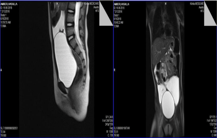
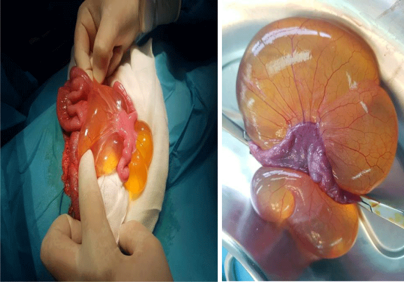
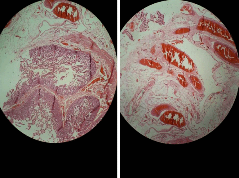
She was the first child of the couple. Pregnancy was normal. It was cesarian birth. Birth weight was 2800g. After birth this girl was nourished with mother and bottle milk. She was vaccinated according to Albanian schedule. Two months ago the little girl complained for frequent vomiting, abdominal and back pain. Her pediatrician considered those symptoms as gastroesophageal reflux. The parents noticed that symptoms are deteriorated and send their girl to regional hospital where an abdominal ultrasound revealed an abdominal mass. At presentation this girl was pale but without visible signs of discomfort. Heart rate was 90/min. Lungs auscultation was normal, both lungs partake uniformly in respiration. Resiratory rate was 30/min. SAT O2=95%. Abdomen was soft and palpable. Liver and spleen were not enlarged. Wheight was 11 kg ( 17% percentile) and height was 84 cm (percentile 25%). Lab studies showed: WBC=12x 103/mm3, RBC=4.51 106/mm3, HgB-11, 4g/dl; HCT-34, 7 MCV-78fL; PLT-275x 103/mm3. In biochemical examinations were noticed a slight hypokalemia. The rest of examinations was normal.We repeated an ultrasound followed by abdominal MRI (Figure 1) which showed a cyst 12x7 cm without infiltration aspect. We planned surgery within 3 days but this girl was returned at our service a day later because of severe pain and abdominal distention making surgery an emergency. Vomiting continued to be the predominant sign. She underwent to intervention and a large cystic formation was removed and send to pathology service (Figure 2a). Follow up was unrevealed. The response of pathology was compatible with a mesenteric cyst lymphangioma (Figure 2b).
Discussion
Lymphangioma account for 5-6% of all benign tumors in children [1-3]. 50% involve the head and neck, only 10 % occurring in internal organs [2] 60% of these masses are present at birth. Abdominal cystic lymphangiomas are very uncommon [4]. Almost 90% are detected by the mean age of 2 years, and most occurs in the mesentery of the small bowel [4]. They result from an embryological failure of the lymphatic system; lack of communication between small bowel lymphatic tissue and the main lymphatic vessels during fetal development result in blind cystic lymphatic spaces lined by endothelial layers. The differential diagnosis mainly includes the other mesenteric cystic lesions such as multicystic mesothelioma, lymphoceles, dermoid cyst, lipomas, teratomas or liposarcomas [5]. We have to discuss some topics according to our case.
Age
In our case the age was 2 year-old equals to the mean age of most cases diagnosed with mesenteric cyst lymphangioma
Sex
According to the literature abdominal cystic lympangiomatosis are more frequent in boys (5:2) whereas our case was a girl.
Image studies
Abdominal ultrasound is the diagnostic procedure of choice [6-9]. On ultrasound, you may see cystic anechoic structures with irregular thin walls often containing septa. On CT scans, they usually appear as a smooth margined unilocular or multilocular cystic mass with homogenous fluid attenuation. The cystic walls show strong enhancement after contrast injection. Magnetic Resonance Imaging (MRI) may be useful for diagnosis but cannot differentiate between dermoid cyst, cystic teratoma, cystic lymphangioma or lymphoceles. It is however, the most useful technique to assess mass extension. The combined use of ultrasonography, CT end MRI is very helpful when there is a high preoperative index of suspicion for abdominal cystic lymphangioma. In our case ultrasonography and RMN was enough for a better orientation before surgery.
Classification
Beahrs et al., [10] classified mesenteric cysts into four groups based on etiology: embryonic or developmental; traumatic or acquired; neoplastic; and infective or degenerative. Recently, a pathological classification is proposed [3]. Type 1 (pedicled) and 2 (sessile) are limited to the mesentery and can be excised completely with or without resection of the involved gut. Type 3 and 4 (multicentric) extend to the retroperitoneum and require complex operations. Based on the contents of the cyst, can be classified into serous, chylous, hemorrhagic and chylolymphatic. In our case as you may see in Figure 2 the cyst was multilocular positioned to the mesenteric side of ileum approximately 50 cm from ileocecal valve. The content was serous.
Histopathology
Mesenteric cyst lymphangioma is characterized by a thin irregular wall covered by endothelium and contains smooth muscle, foam cells and lymphatic tissue. Cystic lymphangioma has a striking resemblance to chylolymphatic mesenteric cyst microscopically. Some authors describe chylolymphatic cyst as a variant of mesenteric cyst [11-13]. The absence of smooth muscle and lymphatic spaces in the wall of the cyst differentiates mesenteric cysts from Cystic lymphangioma. As you may see in Figure 3 our images demonstrate the presence of smooth muscle and lymphatic space which make a certain diagnosis.
Clinical features
Clinical presentation is variable and depend on mass size and location. Although they are usually asymptomatic, they can be complicated with acute intestinal obstruction such as in our case. A painless, soft and moving mass may be seen in examination in 58% of patients [14]. Abdominal swelling, abdominal pain, nausea, vomiting, diarrhea and constipation are the other possible symptoms [15]. Our patient presented recurrent vomiting and abdominal pain. She complained often for back pain which was not a common sign described in literature. We had plan a date for surgery but a day later she presented frequent vomiting and abdominal pain. She was underwent to surgery as an acute abdominal emergency.
Treatment
The main modality of treatment for mesenteric cystic lymphangioma is surgical excision, because their risk of recurrence. During surgery it is often necessary to perform a bowel resection because of the close relation between the cyst and intestinal wall. Laparoscopic management of mesenteric cysts is also being reported [16-19]. In our case the surgeon performed a complete excision of cyst with 15 cm of intestine which was attached very closely.
Conclusion
Mesenteric cystic lymphangioma frequently affect young children and are usually symptomatic making surgery sometime emergency. The diagnosis is well established by ultrasound, CT, MRI. Histopathology plays an important role for an accurate diagnosis. To prevent recurrence, complete excision of the cyst with or without intestinal resection is mandatory.
Consent
Written informed consent was obtained by the parents of our patient.
Authors’ contributions
V.V drafted the manuscript. G.C performed histological examination. A.D, S.H and S.S performed the surgery. P.C review the manuscript. All authors read and approved the manuscript.
- Alqahtani A, Nguyen LT, Flageole H, Shaw K, Laberge JM. 25 years’ experience with lymphangiomas in children. J Pediatr Surg. 1999; 34: 1164-1168. https://goo.gl/2uhrog
- Konen O, Rathaus V, Dlugy E, Freud E, Kessler A, Shapiro M, et al. Childhood abdominal cystic lymphangioma. Pediatr Radiol. 2002; 32: 88-94. https://goo.gl/fgqw4G
- Losanoff JE, Richman BW, El-Sherif A, Rider KD, Jones JW. Mesenteric cystic lymphangioma. J Am Coll Surg. 2003; 196: 598-603. https://goo.gl/nvuoFY
- Goh BK, Tan YM, Ong HS, Chui CH, Ooi LL, Chow PK, et al. Intraabdominal and retroperitoneal lymphangiomas in pediatric and adult patients. World J Surg. 2005; 29: 837-840. https://goo.gl/DR9f7a
- Steyaert H, Guitard J, Moscovici J, Juricic M, Vaysse P, Juskiewenski S. Abdominal cystic lymphangioma in children: benign lesions that can have a proliferative course. J Pediatr Surg. 1996; 31: 677-680.
- Mollitt DL, Ballantine TV, Grosfeld JL. Mesenteric cyst in infancy and childhood. Surgery Gynecol Obsetr. 1978; 147: 182-184. https://goo.gl/y6wiPj
- Kosir MA1, Sonnino RE, Gauderer MW. Pediatric abdominal lymphangiomas: a plea for early recognition. J Pediatr Surg. 1991; 26: 1309-1313. https://goo.gl/CZPejj
- Molander ML, Mortensson W, Udén R. Omental and mesenteric cysts in children. Acta Pediatr Scand. 1982; 71: 227-229. https://goo.gl/dMur5N
- Chou YH, Tiu CM, Lui WY, Chang T. Mesenteric and omental cysts: an ultra-sonographic and clinical study of 15 patients. Gastrointst Radiol. 1991; 16: 311-314. https://goo.gl/kMi7NM
- Beahrs OH, Judd ES Jr, Dockerty MB. Chylous cyst of the abdomen. Surg Clin North Am. 1950; 30: 1081-1096. https://goo.gl/3HFG3c
- Takiff H, Calabria R, Yin L, Stabile BE. Mesenteric cysts and intraabdominal cystic lymphangiomas. Arch Surg. 1985; 120: 1266-1269. https://goo.gl/rKJ6QD
- Bliss DP Jr, Coffin CM, Bower RJ, Stockmann PT, Ternberg JL. Mesenteric cysts in children. Surgery. 1994; 11: 517-577. https://goo.gl/8GTN5W
- Fish JC, Fair WR, Canby JP. Intestinal obstruction in the newborn: an unusual case due to mesenteric cyst. Arch Surg. 1965; 90: 317-318. https://goo.gl/62YC61
- Weeda VB, Booij KA, Aronson DC. Mesenteric cystic lymphangioma: a Congenital and an acquired anomaly? Two cases and review of literature. J Pediatr Surg. 2008; 43: 1206-8. https://goo.gl/dwQg8y
- Egozi EI, Rickets RR. Mesenteric and omental cyst in children. Am Surg. 1997; 63: 287-90. https://goo.gl/aq374V
- Polat C, Yilmaz S, Arikan Y, Mahallesi D, Caddesi KM. Mesenteric cysts. Surg endosc. 2004; 18: 16. https://goo.gl/Xh9Qei
- Theodoridis TD, Zepiridis L, Athanatos D, Tzevelekis F, Kellartzis D, Bontis JN. Laparoscopic management of mesenteric cysts. Surg Endosc. 2003; 17: 2036. https://goo.gl/ZNnC7M
- Bhandarwar AH, Tayade MB, Borisa AD, Kasat GV. Laparoscopic excision of mesenteric cyst of sigmoid mesocolon. J minim Access Surg. 2013; 9: 37-39. https://goo.gl/XwWaf3
- Pampal A, Yagmurlu A. Successful laparoscopic removal of mesenteric and omental cysts in toddler: 3 cases with a literature review. J Pediatr Surg. 2012; 3: e5-8. https://goo.gl/pJ2JLz


Sign up for Article Alerts