The Prevalence and Epidemiology of Ectopic Pregnancies in SMGS: A Tertiary Health Care Hospital in Jammu, India
Supriya Sharma1* and Sumit Sharma2
1Department of Obstetrics and Gynaecology, Government medical college Jammu, Jammu and Kashmir, India
2Department of Radiodiagnosis, Government medical college Srinagar, Jammu and Kashmir, India
*Address for Correspondence: Supriya Sharma, Postgraduate student, Department of Obstetrics and Gynaecology, SMGS Hospital, Government medical college Jammu, Near shiv temple, Narwal bala bye pass Jammu, Jammu and Kashmir 180006, India, Tel: +91-705-126-3330, ORCID ID: http://orcid.org/0000-0002-0729-4780; E-mail: supriyasharma62@yahoo.com
Submitted: 29 August 2019; Approved: 07 September 2020; Published: 10 September 2020
Citation this article: Sharma S, Sharma S. The Prevalence and Epidemiology of Ectopic Pregnancies in SMGS: A Tertiary Health Care Hospital in Jammu, India. Int J Reprod Med Gynecol. 2020;6(1): 025-030.
Copyright: © 2020 Sharma S, et al. This is an open access article distributed under the Creative Commons Attribution License, which permits unrestricted use, distribution, and reproduction in any medium, provided the original work is properly cited
Keywords: Ectopic; Prevalence; Epidemiology
Download Fulltext PDF
Background: Objective of the present study was to evaluate the: prevalence, clinical presentation, USG findings, risk factors and socio-demographic characteristics of ectopic pregnancy
Methods: A retrospective (observational) study. Patients with intrauterine pregnancy were excluded from the study. A total of 282 patients were studied.
Results: Prevalence of ectopic was calculated to be 9.69/1000 pregnancies/year. More common amongst multigravida (77.3%) with mean age of patients being 29.2 ± 5.79 years. Most of the presented as acute (ruptured) ectopic pregnancy (85.1%) with fallopian tubes (95.03%) as the most common site. Right sided ectopics (60.99%) were more common. Symptoms commonly seen amongst these patients were abdominal pain (95.7%), amenorrhoea (85.1%) and bleeding PV (68.44%). No risk factor was identified in 62.06% of the patients and amongst those with risk factors, history of prior abortions followed by D&C (16.31%) was the most common one.
Conclusions: Proper identification of risk factors along with early diagnosis and management of ectopic pregnancy needed to prevent disastrous outcomes.
Introduction
After fertilization, the blastocyst usually implants in the endometrial lining of the uterine cavity. Implantation anywhere else other than the uterine cavity is considered as ectopic pregnancy. It is a major cause of maternal mortality and morbidity all over the world, especially in the developing countries. Ruptured ectopic pregnancy is the leading cause of maternal mortality in the first trimester and accounts for 10-15% of all maternal deaths. It accounts for 3.5-7.1% of all maternal mortality in India [1].
It not only threatens the life of the female, but also has an impact on the future fertility of the women. Nearly 95% of the ectopic pregnancies occur in the fallopian tubes, and out of these 98% occur in the ampullary or isthmic portions of the tube [2]. Other sites seen are ovary, cervix, previous cesarean scar, peritoneal cavity etc.
Over the recent decades, there has been a rise in the incidence of ectopic pregnancy worldwide [3]. This is because of the increased prevalence of risk factors like use of artificial reproductive techniques, conception at older ages and improved diagnostic modalities available at present which lead to early diagnosis. Other risk factors seen to be associated with ectopic pregnancies are pelvic inflammatory disease, previous ectopic pregnancy, previous tubal surgeries, previous pelvic surgeries, previous abortions, dilatation and curettage, use of IUCD, use of progesteron only pills, smoking etc [4-7].
The diagnosis can be made with the help of TVS (USG). However it is usually complicated by a wide spectrum of clinical presentations, varying from asymptomatic cases to acute abdomen and hemodynamic instability. With proper understanding of the risk factors associated with ectopic pregnancies, preventive measures should be taken to reduce the morbidity and mortality associated with ectopic pregnancies.
The present study was conducted to determine the prevalence, clinical presentation and epidemiology of ectopic pregnancies being hospitalized in our tertiary care centre.
Aims and Objectives
• To calculate the prevalence of ectopic pregnancy in our hospital.
• To study the socio demographic characteristics of ectopic pregnancy in our setup (age group, parity, site and side of ectopic).
• To evaluate the clinical and radiological presentation of the patients with ectopic gestation.
• To study various risk factors associated with ectopic gestation.
Methods
This study is a retrospective (observational) study which was conducted in the department of Obstetrics and Gynaecology at SMGS Jammu, India .The data was collected from hospital records, for a duration of 1 year (from April 2018 till March 2019) and was filled on predesigned proformas.
• Inclusion criteria
All cases diagnosed as ectopic pregnancy with ultrasound findings and positive laprotomy/laproscopy findings were included in the study.
• Exclusion criteria
All patients with intrauterine pregnancies
Total number of obstetric admissions and the number of ectopic pregnancies admitted during the above mentioned duration were calculated. A total of 282 patients with diagnosed ectopic pregnancy were studied.
Detailed history, clinical findings and USG findings were noted down from patient’s files. Age of the females presenting with ectopic gestation was noted in 4 groups: ages ≤20 years, 21-30 years, 31-40 years and >40 years. Parity of the patients was studied in 2 groups: primigravida and multigravida. Site of the ectopic pregnancy was studied under 4 groups: tubal, ovarian, and cornual and others. Side involved whether left or right was studied. Presentation of the patients was studied under 3 groups: acute ectopic, unruptured ectopic and chronic ectopic. The symptoms and the risk factors if present in these patients were also noted down.
Statistical Analysis
Statistical analysis was done using Microsoft Excel. After collection of data in tabulated forms, frequency and percentage of each parameter was calculated and represented in form of diagrams.
Results
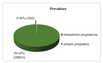
Total obstetric admissions seen during the study period were 29085 and out of these the no of patients diagnosed with ectopic pregnancy (n) were 282. Prevalence of ectopic pregnancy in our setup was calculated to be 9.69/1000 pregnancies/year (8.62-10.87/1000 pregnancies/year) (Figure 1).
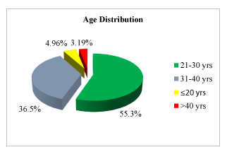
The patients presenting with ectopic pregnancy were between the age groups of 18 years to 42 years with the mean age of 29.2 ± 5.79 years. According to the age groups: ≤ 20 years group had 14/282 (4.96%) patients, 21- 30 years group had 156/282 (55.3%), 31-40 years had 103/282 (36.5%) and >40 years had 9/282 (3.19%) of the patients (Table 1, figure 2).
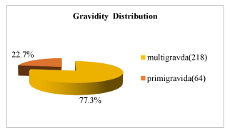
Most of the patients who had presented with ectopic pregnancy were multigravida {218/282 (77.3%)} and only 64/282 (22.7%) were primigravida (Table 1, figure 3).
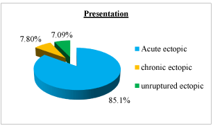
Based on the clinical and USG findings about 85.1% (240 /282) of the patients presented as acute(ruptured) ectopic pregnancy, 7.80% (22/282) as chronic ectopic pregnancy and 7.09% (20/282) as unruptured ectopic pregnancy (Table 1, figure 4).
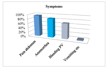
Every single patient had a combination of symptoms with abdominal pain being the most common one seen in 268/282 (95.7%) of the patients followed by amenorrhea in 240/282 (85.1%) patients, bleeding PV in 193/282 (68.44%) and vomitting, syncopal attacks in12/282 (4.25%) of the patients (Table 2, figure 5).
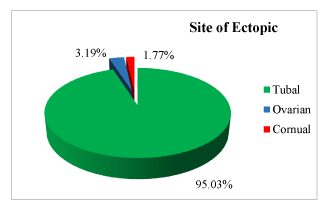
The most common site of ectopic pregnancy in our study was fallopian tubes seen in 268/282 (95.03%) of the patients. Other sites seen were ovaries in 9/282 (3.19%) and cornual pregnancy in about 5/282 (1.77%) of the patients (Table 1, figure 6).
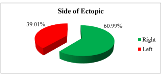
Overall right sided ectopics were more common seen in about 172/282 (60.99%) of the patients. Left sided ectopics were seen in 110/282 (39.01%) of the patients (Table1, figure 7).
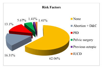
Out of all the patients enrolled in the study no risk factor was identified in 175/282 (62.06%) of the patients. Amongst those with risk factors previous history of abortions followed by D&C was the most common one seen in 46/282 (16.31%) of the patients. Other risk factors seen were Pelvic Inflammatory Disease (PID) in 37/282 (13.1%), history of Prior Pelvic Surgery (LSCS) in16/282 (5.67%), previous ectopic pregnancy in 4/282 (1.41%) and prior IUCD use in 4/282 (1.41%) of the patients (Table 2, figure 8).
Discussion
Ectopic pregnancy is an important issue in the reproductive life of the female and it is on a rising trend as seen over recent decades. It is an important cause and consequence of infertility and its management. Hence needs proper identification of risk factors for prevention and early management to prevent disastrous outcomes.
A total of 29085 obstetric admissions were seen during the study period, out of which 282 cases of ectopic pregnancy were registered. Prevalence of ectopic pregnancy reported in our study was 9.69/1000 pregnancies which is in coherence with the studies by Tahmina, et al. [8] and Bouyer, et al. [9] showing a prevalence of 9.1/1000 and 11.2/1000 pregnancies [8,9]. This result suggests the current trend of late child bearing, use of artificial reproductive techniques, increasing incidence of sexually transmitted diseases, induced abortions and the prevailing lifestyle contributes to the rising prevalence of ectopic pregnancies.
Age group distribution- Majority of the patients were in the age group 21-30 years (55.32%) which is similar to distribution seen in studies by Tahmina, et al. [8], Panchal D, et al. [10] and Mufti, et al. [11] having 51.39%, 71.66% and 75.4% of the patients in the same age group [8,10,11]. This age group is most likely affected as in Indian culture this age group corresponds to the period of peak sexual activity after marriage.
Gravidity distribution -Most of the patients in our study were multigravida (81.66%) similar to studies by Panchal D, et al. [10] and Poonam, et al. [12]. However it is contradictory to studies by Lawani, et al. [13] and Majhi, et al [14], which showed maximum incidence in nullipara [13,14]. Multigravida usually have a previous history of abortions or sexually transmitted diseases, this indicates that these factors might be the cause of more occurrence amongst multigravida.
Most of the patients presented with features of acute (ruptured) ectopic pregnancy (85.1%), it was confirmed radiologically as well, this result was in coherence with study by Shrivastva M, et al. [15] where about 91.5% of the patients presented as ruptured ectopic. Due to a varied presentation of ectopic pregnancies amongst different patients in many it only gets recognized after rupture of the ectopic pregnancy at the abnormal site presenting with features of shock due to hemoperitoneum.
Symptoms of patients- Pain abdomen was the most common symptom, present in about 95.7% of the patients followed by amenorrhea in 85.1% and bleeding PV in 68.44% of the patients. This result is coherent to studies by Prasanna B, et al. [16] and Gupta R, et al. [17] with pain abdomen seen in 90% and 87.5%, amenorrhea in 96% and 90% and bleeding PV in 68% and 67.5% of the patients. However in these studies amenorrhea was more common than pain abdomen. This triad of symptoms was seen in majority of the patients.
Site of Ectopic- 95.03% of the patients in our study had fallopian tube and 3.19% had ovaries as the site of ectopic pregnancy which is coherent to the study by Saeed, et al. [18] where 87% of the patients had ectopic pregnancy in fallopian tubes and 4.2% had it in ovaries.
According to our study the overall most common side involved was right side similar to the results of the study by Sharma R, et al. [19]. This association may be due to presence of appendix leading to an easy spread of infection on right side. However in study by Saeed, et al. [18] equal distribution was seen on both sides.
Risk factors-Many patients had more than one risk factor. Out of the patients having risk factors the most common one was found to be a history of previous abortions seen in about 16.31% of the patients similar to studies by Prasanna B, et al. [16] and Saeed, et al. [18] having this risk factor in 16% and 28.5 % of the patients. Postabortal infections leading to tubal damage explains this occurrence. Other risk factors seen were Pelvic Inflammatory Disease (PID) in 13.1%, history of Prior Pelvic Surgery (LSCS) in 5.67%, previous ectopic pregnancy in 1.41% and prior IUCD use in 1.41% of the patients. Similar risk factors were seen in study by Prasanna B, et al. [16] in 26%, 6%, 6% and 6% of the patients. PID leads to adhesions with distortion of tubal anatomy and most commonly affects the tubes bilaterally, this explains the increased risk of ectopic amongst patients with PID and previous ectopic. IUCD prevents implantation in the uterine cavity however does not interferes with implantation in tubes or ovaries hence pregnancy if seen with IUCD in situ is most likely an ectopic pregnancy. The results of different studies are given in table 3.
Conclusion
Increasing trend in ectopic pregnancy is seen over recent decades with prevalence of 9.69/1000 pregnancies/ year in our setup (previously 3.12/1000 pregnancies) [20]. Mean age group involved is 29.2 ± 5.79 years and multigravida are commonly involved. The most common symptoms are pain abdomen, amenorrhea & bleeding PV. Fallopian tubes being the commonly involved site with more occurrence on right side. Most of the patients in our setup apparently had no risk factor however amongst those who had risk factors, history of previous abortions appears to be most common cause of ectopic pregnancy.
Acknowledgement
Authors acknowledge the immense help received from the scholars whose articles are cited and included in references of this manuscript. The authors are also grateful to authors / editors / publishers of all those articles / journals and books from where the literature for this article has been reviewed and discussed.
- Shah P, Shah S, Kutty RV, Modi D. Changing epidemiology of maternal mortality in rural India: Time to reset strategies for MDG-5.Trop Med Health Sci Res. 2014; 19: 568-575. DOI: 10.1111/tmi.12282
- Zane SB, Kieke BA Jr, Kendrick JS, Bruce C. Surveillance in a time of changing health care practices: Estimating ectopic pregnancy incidence in the United States. Maternal Child Health J. 2002; 6: 227-236. DOI: 10.1023/a:1021106032198
- Storeide O, Veholmen M, Eide M, Bergsjo P, Sandevi R. The incidence of ectopic pregnancy in Horaland country, Norway 1976-1993.Acta Obstet Gynaecol Scand. 1997; 76: 345-349. DOI: 10.1111/j.1600-0412.1997.tb07990.x
- Ankum WM, Mol BW, Vander Veen F, Bossuyt PM. Risk factors for ectopic pregnancy: A meta- analysis.Fertil Steril. 1996; 65: 1093-1099. PubMed: https://pubmed.ncbi.nlm.nih.gov/8641479/
- Sivin I. Alternative estimates of ectopic pregnancy risks during contraception.Am J Obstet Gynaecol.1991; 165: 1900. DOI: 10.1016/0002-9378(91)90065-y
- Job-Spira N, Collet P, Coste J, Bremond A, Laumon B. Risk factors for ectopic pregnancy. Results of a case control study in the Rhone-Alpes region.Contracept Fertil Sex. 1993; 21: 307-312. PubMed: https://pubmed.ncbi.nlm.nih.gov/7951631/
- Bouyer J, Coste J, Shojaei T, Pouly JL, Fernandez H, Gerbaud L, et al. Risk factors for ectopic pregnancy: A comprehensive analysis based on a large case control, population based study in France.Am J Epidemiol. 2003; 157: 185-194. DOI: 10.1093/aje/kwf190
- Tahmina S, Daniel M, Solomon P. Clinical analysis of ectopic pregnancies in a tertiary care centre in Southern India: A six year retrospective study.J Clin Diagn Res. 2016; 10: QC13-QC16. DOI: 10.7860/JCDR/2016/21925.8718
- Bouyer J, Coste J, Fernandez H, Pouly JL, Job-Spira N. Sites of ectopic pregnancy : A 10 year population-based study of 1800 cases. Hum Reprod. 2002; 17: 3224-3230. DOI: 10.1093/humrep/17.12.3224
- Panchal D, Vaishnav G, Solanki K. Study of management in patient with ectopic pregnancy. NJIRM. 2011; 2: 91-94. https://bit.ly/2ZjKuVf
- Mufti S, Rather S, Rangrez AR, Wasiqa, Khalida. Ectopic pregnancy: An analysis of 114 cases.JK-practitioner. 2012; 17: 20-23.
- Poonam, Uprety D, Banerjee B. Ectopic pregnancy- two years review from BPKIHS, Nepal. Kathmandu Uni Med J. 2005; 3: 365-369.
- Lawani OL, Anozie OB, Ezeonu PO. Ectopic pregnancy: A life threatning gynecological emergency. Int J Womens Health. 2013; 5: 515-521.
- Majhi AK, Roy N, Karmakar KS, Banerjee PK. Ectopic pregnancy-- an analysis of 180 cases. J Indian Med Assoc. 2007; 105: 308-312. PubMed: https://pubmed.ncbi.nlm.nih.gov/18232175/
- Shrivastava M, Prashar H, Modi JN. A clinical study of ectopic pregnancy in a tertiary care centre in Central India.Int J Reprod Contracept Obstet Gynaecol. 2017; 6: 2485-2490. https://bit.ly/33fV2FU
- Prasanna B, Jhansi CB, Swathi K, Shaik MV. A study on risk factors and clinical presentation of ectopic pregnancy in women attending a tertiary care centre. IAIM. 2016; 3: 90-96. https://bit.ly/2FjrUWd
- Gupta R, Porwal S, Swarnkar M, Sharma N, Maheshwari P. Incidence,trends and risk factors for ectopic pregnancies in a tertiary care hosital of Rajasthan. J Pharm Biomed Sci. 2012; 16: 1-3.
- Saeed JA, Ogenleye IG, Mahmood M. Epidemiology, risk factors and sites of ectopic pregnancy in Madina maternity and children hospital,Kingdom of Saudi Arabia. JIMDC. 2013; 2: 26-29. https://bit.ly/2ZnTCbw
- Sharma R, Biligi DS. A study of histopathological changes in fallopian tubes in ectopic pregnancy. Int J Res Rev. 2015; 7: 54-58. https://bit.ly/3k0NEVG
- ICMR task force project. J Obst Gynec India.1990; 40: 425.

Sign up for Article Alerts