Clostridioides Difficile beyond the Colon: A Case Report
Yasmine Hussein Agha1*, Garret Seiler1, John Millard2, Joseph Cherabie1 and Maha Assi3*
1Department of Internal Medicine, University of Kansas School of Medicine-Wichita, Robert J. Dole VA Medical Center, Wichita, KS
2Department of Pharmacy, Robert J. Dole VA MC, Wichita KS
3Department of Infectious Disease, University of Kansas School of Medicine-Wichita, Robert J. Dole VA Medical Center, Wichita, Kansas
*Address for Correspondence: Yasmine Hussein Agha, Department of Internal Medicine, University of Kansas School of Medicine-Wichita, Robert J. Dole VA Medical Center, Wichita, KS9911 East 21st Street North, Wichita, KS, Tel: +163-060-106-25; Fax: +316-293-1878; E-mail: yasmineagha@gmail.com
*Address for Correspondence: Maha Assi, Department of Infectious Disease, University of Kansas School of Medicine-Wichita, Robert J. Dole VA Medical Center, Wichita, Kansas, 1010 North Kansas Street, Wichita, KS, 67214, Tel: + 316-213-5627; Fax: + 316-293-1878; E-mail: maassi@hotmail.com
Submitted: 30 May 2019; Approved: 22 June 2019; Published: 25 June 2019
Citation this article: Hussein Agha Y, Seiler G, Millard J, Cherabie J, Assi M. Clostridioides Difficile beyond the Colon: A Case Report. Sci J Immunol Immunother. 2019;3(1): 009-011.
Copyright: © 2019 Hussein Agha Y, et al. This is an open access article distributed under the Creative Commons Attribution License, which permits unrestricted use, distribution, and reproduction in any medium, provided the original work is properly cited
Keywords: Splenic abscess; Clostridioides difficile; Monomicrobial gut infection
Download Fulltext PDF
Extra-colonic Clostridioides difficile infections are rarely reported. Presentations include bacteremia, enteritis, reactive arthritis, skin infections, or visceral abscesses. We present a case of monomicrobial Clostridioides difficile splenic abscess in a patient without colitis, preceding antibiotics, or bacteremia.
Introduction
Clostridioides Difficile (CD) is an anaerobic, spore-forming, gram-positive bacillus. It is a well-known colonizer of the human colon and is transmitted via the fecal-oral route. Clostridioides Difficile Infection (CDI) occurs following disruption of the host’s gastrointestinal microbiota [1]. Risk factors are antibiotic therapy in previous 90 days, elderly, extended hospitalization, inflammatory bowel disease, cirrhosis, and chemotherapy [2]. Since CD is difficult to isolate in culture, the gold-standard diagnostic studies are limited to stool studies: Enzyme Immunoassays (EIA) for Glutamate Dehydrogenase (GDH) or toxin antigens and PCR for the toxin gene. Extra-colonic manifestations of CDI are sparsely documented in the literature. We present a case of an extra-colonic CDI presenting as a monomicrobial splenic abscess.
Case Report
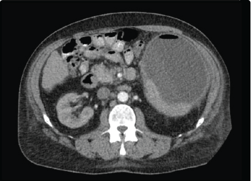
A 54 year old male with history of cirrhosis due to chronic hepatitis B and C was seen in clinic for left upper quadrant pain, feeling febrile, and anorexia. Five weeks prior to presentation, a Transjugular Intrahepatic Portosystemic Shunt (TIPS) revision with splenic artery embolization was performed. He denied associated diarrhea, melena, or hematochezia. His temperature was 98.3 F with a pulse of 79 bpm, respiration rate of 20 breaths/min, and blood pressure of 125/63 mmHg. The physical exam revealed tenderness and guarding in the left upper quadrant along with pain exacerbated by inspiration. Comprehensive metabolic panel, lipase, and complete blood count were unremarkable. Ultrasound with doppler of liver revealed a patent TIPS. A CT of the abdomen was ordered which revealed a subcapsular splenic abscess which measured 17.6 x 15.7 x 11 cm (figure 1). He was admitted to the hospital for further management. Vancomycin and Piperacillin/Tazobactam were empirically administered. Laboratory studies were unremarkable. Blood cultures were negative. CT-guided aspiration with pigtail drain placement yielded 900 mL of purulent drainage. Gram stain revealed white blood cells but no microorganisms. Six days later, Clostridioides difficile was isolated from the culture. He was discharged on oral metronidazole with planned repeat imaging and follow-up with infectious disease. The final CT scan six months after drainage and metronidazole therapy showed a residual collection measuring 4.4 cm x 1.8 cm. The patient stopped following-up, repeat imaging could not be done to document resolution of the abscess.
Discussion
CDI is a disease associated with the colon presenting as diarrhea, pseudomembranous colitis, ileus, or toxic megacolon. Extra-colonic CDI is a phenomenon rarely reported in the literature and usually occurring in the setting of colorectal surgery or colitis [3]. The few documented cases of extra-colonic CDI include: bacteremia [4], enteritis [3,5], reactive arthritis[5], polymicrobial visceral abscesses (splenic abscess, pancreatic abscess, empyema) [5], prosthetic device infections [5], and osteomyelitis [5]. This case involved a patient with cirrhosis, TIPS revision, and recent splenic artery embolization who developed a splenic abscess from which culture grew only CD. Upon chart review, the patient was not given any prophylactic antibiotics prior to TIPS revision. Notably, preceding colonic disease was absent. Over the last 40 years, only 6 cases of CD splenic abscesses have been reported. The first report of splenic abscess due to CD occurred in 1983. The patient had a history of liver cirrhosis and pancreatitis, in the absence of any gastrointestinal colitis or surgery [6]. The second case was seen in 1987 in a patient that had bacteremia with four Clostridioides species (C. difficile, C. fallax, C. perfringes and C. ramosum) 4 months prior to splenic abscess development. Cultures of the abscess contained C. difficile and Pseudomonas paucimobilis j [7]. A third case was seen in 1995 in a patient with atrial fibrillation and congestive heart failure that developed infarction of the colon and spleen. Subsequently, a splenic abscess infected with CD was formed [8]. In 1997, a patient that was given antibiotics for pneumonia following laminectomy surgery developed CD colitis. A few weeks later, a splenic abscess was found. The abscess culture grew CD and coagulase negative staphylococci [9]. Another patient with a history of adenocarcinoma of the cecum, right hemi-colectomy, small bowel infarction, recent Pseudomonas aeruginosa septicemia, treated with multiple antibiotics, presented with CD bacteremia. Upon further evaluation, a splenic abscess growing CD was found [10]. The last case reported occurred in 2003 in a patient with end-stage renal disease who had CD bacteremia and later developed a CD splenic and iliacus muscle abscesses [11]. These cases demonstrated shared preceding events: bacteremia, antibiotic exposure, pancreatitis, colitis, bowel and spleen infarction. Theoretically, these events may cause disruption of the bowel microbiota and mucosa. CD is then able to invade and translocate into the bloodstream then disseminate throughout the body. Splenic abscesses have been reported to follow splenic artery embolization procedures at an incidence of 6.8% [12]. With the absence of preceding colitis, pancreatitis, or antibiotic exposure, the embolization procedure during which colonic translocation of bacteria must have occurred, remains the most likely cause of this patient’s abscess. CD obtained its name by way of being difficult to isolate in culture. Traditionally, the diagnosis was made during endoscopy upon viewing pseudomembranes. The current gold-standard diagnostic studies involve EIA and PCR of stool samples. The lack of a reliable method to diagnose extra-colonic CDI implies that its incidence is underreported. Future areas of research should be dedicated to developing diagnostic tools to detect CD in blood or other body fluids.
- Leffler DA, Lamont JT. Clostridium difficile infection. The New England Journal of Medicine. 2015; 372: 1539-48.
- Kelly CP, LaMont JT. Clostridium difficile - more difficult than ever. N Engl J Med. 2008; 359: 1932-1940. http://bit.ly/2Ku7Cul
- Dineen SP, Bailey SH, Pham TH, Huerta S. Clostridium difficile enteritis: A report of two cases and systematic literature review. World J Gastrointest Surg. 2013; 5: 37-42. http://bit.ly/2xbOqsw
- Doufair M, Eckert C, Drieux L, Amani Moibeni C, Bodin L, Denis M, et al. Clostridium difficile bacteremia: Report of two cases in French hospitals and comprehensive review of the literature. IDCases. 2017; 8: 54-62. http://bit.ly/2Iwiqpw
- Jacobs A, Barnard K, Fishel R, Gradon JD. Extracolonic manifestations of clostridium difficile infections. Medicine (Baltimore). 2001; 80: 88-101. http://bit.ly/2LajQYr
- Saginur R, Fogel R, Begin L, Cohen B, Mendelson J. Splenic abscess due to clostridium difficile. J Infect Dis. 1983; 147: 1105. http://bit.ly/31QZl99
- Studemeister AE, Beilke MA, Kirmani N. Splenic abscess due to Clostridium difficile and Pseudomonas paucimobilis. Am J Gastroenterol. 1987; 82: 389-390. http://bit.ly/31NYTbH
- Stieglbauer KT, Gruber SA, Johnson S. Elevated serum antibody response to toxin a following splenic abscess due to Clostridium difficile. Clin Infect Dis. 1995; 20: 160-162. http://bit.ly/2x6hPEr
- Kumar N, Flanagan P, Wise C, Lord R. Splenic abscess caused by clostridium difficile. Eur J Clin Microbiol Infect Dis. 1997; 16: 938-939. http://bit.ly/2Y1odJ4
- Shedda S, Campbell I, Skinner I. Clostridium difficile splenic abscess. Australian and New Zealand journal of surgery. 2000; 70: 147-148. http://bit.ly/2Y4LFFp
- Bedimo R, Weinstein J. Recurrent extraintestinal clostridium difficile infection. Am J Med. 2003; 114: 770-771. http://bit.ly/2L9c8hi
- Ekeh AP, Khalaf S, Ilyas S, Kauffman S, Walusimbi M, McCarthy MC. Complications arising from splenic artery embolization: a review of an 11-year experience. Am J Surg. 2013; 205: 250-254. http://bit.ly/2WSDcDS


Sign up for Article Alerts