The Role of Transcranial Doppler in the Assessment of Right-to-Left Shunt in Scuba Divers?
Gian P. Anzola1*, Clara Bartolaminelli2, Sofia Fioravanti3, Paolo Lega4, Paolo Limoni5 and Pasquale Longobardi6
1Consultant Neurologist Villa Gemma Hospital Gardone Riviera Italy
2Emergency Department Montichiari Hospital - Asst Degli Spedali Civili Di Brescia, Italy
3Nurse of Hyperbaric Centre Ravenna, Italy
44Consultant Anesthesiologist at Hyperbaric Centre Ravenna, Italy
5Consultant Neurosonologist, Neurologist at Hyperbaric Centre Ravenna, Italy
6Director of Hyperbaric Centre Ravenna, President of SIMSI
*Address for Correspondence: Gian Paolo Anzola, Consultant Neurologist Villa Gemma Hospital, via Zanardelli 101 - Gardone Riviera (Brescia), Italy, Fax: +300-303-755-269; E-mail: gpanzola@numerica.it
Submitted: 15 February 2018; Approved: 03 March 2018; Published: 06 March 2018
Citation this article: Anzola GP, Bartolaminelli C, Fioravanti S, Lega P, Limoni P, et al. The Role of Transcranial Doppler in the Assessment of Right-to-Left Shunt in Scuba Divers. Int J Neurol Dis. 2018;2(1): 001-005.
Copyright: © 2018 Anzola GP, et al. This is an open access article distributed under the Creative Commons Attribution License, which permits unrestricted use, distribution, and reproduction in any medium, provided the original work is properly cited
Download Fulltext PDF
Introduction and Method: The presence of a Right-to-Left Shunt (RLS), most commonly produced by a patent foramen ovale, has been correlated to Decompression Illness (DI) in scuba divers and is suspected to increase the risk of suffering diving accidents. In the present study, RLS was comparatively investigated in a consecutive series of 309 divers with and without DI and in 97 healthy control subjects. All cases were submitted to Transcranial Doppler (TCD) examination. The presence of RLS was confirmed by pulmonary scintigraphy and / or echocardiographic tests if the RLS as assessed with TCD was large.
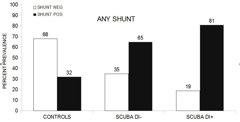
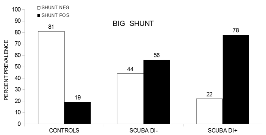
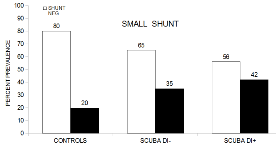
Results: The frequency of DI was the same in recreational and professional divers. DI positive were significantly older than DI negative divers (45 + 10 vs. 42 + 11 year old respectively, p = 0.047). Big shunts were statistically more frequent in DI positive (78%) than in DI negative (56%) divers, in DI positive than in controls (19%) and in DI negative than controls. Small shunts were more frequent in DI positive (42%) divers than in controls (20%), in DI negative (35%) divers than in controls, but not in DI positive compared with DC negative divers. The presence of a big shunt and age turned independent predictors of DI.
Introduction and Aim of the study
Patent Foramen Ovale (PFO) is an embryological remnant that consists in the incomplete postnatal fusion of the cardiac atrial septum primum and secundum hence allowing the shunting of variable amounts of blood from the venous to the systemic circulation. PFO is associated with numerous conditions such as migraine, transient global amnesia, cerebrovascular and coronary ischemia, Obstructive Sleep Apnea (OAS), platypnea/ orthodeoxia syndrome, Decompression Illness (DI) in divers and high-altitude aviators and astronauts [1,2]. However, the high prevalence of PFO itself, on average 27% in the general population, makes its clinical relevance debatable [3]. Neurological decompression illness is one of the principal factors limiting the practice of both recreational and professional diving and since 1986 the Right-to-Left Shunt (RLS), mainly due to PFO, but in a minority of cases also to pulmonary arteriovenous malformations, has been reported to increase the risk of DI [4,5]. In the present study we assessed the prevalence of RLS, using Transcranial Doppler (TCD) ultrasonography technique, in a consecutive series of divers with and without DI in comparison with a sample of normal non-diver subjects. The aim of the study was to evaluate the association of RLS and of other potential important variables with decompression illness in its various sub types.
Material and Methods
Between May 2011 and July 2017, a consecutive series of 309 divers (204 men and 105 women, mean age 44 + 11, range 13 - 79 years) attending the Center of Hyperbaric Oxygen Therapy in Ravenna, Italy, for the assessment of the fitness to diving, underwent TCD sonography with the aim of detecting the presence of an RLS. In this group the occurrence of DI was recorded. In the same period a control group of 97 healthy non-diver volunteers was recruited (32 men and 65 women with a mean age of 39 + 15, range 16 - 88 years). The TCD sonography examination, performed by a certified physician (P. Li.) of the Center of Hyperbaric Oxygen Therapy in Ravenna, Italy, is part of the general evaluation which is routinely performed in all divers. In all subjects a final evaluation was made by a senior physician (P. Lo.) specialized in undersea and hyperbaric therapy. The presence of RLS was confirmed by pulmonary scintigraphy and / or echocardiographic tests if the RLS as assessed withy TCD was of grade 3 or higher (see below).
TCD examination was done in all cases by the same physician, with a DWL Multidop P or, more recently, with a Doppler Box machine. Bilateral monitoring was performed with probes specifically designed to fit in with the LAM-Rack standard probe support (DWL). The first segment of both Middle Cerebral Arteries (MCAs) was recorded at the depth of 50 - 55 mm. A specifically DWL software for emboli detection and count was activated at the start of the examination. The Doppler spectrum and emboli count (microbubbles) were recorded and stored on a hard disk. Patients received a 10 - ml bolus of agitated saline solution (enriched with drops of autologous blood) via an antecubital vein. TCD ultrasonography was considered as positive (indicating the presence of an RLS) when at least one typical High-Intensity Transient Signal (HIT) was recorded on the Doppler spectrum, 5 - 51 secs after the injection. After the injection at rest, the test was repeated with provocative maneuver (Valsalva maneuver). The Valsalva maneuver was documented based on the decrease of blood velocity in the MCAs. An increase of velocity was observed at the release of Valsalva maneuver. The hemodynamic relevance of the RLS was graded following the classification proposed by the 1999 International Consensus Meeting of Venice: 0 = no HITS, 1 = less than 10 HITS, 2 = 10 to 20 HITS, 3 = > 20 HITS shower appearance, 4 = > 20 HITS curtain appearance [6]. The final grading of the RLS was defined after the provocative maneuver when grade 1 or 2 was recorded on normal breathing, whereas in grade 3 or 4 detected during normal breathing Valsalva maneuver was deemed unnecessary. For statistical purpose, we classified “small shunts” those ranked as grade 1 or 2, and “big shunts” those ranked as grade 3 or 4. Data was analyzed with SPSS statistical package. Frequencies were compared with the Chi - square test, numeric variables with t-test and predictors of DI were assessed in a binomial logistic regression analysis.
Results
Among the 309 divers, 200 (65%) reported having had at least one episode of DI (with neurological symptoms in 100 = 50% and non - neurological symptoms in 100 = 50%). The frequency of DI was the same in recreational and professional divers (66% in amateur divers, 59% in instructors, 51% in technical dives, p = 0.229) as well as in males and females (68% in females vs. 63% in males, p = 0.445). The average number of dives per year was likewise not statistically different in divers with (DI +) and without (DI -) decompression illness (60 in DI +, 67 in DI -, p = 0.559). DI + were significantly older than DI - divers (45 + 10 vs. 42 + 11 year old respectively, p = 0.047).The findings of RLS are summarized in (Figures 1,2,3). Compared with controls, overall RLS prevalence was higher both in DI + and in DI – divers (32% in Ctrls, 65% in DI + divers, 81% in DI - divers, p < 0.0001). The difference in RLS prevalence was also statistically significant between DI + and DI - divers (p < 0.0001). Big shunts were statistically more frequent in DI + than in DI - divers (78% vs 56% respectively, p < 0.0001), in DI + than in controls (78% vs. 19% respectively, p < 0.0001) and in DI - than controls (56% vs. 19% respectively, p < 0.0001), whereas small shunts were more frequent in DI + divers than in controls (42% vs. 20% respectively, p = 0.003), in DI - divers than in controls (35% vs. 20%, p = 0.038), but not in DI + compared with DI - divers ((42% vs. 35% respectively, p = 0.412). The presence of a big shunt, age, number of dives per year and type of dives (recreational, instructor and technical) were entered a binomial logistic regression analysis with DI as the dependent variable. The presence of a big shunt and age turned independent predictors of DI (Table 1).
Discussion
DI is caused by the formation of gas bubbles, related to the failure to remove inert gases (nitrogen), in supersaturated blood or tissues during the diver’s ascent. DI can occur even after diving safely, observing decompression times, and this may depend on the physical condition of the diver, his workout, or the external temperature. Most divers with venous emboli remain asymptomatic, because these bubbles are filtered by pulmonary circulation. Symptoms may occur with high bubble load (i.e. pulmonary gas embolism in case of violation of decompression regimen) or from paradoxical embolism (permanent or transient RLS), as RLS can facilitate the passing of the bubbles in the arteries [7,8]. The clinical presentation of DI is heterogeneous, reflecting the number of bubbles and the sites of their formation; there are mild illnesses such as the skin disease characterized by fever, urticarial and itching; the osteoarticular disease characterized by joint pain in the limbs and hands; the lymphatic disease with localized subcutaneous swelling. There are also severe illnesses such as the neurological type (both cerebral and spinal), the otovestibular type (with bubbles forming in the inner ear) or the pulmonary type with gaseous exchange reduction [9]. The connection between DI and PFO was first described in 1980s [5-7]. Since then a number of studies have addressed the relationship between PFO and diving suggesting that RLS increases not only the chance of DI, especially so in cases of neurological impairment, but also the occurrence of clinically silent brain lesions [10]. In earlier studies the assessment of RLS was done with echocardiography, three ultra-sonographic techniques are available for imaging a PFO or detecting a RLS: Transthoracic Echocardiography (TTE), Transesophageal Echocardiography (TEE) and Transcranial Doppler (TCD or TCCD). TTE is still the most widespread initial screening tool for PFO, although it has a much lower sensitivity compared with contrast TCD [11]. On the other hand, the sensitivity and specificity of TCD for RLS detection have been found to vary in different studies [12-14]. In Komar series TCD had an 89% negative predictive value, 98% positive predictive value, 95% sensitivity and 92% specificity. In this study only 4.8% of TEE positive cases did not show the passage of contrast [14]. A recent meta-analysis reported that TCD had a mean sensitivity and specificity of 97% and 93% with TEE as the reference standard [15]. In the above mentioned studies TEE was taken as the gold standard, assuming it had 100% sensitivity in detecting PFO, whereas recent findings have shown that this assumption was flawed. Indeed, Van et al, by simultaneously performing intra cardiac echocardiography and TCD in patients undergoing PFO closure, found that TEE underestimated the shunt by 34% compared with TCD [13]. Likewise, Caputi and colleagues reported that permanent shunts were better identified by TCD than TEE [16]. Based on these findings we deliberated to use TCD as the more sensitive tool for RLS detection as we were particularly interested in assessing the association of RLS with neurological DI. For statistical purpose we therefore classified DI into neurological and non-neurological forms. Overall, confirming earlier reports, we found that divers who had suffered a decompression accident had a prevalence of RLS significantly higher than divers free of DI and then healthy non-diver controls. This incremental prevalence was confirmed when only big shunts were taken into account, whereas for small shunts there was a significantly higher prevalence in divers as compared with controls but not between divers with and without DI. It thus appears that a big shunt increases the risk of suffering a decompression accident, although with no particular propensity for the brain, as there was no difference in RLS prevalence between neurological and non-neurological DI. The higher frequency of small shunts in divers as compared with controls could speculatively result from the continuing Valsalva strains operated during dives that, by increasing right atrial pressure, could keep separated the leaflets of the inter-atrial septum. The same interpretation was proposed as an explanation for the observed increment in RLS in a prospective study on a small cohort of divers [17]. In our cohort of divers with DI we were able to analyze a number of potentially meaningful demographic and procedural variables of interest: DI had the same prevalence in recreational and professional divers and there was no statistical difference in the average annual number of dives between DI + and DI - divers. Decompression accidents were equally frequent in males and females, whereas DI + were older than DI - divers. Indeed, age and the presence of a big shunt were the only predictors of DI, indicating that adding up to the anatomical “flaw”, advancing age increases the risk of decompression illness. It is theoretically possible that not so much age itself, but the presumably higher total number of past dives in older subjects accounts for the finding of DI + being older than DI - divers. Unfortunately, we were unable to record the total number of past dives in all subject and therefore this issue remains unsettled. However, previous reports had highlighted the effect of age on DI suggesting that it may have something to do with endothelial function. Indeed, recent research suggests that the mechanism of tissue damage in decompression illness is multi-faceted; besides mechanical obstruction, nitrogen bubbles have been implicated in causing endothelial dysfunction. Damage to endothelial cells by decompression stress has been reported in a number of studies documenting the synthesis of cytokines and cell adhesion stimulators, and finally systematic inflammation and prothrombotic phenomena. [18]. It is likely that with advancing age vascular endothelium becomes less efficient in repairing mechanical or biochemical insults and hence the effect of the contact with nitrogen bubbles increasingly dangerous. Under this respect the presence of a large shunt, by increasing the amount of bubbles delivered to the brain vessels, might further increment the risk. Privileged targets of bubbles mediated endothelial dysfunctions might be brain and heart vessels. Endothelial damage, likely to be more prevalent in relation to the total number of dives, could thus be the basis of thrombotic events in older divers, especially in those who have had heart complications. This has implications for the functional assessment of divers. In order to prevent cardiovascular accidents, it is important to consider all the risk factors affecting endothelial function (age, diabetes, hypertension, drug intake, cardiovascular disease, smoking habit) and probably also to perform a cardiopulmonary test. This test is a non-invasive diagnostic instrumental examination, used in the appraisal of cardiac patients, bronchi tic patients, neurotic persons or sportsmen; it establishes, by means of the measuring of gases exhaled during a physical exercise, the aerobic capacity and it is able to determine the real working ability of heart, lungs, circulation system or muscles [19]. It seems therefore logical to propose the study of endothelial function as well as the cardiopulmonary test in divers to establish the risk of cardiovascular complications during underwater activity both in recreational and professional diving.
- Kerut EK, Norfleet WT, Plotnick GD, Giles TD. Patent foramen ovale: a review of associated conditions and the impact of physiological size. J Am Coll Cardiol. 2001; 38: 613-623. https://goo.gl/uAzyqS
- Anzola GP. Clinical impact of Patent foramen ovale diagnosis with transcranial Doppler. Eur J Ultrasound. 2002; 16: 11-20. https://goo.gl/ZJbwfm
- Hagen PT, Scholz DG, Edwards WD. Incidence and size of patent foramen ovale during the first 10 decades of life: an autopsy study of 965 normal hearts. Maio Clin Proc. 1984; 59: 17-20. https://goo.gl/zCAVG8
- Lairez O, Cournot M, Minville V, Roncalli J, Austruy J, Elbaz M, et al. Risk of neurological decompression sickness in the diver with a rigt-to-left shunt: Literature review and meta-analysis. Clin J Sport Med. 2009; 19: 231-235. https://goo.gl/cfYdhG
- Wilmshurst PT, Ellis BG, Jenkins BS. Paradoxical gas embolism in a scuba diver with an atrial septal defect. Brit Med J. 1986; 293: 1277. https://goo.gl/3QpEQU
- Jauss M. Zanette E. Detection of right-to-left shunt with ultrasound contrast agent and transcranial doppler sonography. Cerebrovasc Dis. 2000; 16: 490-496. https://goo.gl/suxWEA
- Moon RE, Camporesi EM, Kisslo JA. Patent foramen ovale and decompression sickness in divers. Lancet. 1989; 1: 513-514. https://goo.gl/evQnPt
- Honek J, Sefc L, Honek T, Sramek M, Horvath M, Veselka J. Patent foramen ovale in recreational and professional divers: an important and largely unrecognized problem. Can J Cardiol. 2015; 31: 1061-1066. https://goo.gl/C6yKzY
- Sykes O, Clark JE. Patent foramen ovale and scuba diving: a practical guide for physicians on when to refer for screening. Extrem Physiol Med. 2013; 2: 10. https://goo.gl/5V5v6g
- Koch AE, J Kampen, K Tetzlaff, M Reuter, P McCormack, P W Schnoor, et al. Incidence of abnormal cerebral finding in the MRI of clinically healthy divers: role of patent foramen ovale. Undersea Hyper Med. 2004; 31: 261-268. https://goo.gl/cNv1b5
- Zhao E, Wei Y, Zhang Y, Zhai N, Zhao P, Liu B. A comparison of transthoracic echocardiography and transcranial Doppler with contrast agent for detection of patent foramen ovale with and without the Valsalva maneuver. Medicine (Baltimore). 2015; 94: 1937. https://goo.gl/oj3rud
- Klotzsch C, Janssen G, Berlit P. Transesophageal echocardiography and contrast TCD in the detection of a patent foramen ovale: experiences with 111 patients. Neurology. 1994; 44: 1603-1606. https://goo.gl/S5sKuc
- Van H. Poommipanit P, Shalaby M, Gevorgyan R, Tseng CH, Tobis J. Sensitivity of transcranial Doppler versus intracardiac echocardiography in the detection of right-to-left shunt. JACC Cardiovasc Imaging. 2010; 3: 343-348. https://goo.gl/cXL39o
- Komar M, Olszowska M, Przewłocki T, Podolec J, Stępniewski J, Sobień B, et al. Transcranial Doppler ultrasonography should it be the first choice for persistent foramen ovale screening?. Cardiovasc Ultrasound. 2014; 12: 16. https://goo.gl/XqAiHX
- Mojadidi MK, Roberts SC, Winoker JS, Romero J, Goodman-Meza D, Gevorgyan R, et al. Accuracy of transcranial Doppler for the diagnosis of intracardiac right-to-left shunt: a bivariate meta-analysis of prospective studies. JACC Cardiovasc Imaging. 2014; 7: 236-250. https://goo.gl/ZNgbAF
- Caputi L, Carriero MR, Falcone C, Parati E, Piotti P, Materazzo C, et al. Transcranial doppler and transesophageal echocardiography: comparison of both techniques and prospective clinical relevance of transcranial doppler in patent foramen ovale detection. J Stroke Cerebrovasc Dis. 2009; 18: 343-348. https://goo.gl/28ZozC
- Germonpre P, Hastir F, Dendale P, Marroni A, Nguyen AF, Balestra C. Evidence for increasing patency of the foramen ovale in divers. Am J Cardiol. 2005; 95: 912-915. https://goo.gl/fNTgUa
- Zhang K, Wang M, Wang H, Liu Y, Buzzacott P, Xu W. Time course of endothelial dysfunction induced by decompression bubbles in rats. Front Physiol. 2017; 8: 181. https://goo.gl/B8oQqU
- Milani RV, Lavie CJ, Mehra MR, Ventura HO. Understanding the basic of cardiopulmonary exercise testing; Mayo Clin Proc. 2006; 81: 1603-1611. https://goo.gl/dMDTt6


Sign up for Article Alerts