Heterotopic Ossification in a Patient with Cerebral Palsy and Stroke?
Avraam Ploumis*, Areti Theodorou, Kleio T. Kappatou, Lamprina G. Magkou, Paraskevas S. Tseniklidis and Georgios I. Vasileiadis
Department of Physical Medicine and Rehabilitation, Division of Surgery, Faculty of Medicine, University of loannina, Greece
*Address for Correspondence: Avraam Ploumis, Department of Physical Medicine and Rehabilitation, Division of Surgery, University of loannina, Greece, Tel: +30-693-208-0701; ORCID ID: orcid.org/0000-0002-6828-3196; E-mail: aploumis@uoi.gr
Submitted: 09 February 2020; Approved: 27 February 2020; Published: 28 February 2020
Citation this article: Ploumis A, Theodorou A, Kappatou KT, Magkou LG, Tseniklidis PS, et al. Heterotopic Ossification in a Patient with Cerebral Palsy and Stroke. Int J Ortho Res Ther. 2020;4(1): 001-003.
Copyright: © 2020 Ploumis A, et al. This is an open access article distributed under the Creative Commons Attribution License, which permits unrestricted use, distribution, and reproduction in any medium, provided the original work is properly cited
Keywords: Heterotopic ossification; Myositis ossificans; Ectopic ossification; Stroke; Cerebral palsy
Download Fulltext PDF
Heterotopic Ossification (HO) is defined as pathological bone formation at locations where bone normally does not exist. The presence of HO has been found to be a rare complication after stroke in several studies, whereas there are only sporadic references relating HO to Cerebral Palsy (CP) and few for CP and stroke. No effective treatment for HO has yet been found, whereas the cellular and molecular mechanisms have not been completely understood. Therefore, increased awareness among physicians is required, as a challenge for early diagnosis and treatment. A case of a male patient with CP, who developed HO on the paretic hip joint following an ischemic stroke is presented.
Introduction
Heterotopic Ossification (HO) is ectopic bone formation in non-osseous tissues. The prevalence of HO in patients with stroke is 1.3% [1,2]. Additionally, the risk of developing stroke is almost doubled among patients with Cerebral Palsy (CP) in comparison with the general population and five times higher in patients under 50 years old [3]. Our aim is to present a case in which a CP patient underwent stroke and his medical condition was complicated by HO at the hip. We strongly suspect that there is an additive causative effect in the HO pathogenesis and highlight their possible interaction is highlighted.
Case History
A 35-year-old male with CP was urgently admitted to University Hospital of Ioannina, after developing sudden left hemiplegia and being lethargic during awakening. Brain Computed Tomography scan (CT) showed a right large low density area at the parietal and temporal lobe of the brain, compatible with right Middle Cerebral Artery (MCA) stroke pressing the frontal horn of the right lateral ventricle. The patient was admitted to the neurological clinic presenting with left hemiplegia, inability to speak and signs of neglect. His treatment included anti-edematous and protective dose of anti-coagulant agents, sultamicillin 3x3 gr for the risk of aspiration pneumonia and nasal O2 administration. The patient underwent biochemical, coagulation, immunological (including antiphospholipid antibodies), viral testing and carotid ultrasound imaging as well, with no pathological findings. After two weeks of hospitalization, his clinical condition started to deteriorate, and Magnetic Resonant Imaging and Arteriography (MRI-MRA) scan revealed hemorrhagic conversion of the stroke and thin MCA, vertebral and basilar arteries. Furthermore, transthoracic heart ultrasound and ambulatory electrocardiography showed the presence of tachycardia and due to strong suspicion; trans-esophageal heart ultrasound was also conducted, revealing patent foramen ovale. Finally, due to possible cardiovascular origin of the stroke, rivaroxaban 1x15 mg was administered.
During the third week of hospitalization, the patient’s level of consciousness was improved and he was responsive to his name. Although signs of neglect for the hemiplegic side were no more observed, there was no improvement to his movement.
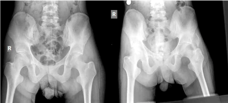
After a month of hospitalization, the patient was transferred to the Physical Medicine and Rehabilitation clinic, where physical therapy was pursued. Two weeks later limited movement and pain in the vicinity of the left hip were noticed and radiography revealed HO in the hip joint. Treatment with indomethacin 75 mg per os once daily and diclofenac cut. Sol. 1.5% locally was initiated. The dose of indomethacin used was low, because of the high risk of brain haemorrhage in the patient. Also, major importance was given for the physiotherapy to be performed within the pain-free range of motion. After a week, etidronate 20 mg/kg p.o. for three months was administered. One year later, in the follow up examination of the patient, x-rays showed that the formation of ectopic bone had stopped, probably because of the use of etidronate.
Discussion
Our aim is to present a rare case of CP, stroke and HO coexistence. Risk factors for HO development are haemorrhagic stroke (55-70%, rather than 30% in ischemic stroke), male sex, young age, spasticity, prolonged immobilization or forcible mobilization, ventilator support and the presence of pressure sores [1,2,4]. In our case, the patient was a young male with spasticity due to CP and suffered an ischemic stroke with hemorrhagic conversion. Therefore, there was significant risk for HO development due to add-on phenomenon (old CP and current stroke). To the best of our knowledge, the probability of these combined factors in appearance of HO has not been studied.
The most common location for HO formation is in the vicinity of the hip, followed by elbow, knee and shoulder [1,2,5,6]. It is more likely to be presented in the paretic extremity, but there are also cases where the non-paretic side or both sides developed HO [1,2]. Its formation usually occurs within two months and its symptoms include limited range of motion, pain, palpable mass, fever and swelling [1,5-7]. Those symptoms are also present in various other conditions such as thrombophlebitis, deep venous thrombosis, cellulitis and osteomyelitis, making the recognition more challenging [7]. Furthermore, sensory loss, severe aphasia and cognitive impairment or neglect of the paretic side, can complicate the situation even more [1]. In our case, HO was formed on the paretic hip joint within a month after stroke and became clinically apparent with pain and limited movement during physical therapy.
For the detection of HO formation, methods such as radiography, CT, ultrasound and bone scintigraphy can be used [5,7]. As for the initial evaluation, radiography can reveal a soft-tissue mass with calcified peripheral zone. Similarly, CT images can show the bone cortex more distinct as the bone matures. Also ultrasound can detect HO sooner than radiography and can be used during the operative process to visualize the lesion before its excision. Finally, three-phase bone scintigraphy still remains the most sensitive imaging modality for the early detection of HO, the development and maturity of the ossification and the determination of the optimal time for surgical resection [5,7]. In our patient, radiography was conducted twice with the first revealing a suspicious radiopaque lesion and the second confirming HO formation.
It is suggested that brain or spinal cord injury combined with local peripheral tissue microtrauma, caused by prolonged immobilization, can lead to inflammatory factors release [5-7]. This can stimulate cell differentiation, collagen deposition and create hypoxic microenvironment at the peripheral injured location. In such an environment, the cells with chondroosseous differentiation ability differentiate to chondrogenic cells, leading to cartilage deposition [4,5-7]. Also, due to hypoxic microenvironment and matrix remodeling, angiogenic factors are released to provoke neovascularization. This process provides sufficient oxygen tension, promoting osteoblastic differentiation and deposition of osseous tissue. Over time, the initially formed bone undergoes remodeling and maturation, resulting in a mature lamellar bone with Haversian canals, blood vessels and marrow cavity [5-7].
Medication, irradiation, physical therapy and surgical excision, can be used for the prevention and treatment of HO [5,7]. Drugs that are most frequently administered are indomethacin, which inhibits the formation of prostaglandin-E2, and bisphosphonates, by delaying calcium deposition at the osteoid [5-7]. Radiation therapy can prevent HO formation by interfering in the differentiation process of the mesenchymal cells to osteoprogenitor cells. As for the physical therapy, there is much controversy regarding its efficiency. Forcible manipulation of the extremities may induce HO formation, [8] whereas gentle exercising may have no effects or even provoke its development, due to lack of movement [4,5,7]. Finally, surgical excision is the only effective method to treat HO and it is mostly suggested that the operation should proceed after bone has reached maturity, so that HO recurrence is prevented. While its recurrence reduces the effectiveness of surgical therapy, post-operative administration of indomethacin or irradiation is necessary for its prevention [5-7]. In our case, the patient was treated with indomethacin, local anti-inflammatory and etidronate in combination with active assisted kinesiotherapy.
Conclusions
The significant increase risk for HO development due to add-on phenomenon (old cerebral palsy lesion and current brain stroke) of central nervous system injury must be suspected by physicians and therapists in this particular subset of CNS patients. Further research in the area of causative factors of HO formation is warranted.
- Cunha DA, Camargos S, Passos VMA, Mello CM, Vaz LS, Lima LRS. Heterotopic ossification after Stroke: Clinical profile and severity of ossification. J Stroke Cerebrovasc Dis. 2019; 28: 513-520. PubMed: https://www.ncbi.nlm.nih.gov/pubmed/30466894
- Genet F, Minooee K, Jourdan C, Ruet A, Denormandie P, Schnitzler A. Troublesome heterotopic ossification and stroke: Features and risk factors. A case control study. Brain Inj. 2015; 29: 866-871. PubMed: https://www.ncbi.nlm.nih.gov/pubmed/25915823
- Wu CW, Huang SW, Lin JW, Liou TH, Chou LC, Lin HW. Risk of stroke among patients with cerebral palsy: A population-based cohort study. Dev Med Child Neurol. 2017; 59: 52-56. PubMed: https://www.ncbi.nlm.nih.gov/pubmed/27346658
- Bargellesi S, Cavasin L, Scarponi F, De Tanti A, Bonaiuti D, Bartolo M, et al. Occurrence and predictive factors of heterotopic ossification in severe acquired brain injured patients during rehabilitation stay: Cross-sectional survey. Clin Rehabil. 2018; 32: 255-262. PubMed: https://www.ncbi.nlm.nih.gov/pubmed/28805078
- Ranganathan K, Loder S, Agarwal S, Wong VW, Forsberg J, Davis TA, et al. Heterotopic ossification: Basic-science principles and clinical correlates. J Bone Joint Surg Am. 2015; 97: 1101-1111. PubMed: https://www.ncbi.nlm.nih.gov/pubmed/26135077
- Brady RD, Shultz SR, McDonald SJ, O'Brien TJ. Neurological heterotopic ossification: Current understanding and future directions. Bone. 2018; 109: 35-42. PubMed: https://www.ncbi.nlm.nih.gov/pubmed/28526267
- Vanden Bossche L, Vanderstraeten G. Heterotopic ossification: A review. J Rehabil Med. 2005; 37: 129-136. PubMed: https://www.ncbi.nlm.nih.gov/pubmed/16040468
- Crawford CM, Varghese G, Mani MM, Neff JR. Heterotopic ossification: Are range of motion exercises contraindicated? J Burn Care Rehabil. 1986; 7: 323-327. PubMed: https://www.ncbi.nlm.nih.gov/pubmed/3117800


Sign up for Article Alerts