An Unusual Presentation of a Sinonasal Mass in Infancy
Christophe Abi Zeid Daou1, Shadi Bsat2, Ramzi T Younis1, Hussein Darwish2 and Zeina R Korban1*
1Department of Otolaryngology and Head and Neck Surgery, American University of Beirut Medical Center, Lebanon
2Department of Neurosurgery, American University of Beirut Medical Center, Lebanon
*Address for Correspondence: Zeina Korban, Department of Otolaryngology and Head and Neck Surgery, American University of Beirut Medical Center Riad El Solh, 1107 2020, Beirut, Lebanon, Tel: +961-373-7264; E-mail: zk27@aub.edu.lb
Submitted: 07 January 2020; Approved: 22 January 2020; Published: 24 January 2020
Citation this article: Zeid Daou CA, Bsat S, Younis RT, Darwish H, Korban ZR. An Unusual Presentation of a Sinonasal Mass in Infancy. Int J Rhinol Otological. 2020;3(1): 001-003.
Copyright: © 2020 Zeid Daou CA, et al. This is an open access article distributed under the Creative Commons Attribution License, which permits unrestricted use, distribution, and reproduction in any medium, provided the original work is properly cited
Download Fulltext PDF
NHL (Non-Hodgkin’s Lymphoma) is very rapidly growing and can eventually cause a mass effect on surrounding structures and facial asymmetry. Because of its fast growth and ambiguous clinical presentation, it is crucial to get a diagnosis early to start the appropriate and targeted therapy. There are limited cases in the literature reporting the incidence and survival of children with sinonasal tumors so the data evaluating tumor types, treatment and survival rates is lacking. Treatment includes Chemotherapy or concomitant chemoradiation. We hereby report a rare presentation of blindness due to a large sinonasal mass in a six-month-old boy.
Introduction
Sinonasal lymphomas are rarely extra-nodal and are in the majority of cases Non-Hodgkin’s Lymphoma (NHL) involving most frequently the maxillary sinus [1]. Sinonasal NHL accounts for 1.5% of all NHL cases in the United States. Globally, there exists racial and geographical variations in the incidence of NHL with B-cell subtype more prevalent in the eastern population and the NK/T-cell subtype more common in the Asian and Latin populations [2]. NHL is very rapidly growing and can eventually cause a mass effect on surrounding structures and facial asymmetry. Because of its fast growth and ambiguous clinical presentation it is crucial to get a diagnosis early to start the appropriate and targeted therapy [3].
Long term survival is attributed to age, stage at presentation and histopathology. Rhabdomyosarcoma is the most common sinonasal tumor of childhood and adolescence with an overall 5-year survival of around 62% [4]. Lymphomas are less common as well as sarcomas and neuroblastomas [5].
There are limited cases in the literature reporting the incidence and survival of children with sinonasal tumors so the data evaluating tumor types, treatment and survival rates is lacking. We hereby report a rare presentation of blindness due to a large sinonasal mass in a six-month-old boy.
Case Presentation
A 6-month-old baby boy, previously healthy, presented to our medical center with progressive exophthalmos and ptosis in the left eye that was noticed by the mother of one-week duration. She also reported that her son stopped following her with both eyes few days prior. Patient did not have an associated fever or recent upper respiratory infection. There were no other complaints at presentation. He was born by C-section, full-term, after failure to descend and is neurodevelopmentally normal. Ophthalmic examination showed bilateral vision loss and absent light perception in both eyes. The patient had a normal laboratory work up.
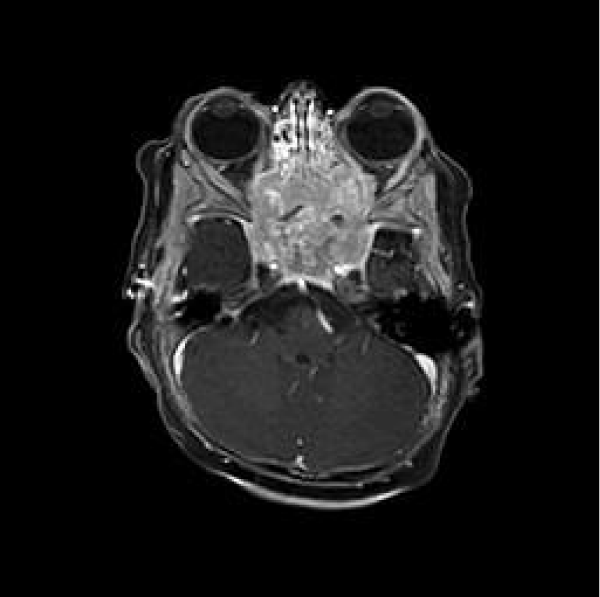
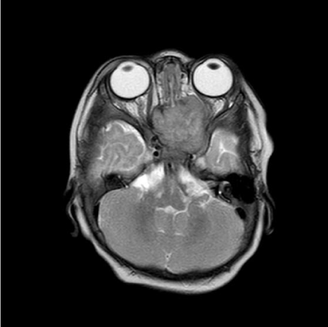
MRI brain and orbits revealed a large destructive sinonasal mass, 3.1x2.3x3.4 cm in size, involving the ethmoid bone and sinuses posteriorly, sphenoid body and portion of the clivus (basisphenoid) with preservation of the sphenooccipital synchondrosis.
The mass is isointense on T1 and T2-weighted images. It shows heterogeneous enhancement after IV contrast administration and restricted diffusion with central areas of necrosis (Figure 1). The soft tissue component is invading the orbital cavity bilaterally with mass effect and displacement of the medial rectus muscle bilaterally, more so on the left exerting mass effect on the optic nerves with lateral displacement of the left optic nerve (Figure 2). It is involving the cavernous sinuses bilaterally with narrowing of the cavernous MCA. The differential diagnosis entailed sarcoma, neuroblastoma, lymphoproliferative disease or esthesioneuroblastoma.
Due to the aggressive and invasive nature of this mass, the patient was taken to the operating room for an endoscopic endonasal transphenoidal biopsy to establish a diagnosis.
Under general anesthesia, using the 30 degree endoscope, the right and left sphenoid sinuses were accessed and an exophytic soft, friable mass was encountered and multiple biopsies were taken and sent for permanent pathology analysis.
The pathology report was consistent with large B-cell lymphoma positive for CD20, CD45 and MUM1 while being negative for CD5 and CD10. There was no significant EBV expression.
Post-operatively, in light of the diagnosis, a work-up for immunodeficiencies was completed and the patient was found to have Severe Combined Immunodeficiency Disease (SCID).
Discussion
We present a rare case of sinonasal large B cell lymphoma in a 6-month-old boy presenting with orbital involvement in the setting of SCID. The initial presentation of rapidly progressive visual loss, exophthalmos and proptosis in the absence of other clinical clues urges the exclusion of an underlying malignant process. Clinical presentations of sinonasal NHL are very nonspecific and can vary from epistaxis to nasal obstruction, facial swelling, hearing impairment or visual disturbances [6]. Due to the highly malignant and aggressive profile of NHL it is important to not delay imaging and diagnosis in order to prolong survival [2].
The MRI spotted a sinonasal mass, but the radiological clues were not enough to correctly diagnose the type of tumor. Indeed, in a case series by Yi, et al. imaging modalities (CT or MRI) were found to be only 57% accurate in the setting of sinonasal lymphomas as compared to 83% in rhabdomyosarcoma [5]. Nonetheless, Diffusion-Weighted Imaging (DWI) has been more recently used to provide functional and morphologic information on the lesion in question as this technique quantifies the random motion of water protons in tissues [7]. The Apparent Diffusion Coefficient (ADC) obtained from the DWI itself is helpful in differentiating squamous cell carcinoma from malignant lymphoma and can therefore guide diagnosis and treatment plan early on while avoiding invasive procedures in some cases [1,7]. The diagnostic gold standard hence relies on getting a tissue biopsy of the mass after ruling out a vascular origin [8]. Lymphomas of sinonasal origin in children include B-cell lymphomas and NK/T-cell lymphomas with NK/T-cells being the more common. Clinically B-cell lymphomas tend to involve the nasal cavity along with one or more paranasal sinuses, similar to the present case, why the latter is usually restricted to the nasal cavity [5]. Pathologic examination of the tissue specimen is needed to correctly diagnose the tumor subtype along with chemical immunostaining [9].
Treatment of sinonasal lymphomas mainly includes chemotherapy or chemo radiation. Radiation alone is only favored in early stage disease. Surgery plays only a minimal role in these patients and is only needed to aid in diagnosis or reduce disease burden rather than treatment [2]. Despite advances in diagnosis, treatment options and imaging, survival rates remain as low as 50% [10].
In children, the differentiation between the subtypes of B-cell lymphomas especially Burkitt’s and diffuse large B-cell is not as important as in adults since it does not alter the management or treatment course. It is important to note that both of the above mentioned neoplasms are aggressively malignant and should be promptly treated [11].
Cases previously reported only involved children older than 1 year of age. Earlier presentation is unusual, such as that reported in our case making it the first reported in the literature, which lead the team to suspect an underlying disease mechanism favoring lymphoma formation. The patient was further diagnosed with SCID and indeed SCID is associated with an increased incidence of B-cell lymphomas especially NHL not associated with EBV as in our case, but rather caused by gene rearrangement [12].
Conclusion
It is crucial to have a high clinical suspicion, regardless of age once dealing with rapidly progressive sinonasal and orbital symptoms. We emphasize the significance of obtaining a tissue diagnosis rather than leaping into surgical resection to ensure an accurate diagnosis and management accordingly.
- Nagafuji H, Yokoi H, Ohara A, Fujiwara M, Takayama N, Saito K. Primary diffuse large B-cell lymphoma of the frontal sinus: A case report and literature review. Radiol Case Rep. 2018; 13: 635-639. PubMed: https://www.ncbi.nlm.nih.gov/pubmed/30167025
- Antonios N Varelas, Michael Eggerstedt, Ashwin Ganti, Bobby A. Tajudeen. Epidemiologic, prognostic, and treatment factors in sinonasal diffuse large B -cell lymphoma. Laryngoscope. 2019; 129: 1259-1264. https://bit.ly/2RnUr0e
- Reichman OS, Goldin AD, Bayles SW, Donnelly KJ. Non-Hodgkin's lymphoma presenting as facial swelling and nasal obstruction in a pediatric patient. Otolaryngol Head Neck Surg. 1999; 121: 127-129. PubMed: https://www.ncbi.nlm.nih.gov/pubmed/10388894
- Gerth DJ, Tashiro J, Thaller SR. Pediatric sinonasal tumors in the United States: incidence and outcomes. J Surg Res. 2014; 190: 214-220. PubMed: https://www.ncbi.nlm.nih.gov/pubmed/24793449
- Yi JS, Cho GS, Shim MJ, Min JY, Chung YS, Lee BJ. Malignant tumors of the sinonasal tract in the pediatric population. Acta Otolaryngol. 2012; 132: 21-26. PubMed: https://www.ncbi.nlm.nih.gov/pubmed/22582777
- Hong X, Khalife S, Bouhabel S, Bernard C, Daniel SJ, Manoukian JJ, et al. Rhinologic manifestations of Burkitt Lymphoma in a pediatric population: Case series and systematic review. Int J Pediatr Otorhinolaryngol. 2019; 121: 127-136. PubMed: https://www.ncbi.nlm.nih.gov/pubmed/30897372
- Maeda M, Kato H, Sakuma H, Maier SE, Takeda K. Usefulness of the apparent diffusion coefficient in line scan diffusion-weighted imaging for distinguishing between squamous cell carcinomas and malignant lymphomas of the head and neck. AJNR Am J Neuroradiol. 2005; 26: 1186-1192. PubMed: https://www.ncbi.nlm.nih.gov/pubmed/15891182
- Holsinger FC, Hafemeister AC, Hicks MJ, Sulek M, Huh WW, Friedman EM. Differential diagnosis of pediatric tumors of the nasal cavity and paranasal sinuses: a 45-year multi-institutional review. Ear Nose Throat J. 2010; 89: 534-540. PubMed: https://www.ncbi.nlm.nih.gov/pubmed/21086277
- Khan NR, Lakicevic G, Callihan TR, Burruss G, Arnautovic K. Diffuse large B-cell lymphoma of the frontal sinus presenting as a pott puffy tumor: Case report. J Neurol Surg Rep. 2015; 76: 23-27. PubMed: https://www.ncbi.nlm.nih.gov/pubmed/26251804
- Steele TO, Buniel MC, Mace JC, El Rassi E, Smith TL. Lymphoma of the nasal cavity and paranasal sinuses: A case series. Am J Rhinol Allergy. 2016; 30: 335-339. PubMed: https://www.ncbi.nlm.nih.gov/pubmed/27657899
- Allen CE, Kelly KM, Bollard CM, Pediatric lymphomas and histiocytic disorders of childhood. Pediatr Clin North Am. 2015; 62: 139-165. PubMed: https://www.ncbi.nlm.nih.gov/pubmed/25435117
- Garcia CR, Brown NA, Schreck R, Stiehm ER, Hudnall SD. B-cell lymphoma in severe combined immunodeficiency not associated with the Epstein-Barr virus. Cancer. 1987; 60: 2941-2947. PubMed: https://www.ncbi.nlm.nih.gov/pubmed/2824020


Sign up for Article Alerts