Stress Fracture of The Lamina: A Diagnosis of Suspicion?
Jorge Lopez-Subias1*, Jorge Gil-Albarova2, Marina Lillo-Adan3, Victoria-Eugenia Gómez Palicio4
1Jorge Lopez Subias, Tel: +34 976765605; e-mail: jjsubias@gmail.com
2Jorge Gil-Albar ova, Tel:+34 976765605; e-mail: jgilalba@unizar.es
3Marina Lillo-Adan, Tel:+34 976765605; e-mail: mlilloadan@gmail.com
4Victoria-Eugenia Gomez Palacio, Tel:+34 976765605; e-mail: vegomez@salud.aragon.es
*Address for Correspondence: Jorge Lopez Subias, Tel: +34 976765605; jjsubias@gmail.com
Submitted: 25 April 2017; Approved: 28 August 2017; Published: 29 August 2019
Citation this article: Lopez-Subias J, Gil-Albarova J, Lillo-Adan M, Gómez Palicio VE. Stress Fracture of The Lamina: A Diagnosis of Suspicion. Open J Pediatr Neonatal Care. 2017;2(2): 038-041.
Copyright: © 2019 Lopez-Subias J, et al. This is an open access article distributed under the Creative Commons Attribution License, which permits unrestricted use, distribution, and reproduction in any medium, provided the original work is properly cited
Keywords: Retroishmic cleft; Laminolysis; Stress fracture
Download Fulltext PDF
Lumbar laminolysis, a stress fracture of the lamina, is one of the causes of chronic low back pain in adolescents. Its prevalence is very low, and it is described as an incidental finding in most cases. The authors report the case of a 14-year-old girl with a symptomatic stress fracture of the left lamina of the fifth lumbar vertebra. Recent advances in diagnostic tools and techniques enable early diagnosis. Multimodal radiological examinations of the whole lumbar spine are recommended in cases of symptomatic patients with low back pain who do not respond to initial treatment with basic physiotherapy or analgesic medical treatment. Knowledge of laminolysis in adolescent patients with low back pain is necessary to prevent it from being overlooked or late diagnosis. In almost all cases, conservative treatment may be sufficient to achieve bone healing.
Introduction
Low back pain in adolescents is a growing public health problem, and their presence increases the risk of back pain in the future. Some studies have shown that low back pain can lead to disability and limit daily activities for between 13% and 38% of adolescents [1, 2].
The differential diagnosis of low back pain in this population is broad, and etiologic factors are most often associated with musculoskeletal overuse or trauma [3].
However, some cases are caused by bony defects in the vertebral arch, which can appear in several locations. We may find clefts in the pars-interarticulais (spondylolysis), which affect around 40% of the pediatric patients, with low back pain persisting for longer than 2 weeks [4]. These are sometimes in the pedicle (pediculolysis) [5] and on rare occasions, in the lamina (laminolysis) [6].
The Retroishmic cleft in the spine lamina is rarely detected. This term was introduced by Broacher [7]. Who was the first to describe its radiographic appearance For years, it was considered as a congenital deformity. However, Wick et al [8].
Proposed the term laminolysis to describe the Retroishmic cleft by analogy to the nomenclature of the applied stress fractures of the pars interarticulais (spondylolysis) and the pedicle (pediculolysis) being the fi rst to suggest that laminolysis was a nonunion after a stress fracture.”
Although laminolysis is very rare, we should consider this diagnostic suspicion in adolescent patients with low back pain. Finally, the correct diagnosis will be confirmed by means of image studies, such as plain radiography in oblique views, computed tomography or MRI (Magnetic Resonance Imaging).
Case Report
A 14 years old girl, without previous disease, reported severe low pain for five months. She practiced gymnastics four days per week. She denied any history of acute trauma.
Physical examination showed notable tenderness at L5 spinous process. The pain was exacerbated by lumbar hyperextension during back somersault. Lumbar flexion was moderately restricted. She had no associated limb par aesthesia or numbness. No neurological abnormality of tone, sensation, reflexes or power was identified.
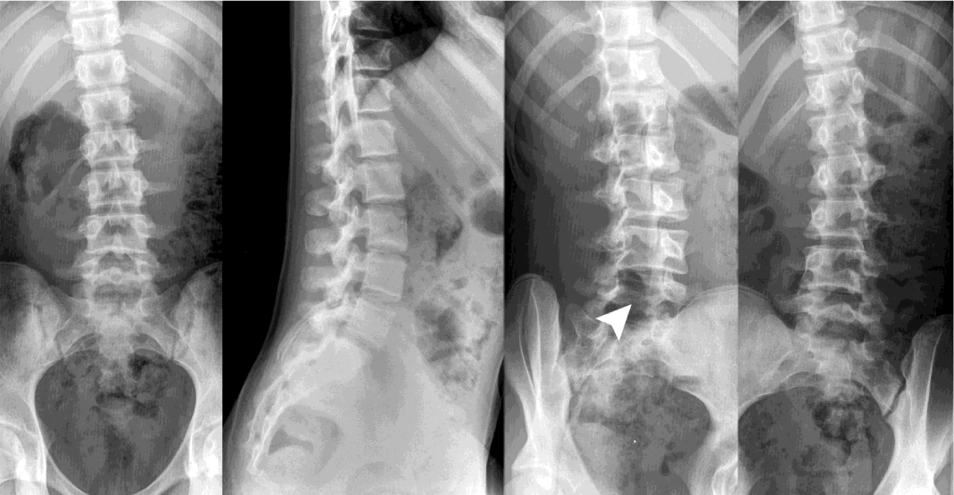
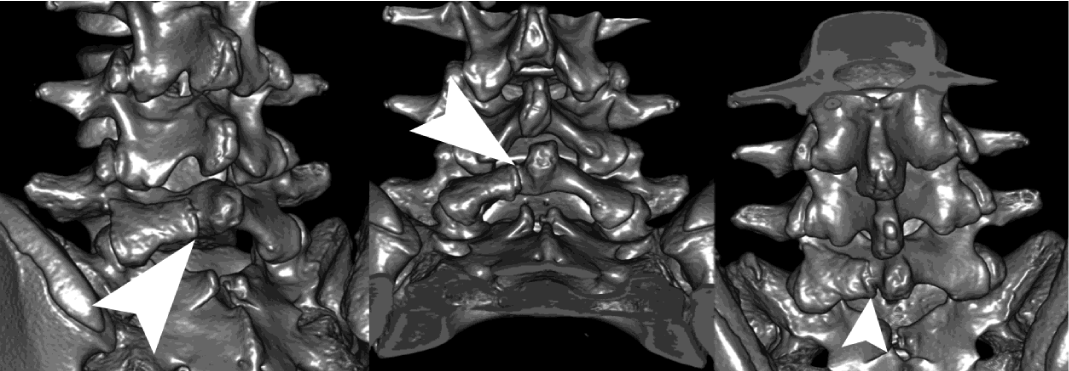
Plain radiographs were performed in anteroposterior, lateral and oblique views. The oblique view showed a possible cleft image in the L5 left lamina (Figure 1). The computed tomography study revealed a complete cleft of the L5 left lamina in the coronal orientation, which suggests a stress fracture of the L5 lamina (laminolysis) (Figure 2-3).
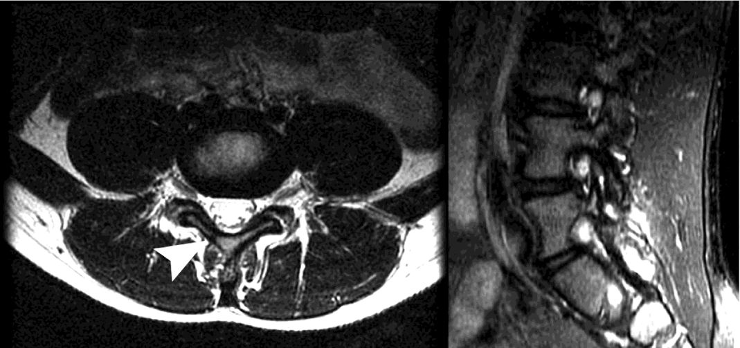
Moreover, MRI study showed inflammatory changes in the soft tissues, with edema in the par spinal musculature (Figure 4).
We proposed a conservative treatment with analgesia, avoiding sports practice, a rehabilitation program focused on core stabilization, and strengthening and flexibility exercises. At the three month follow up, the low back pain had disappeared and she was allowed to resume all her activities progressively, avoiding repetitive hyperextension exercises of the lumbar spine. After one year, the patient is asymptomatic and has returned to her usual sporting activities.
Discussion
Spondylolysis is a defect of the pars interarticulais with an incidence in the general population of approximately 6% [9, 10]. However an incidence of more than 40% has been reported in adolescents who participate heavily in sport, especially gymnasts, soccer players or ballet dancers [11]. Spondylolysis is the most frequently cleft found in the vertebral arch, although sometimes it may be observed in the pedicle (pediculolysis) [12] and rarely in the lamina (laminolysis) [13].
Previously, they were all considered as congenital abnormalities [7-15], but the most recent literature shows that sometimes they may be a consequence of stress fractures [13-18]. Laminolysis typically concerns a unique lamina, although there are cases reported with bilateral lamina fractures [19].
The incidence of these fractures is increasing in recent years in relationship with intense physical activity of medium-high level in adolescents. Chronic low back pain in adolescents, without a history of trauma, should suggest the suspicion of these injuries. Usually these injuries occur in adolescents [9, 10] with a male predominance, although Wick et al [8] describe cases in adults.
Radiography, scintigraphy, computed tomography and MRI have been used in different combinations for diagnosis. Although scintigraphy is very sensitive for detecting early stress reaction lesions, and radiography is capable of showing complete fractures in oblique view, computed tomography has been the reference standard for the diagnosis, monitoring and fracture healing.
Moreover, the combination with MRI, allows us to assess the healing stage of the fracture, differentiating between acute and chronic injuries. However, recent studies have suggested that MRI should be the first diagnostic tool and combined with computed tomography to clarify fracture morphology and follow-up bone healing [20, 21]. In this case report, the suspicion of laminolysis in plain radiographs, in combination with computed tomography and MRI was successful for accurate diagnosis.
Biomechanically, extension and rotation movements of the lumbar spine are considered capable of inducing stress fracture at the pars interarcularis and at the lamina [22].
Saury et al [23] analyzed the lines of highest stress at the pars interarticulais using the finite element model. They concluded that high stress, in extension at the coronal orientation and in rotation at the Saigal orientation, may cause spondylolysis in the pars interarticulais. Therefore, laminolysis may be a result of stress fracture due to repetitive extension loading. The cases described in the literature so far agree that the cause of the stress fracture of the lamina is associated with repeated hyperextension mechanisms [8-16].
Laminolysis was thought to be associated with unilateral or contralateral spondylolysis. Some authors have suggested that, in the presence of unilateral spondylolysis, the remainder of the neural arch is exposed to abnormal stresses [14-26]. However, the present case did not suffer unilateral spondylolysis.
It is well known that acute early fractures, especially those that are incomplete and unilateral, have a better healing potential with conservative measures, in comparison to chronic, bilateral complete fractures [5, 27]. According to these points, conservative treatment for laminolysis should be performed as for spondylolysis. This will be, basically, relative rest, abstention from sports, and exercises for strengthening abdominal and lumbar musculature [13, 16]. Sometimes the use of a trunk brace is recommended to obtain pain relief and reduce the risk of worsening symptoms. Return to sport is allowed once the patient is pain free, is normal on examination, and has undergone rehabilitation.
In conclusion, laminolysis is not known as a common cause of low back pain. It is hardly ever seen in routine daily practice and therefore, it is important to emphasize its inclusion in the differential diagnosis of lower back pain, to help avoid misdiagnosis. Correct diagnosis is based on plain radiographs including oblique views, computed tomography and MRI. Conservative treatment is indicated in almost all cases.
- Calvo-Munoz I, Gomez-Conesa A, Sanchez-Meca J. Prevalence of low back pain during childhood and adolescence: a systematic review. Rev Esp Salud Publica. 2012; 86: 331-56. https://goo.gl/4FcqPu
- Stallknecht SE, Strandberg-Larsen K, Hestbæk L, Andersen AN. Spinal pain and co-occurrence with stress and general well-being among young adolescents: a study within the Danish National Birth Cohort. Eur J Pediatr. 2017; 176: 807-814. https://goo.gl/M5QwLH
- MacDonald J, Stuart E, Rodenberg R. Musculoskeletal Low Back Pain in School-aged Children: A Review. JAMA Pediatr. 2017; 171: 280-287. https://goo.gl/kZaGpQ
- Nitta A, Sakai T, Goda Y, Takata Y, Higashino K, Sakamaki T, et al. Prevalence of Symptomatic Lumbar Spondylolysis in Pediatric Patients. Orthopedics. 2016; 39: e434-7. https://goo.gl/9eirvd
- Sairyo K, Katoh S, Takata Y, Terai T, Yasui N, Goel VK, et al. MRI signal changes of the pedicle as an indicator for early diagnosis of spondylolysis in children and adolescents: a clinical and biomechanical study. Spine (Phila Pa 1976). 2006; 31: 206-11. https://goo.gl/hL4VUA
- Abraham T, Holder L, Silberstein C. The retroisthmic cleft. Scintigraphic appearance and clinical relevance in patients with low back pain. Clin Nucl Med. 1997; 22: 161-5. https://goo.gl/wVrE5V
- Brocher JEW. Konstitutionell bedingte Veranderungen des wirbelbogens. Fortschr Rontgenstr 1960; 92: 363-380. https://goo.gl/Y3CcZa
- Wick LF, Kaim A, Bongartz G. Retroisthmic cleft: a stress fracture of the lamina. Skeletal Radiol. 2000; 29: 162-4. https://goo.gl/Ai1ikc
- Fredrickson BE, Baker D, McHolick WJ, Yuan HA, Lubicky JP. The natural history of spondylolysis and spondylolisthesis. J Bone Joint Surg Am. 1984; 66: 699-707. https://goo.gl/CxLD4U
- Sakai T, Sairyo K, Takao S, Nishitani H, Yasui N. Incidence of lumbar spondylolysis in the general population in Japan based on multidetector computed tomography scans from two thousand subjects. Spine (Phila Pa 1976). 2009; 34: 2346-50. https://goo.gl/kUFc6q
- Micheli LJ, Wood R. Back pain in young athletes. Significant differences from adults in causes and patterns. Arch Pediatr Adolesc Med. 1995; 149: 15-8. https://goo.gl/4Q6KXS
- Sairyo K, Katoh S, Sasa T, Yasui N, Goel VK, Vadapalli S, et al. Athletes with unilateral spondylolysis are at risk of stress fracture at the contralateral pedicle and pars interarticularis: a clinical and biomechanical study. Am J Sports Med. 2005; 33: 583-90. https://goo.gl/BPuA3k
- Sakai T, Sairyo K, Takao S, Kosaka H, Yasui N. Adolescents with symptomatic laminolysis: report of two cases. J Orthop Traumatol. 2010; 11: 189-93. https://goo.gl/gxfvSf
- Wortzman G, Steinhardt MI. Congenitally absent lumbar pedicle: a reappraisal. Radiology. 1984; 152: 713-8. https://goo.gl/FRnTDZ
- Durrigl P. Ein Fall von angeborener retroisthmischer Wirbelbogenspalte. Z Orthop 1965; 100: 374–376. https://goo.gl/Y61dWs
- Sairyo K, Katoh S, Sakamaki T, Komatsubara S, Endo K, Yasui N. Three successive stress fractures at the same level in an adolescent baseball player. Am J Sports Med. 2003; 31: 606-10. https://goo.gl/gshi6e
- Wiltse LL, Widell EH Jr, Jackson DW. Fatigue fracture: the basic lesion in isthmic spondylolisthesis. J Bone Joint Surg Am. 1975; 57: 17-22. https://goo.gl/fGQptN
- Gunzburg R, Fraser RD. Stress fracture of the lumbar pedicle. Case reports of “pediculolysis” and review of the literature. Spine (Phila Pa 1976). 1991; 16: 185-9. https://goo.gl/zje2KW
- Sakai T; Sairyo K; Mase Y; Dezawa A. Synchronic multiple stress fractures of L5 left hemilamina: a case of an unusual type of lumbar spondylolysis. Eur J Orthop Surg Traumatol. 2012; 22 Suppl 1: 41-3. https://goo.gl/jXAAXL
- Dunn AJ, Campbell RS, Mayor PE, Rees D. Radiological findings and healing patterns of incomplete stress fractures of the pars interarticularis. Skeletal Radiol. 2008; 37: 443-50. https://goo.gl/UDyG8U
- Campbell RS, Grainger AJ, Hide IG, Papastefanou S, Greenough CG. Juvenile spondylolysis: a comparative analysis of CT, SPECT and MRI. Skeletal Radiol. 2005; 34: 63-73. https://goo.gl/3H1dtZ
- Sakai T, Yamada H, Nakamura T, Nanamori K, Kawasaki Y, Hanaoka N, et al. Lumbar spinal disorders in patients with athetoid cerebral palsy: a clinical and biomechanical study. Spine (Phila Pa 1976). 2006; 31: E66-70. https://goo.gl/PxJUBG
- Sairyo K, Katoh S, Komatsubara S, Terai T, Yasui N, Goel VK et al. Spondylolysis fracture angle in children and adolescents on CT indicates the facture producing force vector: a biomechanical rationale. Internet J Spine Surg 2004; 1. https://goo.gl/aGZEkb
- Kim KS, Kim YW, Kwon HD. Unilateral spondylolysis combined with contralateral lumbar pediculolysis in a military parachutist. J Spinal Disord Tech. 2006; 19: 65-7. https://goo.gl/TWwNBa
- O’Beirne JG, Horgan JG. Stress fracture of the lamina associated with unilateral spondylolysis. Spine (Phil Pa 1976) 1998; 13: 220–222. https://goo.gl/b1NaCa
- Vialle R, Mary P, de Carvalho A, Ducou le Pointe H, Damsin JP, Filipe G. Acute L5 pedicle fracture and contralateral spondylolysis in a 12-year-old boy: a case report. Eur Spine J. 2007; 16 Suppl 3: 316-7. https://goo.gl/j3DhKU
- Dunn AJ, Campbell RS, Mayor PE, Rees D. Radiological findings and healing patterns of incomplete stress fractures of the pars interarticularis. Skeletal Radiol. 2008; 37: 443-50.


Sign up for Article Alerts