Congenital Candidiasis: A Rare and Detrimental Disease?
Wai Quen Lee1*, Lip Yuen Teng2, Pauline Poh Ling Choo3 and Kah Kee Tan4
1,5Hospital Tuanku Ja’afar, Jalan Rasah, 70 300, Seremban, Negeri Sembilan Darul Khusus, Malaysia
2Hospital Port Dickson, Ministry of Health, Malaysia
3,4Hospital Tuanku Ja’afar, Seremban, Ministry of Heath, Malaysia
*Address for Correspondence: Wai Quen Lee, Hospital Tuanku Ja’afar, Jalan Rasah, 70 300 Seremban, Negeri Sembilan Darul Khusus, Malaysia,, Tel: +60 6 768 4000; E-mail: iwaiquen@gmail.com
Submitted: 13 August 2017; Approved: 02 September 2017; Published: 05 September 2019
Citation this article: Lee WQ, Teng LY, Pauline Choo PL, Tan KK. Congenital Candidiasis: A Rare and Detrimental Disease. Open J Pediatr Neonatal Care. 2017;2(2): 046-050.
Copyright: © 2017 Lee WQ, et al. This is an open access article distributed under the Creative Commons Attribution License, which permits unrestricted use, distribution, and reproduction in any medium, provided the original work is properly cited
Keywords: Congenital; Candidiasis; Preterm
Download Fulltext PDF
Congenital Candidiasis is extremely rare, with clinical manifestations ranging from localized skin disease to systemic involvement. Firm recommendations for the management of congenital Candidiasis are difficult to be made due to rarity of the disease. We are reporting a preterm infant who was diagnosed with invasive congenital Candidiasis with no mucocutaneous involvement but rapid clinical deterioration, resulting in early neonatal death. A baby girl, the first twin of a monochorionic diamniotic pregnancy with a gestation of 29 weeks and 5 days, was born not vigorous and intubated at birth. The child had hepatomegaly and pancytopenia from birth. At 15 hours of life, she was transfused with packed cells as haemoglobin was only 7.4 g/dl. She further deteriorated at 21 hours of life, with frequent desaturation and poor perfusion that required high ventilator settings and multiple crystalloid boluses. She developed coagulopathy, hemodynamic instability and succumbed at 32 hours of life. The baby’s blood culture taken at 27 hours of life, peripheral and intracardiac post-mortem samples all showed pure growth of albicans. Mother’s vaginal swab also showed pure growth of albicans but never treated with topical or systemic anti-fungal therapy. One of the most striking features of this infant is the rapid deterioration and pancytopenia from birth. Retrospectively, this made us consider whether systemic anti-fungal should have been started at birth.
Introduction
Congenital Candidiasis is extremely rare in term and preterm infants, with <100 cases reported in medical literature [1,2] . It presents with in the first six days of life with varied clinical manifestations ranging from localized skin disease to invasive disease i.e. pneumonia, meningitis, sepsis, and death [3]. About 10-35% of women have candidial valginitis during pregnancy, but only < 1% develop candidial chorioamnionitis [1-4]. Candidial chorioamnionitis has been associated frequently with preterm labour and intrauterine fetal death than with congenital Candidiasis [3,4].
Due to its rarity, firm recommendations on the management of congenital Candidiasis are difficult to be made. It is mainly based on anecdotal experience.
Here, we have a premature infant, who is the first twin of a Mono Chorionic, Diamniotic (MCDA) pregnancy with severe congenital Candidiasis leading to septicaemia shock and death.
Case Report
Randomized clinical trial conducted in November and December 2014 in a 3C neonatal unit that cares for an annual mean of 140 newborns under 1500 g or under 32 weeks.
A preterm girl, 1.65kg was born via spontaneous vaginal delivery at 29weeks 5 days to a Para 1 mother with MCDA pregnancy. Her second twin was a 1.21kg girl. The mother was admitted at 28 weeks of gestation for premature contractions and completed two doses of intramuscular dexamethasone and intravenous magnesium sulphate. High vaginal swab showed pure growth of Candida albicans but she was not treated with any antifungal. She subsequently complained of whitish vaginal discharge 3 days before delivery.
She went into spontaneous labour at 29 weeks 5 days. Intramuscular dexamethasone was given before delivery. The first twin (index patient) had secondary apnea shortly after birth with Apgar scores of 31/55 and was incubated for poor respiratory effort. Apgar scores improved to 610/915.
The baby was put on conventional mechanical ventilation but subsequently had multiple episodes of desaturation. Her abdomen was distended with hepatomegaly (2cm below the costal margin). There were no mucocutaneous manifestations. However, she had rapidly worsening pancytopenia within 12 hours after birth (Table 1).
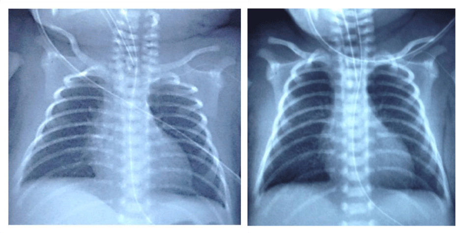
She further deteriorated at 21 hours of life. She developed shock, requiring multiple crystalloid boluses and inotropes. She also had coagulopathy with left grade II Intraventricular haemorrhage requiring packed cell and fresh frozen plasma transfusions. Bedside echocardiogram showed poor cardiac contractility with tricuspid regurgitation. Despite absence of pneumothorax or worsening pneumonia (Figure 1), the baby was unable to maintain saturation with either conventional or high frequency ventilation. She finally succumbed to her illness at 32 hours of life.
Blood culture taken at 27 hours of life and post-mortem blood from intracardiac and peripheral samples all showed pure growth of Candida albicans. The mother’s antenatal and postnatal vaginal swabs also showed pure growth of Candida albicans.
The second twin was relatively well. Because of the first twin’s culture positivity, she was empirically covered with IV Fluconazole for 2 weeks. Both her blood and urine cultures had no growth. She was discharged well on day 16 of life with a weight of 1.79kg.
Discussion
Invasive Candidiasis is a frequent nosocomial infection in neonatal intensive care units11 while congenital Candidiasis is a rare entity for which no treatment protocol is available to date.
Congenital Candidiasis manifests widely, ranging from diffuse skin eruptions to systemic disease, causing intrauterine or early neonatal death [12]. Diana et al described the typical appearance of congenital cutaneous Candidiasis as “white dots on the placenta and red dots on the baby” [11-15]. This refers to white micro abscesses on placenta and umbilical cord which can be diagnosed microscopically and are suggestive of Candida placentitis, and generalized eruption of erythematous macules, papules and/or pustules on the newborn [2]. Skin scraping microscopy will show pseudohyphae [2].
The condition is acquired via ascending infection or during delivery. Like this patient, most cases have been reported with intact amniotic membranes. There is evidence that Candida albicans can penetrate intact membranes causing vertical transmission [14]. For our patient, the mother had premature contractions and significant vaginal discharge caused by Candida infection. She was never treated with anti-fungal prior to delivery as there was no clinical Chorioamnionitis and Candida albicans is a common commensal in the female genitalia.
To date, < 100 cases of congenital Candidiasis have been reported [11-16]. In addition to our patient, we found another 10 case reports describing invasive congenital Candidiasis through Pub Med search as far back as year 2000 (Table 2). Six literatures reported twin pregnancies, while 4 reported singleton pregnancies. There were 9 preterm deliveries (82%) at 26-34 weeks and birth weights ranging 425-2362g [6-14]. Among the 15 preterm babies, 9 cases including one intrauterine death had candidemia sepsis and 6 of them died (5 died on day 1-4 of life) [6-14]. For the 9 cases of systemic Candidiasis, one presented with intrauterine death, six with sepsis, one with skin rash only and one was asymptomatic [6-14]. For the 6 deaths, 3 had rapid clinical deterioration and died before systemic anti-fungal was initiated [6,9,10], just like our patient. Two cases were started on systemic anti-fungal after blood culture showed Candida [14]. For our patient, the blood culture was reported as Candida 12 hours after her demise. One patient who died at day 128 of life did not have detailed documentation on when anti-fungal was initiated [12]. For the 3 survivors of Candida septicaemia, two received early anti-fungal therapy as the mothers had candidial Chorioamnionitis [7] and candidemia sepsis [13] prior to delivery. One patient presented with typical rash at birth, relatively without features of candidemia sepsis [11]. In these reports, babies with only candiduria or cutaneous Candidiasis all survived [1,8,10,14].
Chen, et al described that for premature twins, first twins have higher risk of invasive disease compared to second twins [14]. First twins are the first to be affected by ascending infection while second twins are usually infected during delivery [14]. Thus, second twins are generally less severely affected than first twins, as observed in 4 out of 6 twin case reports [6,7,10,14] and in our patient.
For our index patient, a striking feature was pancytopenia from birth with rapid clinical deterioration. The baby did not have mucocutaneous involvement, but presented with fulminant systemic infection, leading to multi-organ failure. Chest radiographs did not show changes likely to have caused secondary apnoea and the subsequent clinical deterioration. This made us consider retrospectively whether systemic anti-fungal should have been started when she deteriorated, with decreasing white cell and platelet counts.
From our review, we noticed that babies with positive candidial growth on blood cultures generally had worse outcomes than those with cutaneous Candidiasis or candiduria. Early recognition of congenital Candidiasis with prompt systemic anti-fungal was crucial [14]. The clinical course became fulminant and anti-fungal therapy was ineffective when there were features of candidemia sepsis [14]. Therefore, it is important to have a high index of suspicion for congenital Candidiasis especially if there is significant maternal fungal infection and invasive procedures like cervical cerclage, intrauterine device and amniocentesis [14]. In the presence of risk factors, we should consider systemic anti-fungal early if there is no clinical improvement with antibiotics [14].
Conclusion
We report a case of invasive congenital Candidiasis with no mucocutaneous involvement in a preterm twin pregnancy, who had rapid clinical deterioration within the first two days of life, resulting in early neonatal death. We postulate that the patient’s outcome may have been improved if systemic anti-fungal was initiated early. However, due to the uncertainty of this rare congenital infection, more evidence is required to change current guidelines.
Acknowledgements
This work was supported by the Department of Paediatrics, Hospital Tuanku Ja’afar, Seremban.
- Carlos Aldana-Valenzuela, Margarita Morales-Marquec, Javier Castellanos-Martinez & Manuel DeAnda-Gómez. Congenital Candidiasis: A Rare and Unpredictable Disease. J Perinatol. 2005; 25: 680-2. https://goo.gl/Mueii6
- Darmstadt GL, Dinulos JG & Miller Z. Congenital cutaneous candidiasis: clinical presentation, pathogenesis, and management guidelines. Pediatrics. 2000; 105: 438-44. https://goo.gl/KTmkT7
- Cotch MF, Hillier SL, Gibbs RS & Eschenbach DA. Epidemiology and outcomes associated with moderate to heavy Candida colonization during pregnancy. Vaginal Infections and Prematurity Study Group. Am J Obstet Gynecol. 1998; 178: 374-80. https://goo.gl/TFp3uu
- Roque H, Abdelhak Y & Young BK. Intra amniotic candidiasis. J Perinat Med 1999; 27: 253−262.
- Qureshi F, Jacques SM, Bendon RW, Faye-Peterson OM, Heifetz SA, Redline R, et al. Candida funisitis. A clinicopathologic study of 32 cases. Pediatr Dev Pathol. 1998; 1: 118-24. https://goo.gl/3njrMR
- Friebe-Hoffmann U, Bender DP, Sims CJ, Rauk PN. Candida albicanschorioamnionitis associated with preterm labour and sudden intrauterine demise of one twin. A case report. J Reprod Med. 2000; 45: 354-6. https://goo.gl/gB9Xbx
- Arai H, Goto R, Matsuda T, Saito S, Hirano H, Sanada H, Sato A, Takada G. Case of Congenital Infection with Candida Glabrata In One Infant In A Set Of Twins. Pediatr Int. 2002; 44: 449-50. https://goo.gl/1GXqRj
- Wang SM, Hsu CH, Chang JH. Congenital Candidiasis. PediatrNeonatol. 2008; 49: 94-96. https://goo.gl/oAyn5c
- Krallis N, Tzioras S, Giapros V, Leveidiotou S, Paschopoulos M, Stefanou D, et al. Congenital candidiasis caused by different Candida species in a dizygotic pregnancy. Pediatr Infect Dis J. 2006; 25: 958-9. https://goo.gl/Wea98t
- Carmo KB, Evans N, Isaacs D. Congenital candidiasis presenting as septic shock without rash. Arch Dis Child. 2007; 92: 627-8. https://goo.gl/veYWtS
- Tiraboschi IC, Niveyro C, Mandarano AM, Messer SA, Bogdanowicz E, Kurlat I, et al. Congenital candidiasis: confirmation of mother-neonate transmission using molecular analysis techniques. Med Mycol. 2010; 48: 177-81. https://goo.gl/da6VHz
- Li MJ, Hsueh PR, Lu CY, Chou HC, Lee PI, Chang LY, et al. Disseminated candidemia refractory to caspofungin therapy in an infant with extremely low birth weight J Formos Med Assoc. 2012; 111: 46-50. https://goo.gl/bnpCN9
- Pineda C, Kaushik A, Kest H, Wickes b, Zauk A. Maternal sepsis, chorioamnionitis, and congenital Candida Kefyr infection in premature twins. Pediatr Infect Dis J. 2012; 31: 320-2. https://goo.gl/pt3Kbw
- Chen WY, Chen SJ, Tsai SF, Tsao PC, Tang RB, Soong WJ. Congenital systemic fungus infection in twin prematurity – a case report and literature review. AJP Rep. 2015; 5: e46-50. https://goo.gl/Y6M3Dn
- Diana A, Epiney M, Ecoffey M, Pfister RE. “White dots on the placenta and red dots on the baby”: congenital cutaneous candidiasis – a rare disease of the neonate. Acta Paediatr. 2004; 93: 996-9. https://goo.gl/cjWqgM
- Iwatani S, Murakami Y, Mizobuchi M, Fujioka K, Wada K, Sakai H, et al. Successful management of an extremely premature infant with congenital Candidiasis. AJP Rep. 2014; 4: 5-8. https://goo.gl/wZBULJ


Sign up for Article Alerts