Does the Nervous System Constrain Lower Extremity Force Output of the Uninvolved Limb during Running in Patients after anterior Cruciate Ligament Reconstruction?
Hackney James M*, Wade Michael G and Larson Christopher M
Associate Professor of Physical Therapy, Missouri State University, Department of Physical Therapy, 901 South National Avenue
*Address for Correspondence: James Hackney, Associate Professor of Physical Therapy, Missouri State University, Department of Physical Therapy, 901 South National Avenue, USA, Fax: 417-836-6229; Tel: + 417-836-6239; E-mail: jameshackney@missouristate.edu
Submitted: 21 July 2017; Approved: 04 September 2017; Published: 05 September 2017
Citation this article: James MH, Michael GW, Christopher ML. Does the Nervous System Constrain Lower Extremity Force Output of the Uninvolved Limb during Running in Patients after anterior Cruciate Ligament Reconstruction? Int J Sports Sci Med. 2017;1(1): 006-009.
Copyright: © 2017 James MH, et al. This is an open access article distributed under the Creative Commons Attribution License, which permits unrestricted use, distribution, and reproduction in any medium, provided the original work is properly cited
Download Fulltext PDF
This study was designed to test whether Maximum Vertical Ground Reaction Force (MVGRF) of the nonsurgical limb of patients after ACLR could be predicted from the MVGRF of the surgical limb while running with a stiff surface underfoot. We measured the MVGRF of 6 patients who were 5 to 14 weeks post ACLR while they ran for 60 seconds in shoes modified with stiff, 1 cm. thick outsoles. The MVGRF measured from the patients’ surgical limbs accounted for .952 of the variance of their non-surgical limbs. One explanation for this finding is that the central nervous system constrained the MVGRF of the non-surgical side in order to maintain an equivalent vertical forces during gait and prevent asymmetry.
Introduction
Serious injury of the Anterior Cruciate Ligament (ACL) of the knee is a widespread problem, especially among participants in recreational or competitive sports. According to [1], about 1 of every 3000 individuals in the U.S. suffers an ACL injury at some point; with over 100,000 occurrences reported annually. For example, in 2002, at least 7,000 ACL injuries were reported in high school female basketball players, which accounted for 1 out of every 65 participants. Rates of ACL injury in soccer have been estimated at up to 3.7 for every 1000 hours of active participation [2], accounting for thousands of additional ACL injuries yearly.
Isokinetic studies have demonstrated that individuals who have recently undergone Anterior Cruciate Ligament Reconstruction (ACLR) generally demonstrate significant deficits in knee joint torque relative to the non-surgical side, especially in knee extension [3-7] found that only 15% of adolescents who had undergone ACLR had recovered 85% of knee extension torque when measured at 180o per second, compared to the non-surgical side, 3 months post-operatively. [5] Found that patients, a year postoperatively, had recovered an average of 82% of isokinetic knee extension torque measured at 180o.
Based upon the greater knee joint torque capacity, one would expect that the non-surgical limbs on these patients to be able to generate a higher ground reaction force in running than the surgical limb, since ground reaction force is the net results of the total joint moments of the lower extremities [8]. The relationship between knee joint torques and maximum ground reaction force was demonstrated in research from [9] who showed that athletes with lower isokinetic strength and power values also had lower ground reaction forces and lower LE stiffness values in jumping. [10] Also showed both lower knee isokinetic torques and maximum ground reaction force in jumping in the non-dominant compared to dominant LE. However, asymmetry of ground reaction force in running would ill-serve the patients if this took place because it would result in a significantly greater energy requirement for running [11]. It would therefore be a logical strategy for the Central Nervous System (CNS) to constrain the stronger limb to produce a submaximal vertical force in order to maintain force output symmetry during running gait. This strategy is more feasible than it would be to somehow facilitate a limb impaired by injury, surgery, and disuse to produce a supramaximal force. Research published by [12] suggested that this may be taking place in patients after ACLR, when they showed that isokinetic knee torque was not predictive of external knee moments during walking (although [12]. measured ground reaction forces for the calculation of the external moments, they did not report them).
With this study, we intended to investigate whether the Maximal Vertical Ground Reaction Force (MVGRF) generated by the surgical limb in patients 14 weeks or less status post unilateral ACL reconstruction can predict the force output of nonsurgical limbs during running with a hard surface underfoot. Evidence of this relationship will imply that potential force output of the stronger (non-surgical) limb is very accurately constrained by the CNS in order to avoid asymmetry of vertical force in running gait.
Materials and Methods
Participants
Six patients (4 female, 2 male) participated in this study. Their ages ranged from 15 to 30 years (mean = 20.0 ± 5.48), and their body mass ranged from 42 to 94 kg (mean = 63.42 ± 17.64). The patients had all undergone ACLR (and in one case minor meniscectomy) for unilateral anterior cruciate ligament rupture. In four cases, the graft source was patellar tendon allograft, in one case, it was patellar tendon autograft, and in one case, it was hamstring tendon autograft. By the time of experimental trial, all patients had met the criteria to return to running as part of their rehabilitation. These criteria included being free of pain, having full knee extension and full or close to full knee flexion range of motion, and having manual muscle test grade of the post-surgical limb for knee extension which was categorically the same as that of the non-surgical limb (e.g., 4+/5), and like-wise for knee flexion. The range of time between surgery date and experimental trial was 35 to 98 days (mean = 66.5 days ± 21.63).
Instrumentation
Pedar insole foot vertical force measurement system: The Pedar insole system (Novel Electronics, Munich, Germany) was used to measure MVGRF. The data gathering components of this instrument are insoles that are shaped like the insole of a shoe, and are constructed of a matrix of 99 sensors, each with an effective sensor area of approximately 1.5 cm2. These insoles were placed between the subjects’ feet and the commercial insoles of the modified shoes, and were cable connected to mobile data gathering box, which was in turn cable connected to a computer. Because of the mathematical relationship between force and pressure (force = pressure X area) a researcher is easily able to analyze data either as pressure or as vertical force.
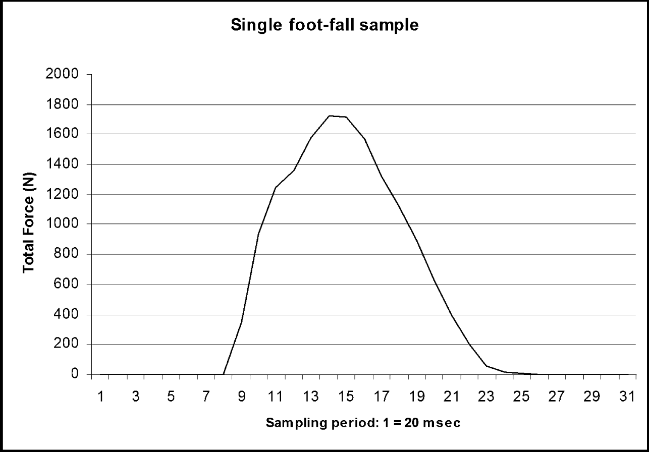
The sampling rate of the Pedar used in this study was 50 Hz (once every 20 milli-seconds). Researchers have previously demonstrated that the integrals of vertical force profiles generated by the Pedar at 50 Hz correlated closely to those generated by force plates sampling at 99 Hz, r = .99 [13] and at 1000 Hz, r = .95 [14]. We expected that at 50 Hz, the foot contact force profile (an example of which is illustrated in Figure 1) would be sampled at least 4 times before it reached the propulsive peak vertical force, and at least 10 times for even the shortest foot contact period. As described in the article by [15] the propulsive peak ground reaction force reaches its maximum at about 85 milliseconds. The propulsive peak is the parameter of interest in this study because it reflects (among other factors) the output of muscular tensions.
Modified shoes: Shoes (‘Athletic Works, Major’, Walmart, Bentonville, AR, USA) were modified by gluing an outsole attachment to the factory outsole (see Figure 2). The outsole attachment was 1 cm thick pad of ethyl vinyl-acetate, glued onto each of 6 sets of identical shoes of successive sizes (American men’s sizes 6 ½, 7 ½, 8 ½, 9 ½, 10 ½, and 12). The outsole had a durometer rating of 55 Shore. The use of these shoes insured that stiffness underfoot was uniform between all patient participants. The modified shoes had a semi-curved last to accommodate the maximum number of participants.
Procedure: Volunteer participants read and signed consent forms and were then weighed for body mass. The Pedar insoles of the appropriate size were placed inside the shoes by one of the investigators prior to the subjects donning them. These data were collected initially as part of another experiment investigating the ability of patients who had undergone ACLR to adjust MVGRF to change in hardness underfoot during running [16].
During each trial, each patient walked on the treadmill for 60 seconds as a warm up, and then advanced the treadmill speed to a self-selected, comfortable running speed. Once they had reached their self-selected speed, he or she signaled the investigator to begin data collection, which continued for 60 seconds.
During the running trials, the mobile data gathering unit was supported by the investigator standing beside the participant. The vertical ground reaction force data collection took place in real time via cable connection from the mobile data gathering unit to a computer. The data were normalized by dividing resultant Newtons of force by kilograms body mass.
Figure 1 portrays an example of the output of the Pedar from a single foot contact. In the vertical force profile generated, the passage of time is represented on the x-axis, and force underfoot is represented on the y-axis. For each force profile, the 60 consecutive msec. with the highest force for each foot contact (representing the interval enclosed by 4 data points per each foot contact) was averaged to generate a MVGRF value for that foot contact, and then all resulting force values from every foot contact within the 60 second trial were averaged to generate a mean MVGRF value for that limb. This ranged from 60 to 100 foot contacts per limb, depending upon the cadence of the participant.
Statistical Analysis: Trial means were used to perform linear regression analysis on the body-mass normalized MVGRF in running between the surgical and non-surgical side. The surgical limb trial mean values for each participant served as the predictor variable, and the non-surgical trial mean values as the response variable.
Results
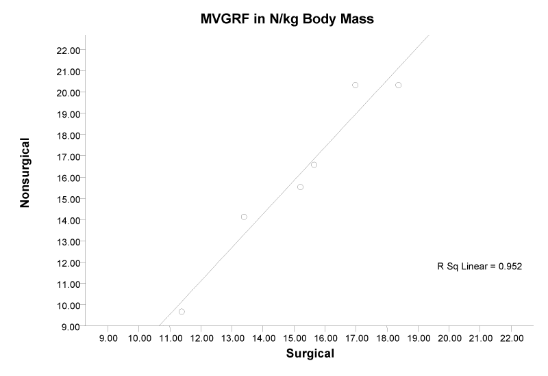
Coefficient of regression (r2) indicated that the MVGRF of the surgical limb accounted for .952 of the variance in MVGRF of the non-surgical limb, P = .001. See Figure 3 for the scatter plot and regression line of the surgical vs. non-surgical limb MVGRF for each patient.
Discussion
The finding of this study is that with patients who have had recent (5 to 14 weeks previous) ACLR, while running in shoes modified with a hard outsole, MVGRF on the nonsurgical side can be predicted from MVGRF on the surgical side. Because previous studies describe that the non-surgical limb can generate greater knee muscle torque compared to the surgical side, it stands to reason that the nonsurgical side is easily able to exceed the MVGRF of the surgical side. The findings suggest that the central nervous system may be constraining the MVGRF of the non-surgical side in order to maintain symmetry in running gait during running with a hard surface underfoot.
The study we have described was novel in examining the effect of an anatomical impairment upon the phenomenon of regulation of support vector, i.e. the force component of the stiffness ratio; k = f/∆l. The equality of force demonstrated between the surgical and non-surgical limb during running with these patients suggests that the regulation of stiffness is a robust phenomenon, and that the CNS is able to use neural input regarding force output to maintain inter-limb coordination of the support vector during running.
There were three potential limitations to this study. Since no kinematic data was collected, we cannot show for certain that, although the MVGRF of the non-surgical limb during running was very highly predictable from that of the surgical limb, stiffness did not vary between limb conditions. Second, since we did not collect quantitative knee joint torque with these patients, we cannot demonstrate for certain that there was a significant knee joint torque deficit for the surgical limb compared to the non-surgical limb with these particular patients. Third, there is a question regarding whether the 50 Hz sampling rate of MVGRF used in this study was adequate to capture the data relevant to the experimental question.
First, we will attempt to address the concern that limb stiffness during running was not measured, only MVGRF. In future studies, it will be important to measure kinematic as well as ground reaction force data, in order to be able to make conclusions about lower limb stiffness less equivocally. However, since variation of stiffness in the presence of equal force would have such a profound and maladaptive effect on the vertical excursion of COM, it is reasonable to assume that lower limb stiffness as well as MVGRF was coordinated between the surgical and non-surgical limbs with these patient participants.
The second potential concern was regarding the lack of data regarding knee joint torque differences between surgical and non-surgical limbs. It is conceptually possible that since knee joint torque was not quantifiably measured with these patients, that knee joint torque production was close to equal between the surgical and nonsurgical limbs with these individuals. However, because the literature has been so reliable in documenting it [4-7] torque deficit on the surgical side may also be reasonably assumed with these patients.
The issue of sampling rate presents a potential limitation in the interpretation of the data in this study. Although empirical data show that ground reaction force sampled at 50Hz is strongly correlated to data sampled at higher frequencies [13,14], theoretically it can be argued that this sampling rate may not be adequate. If the force profile is considered as the top phase of a sine wave, the highest frequency of such a wave generated by our data was 2.5 Hz. The 50 Hz sampling rate samples this wave every 18 degrees, and therefore, the greatest deviation from the actual peak may be sine of 72 degrees, the value of which is .951. Therefore, the greatest amount of potential error from the actual peak with the shortest duration force profile is almost. 05 [17], implying that the actual peak force may not have been sampled in several of the force profiles of the data. At least one characteristic of the data which helped to protect it from this limitation was the number of data points that contributed to each trial mean. In the method of sampling used in this study, each foot contact in the trial contributed a data point, as described in the “methods”. If the conclusions of our study were based upon one or a few data points, then the potential error caused by the low sampling rate might have a large impact on the trial means analyzed. However, since between 60 and 100 data points were averaged for each trial mean, the potential error as a result of low sampling rate is mitigated.
An important limitation of this study is also the small sample size. Although the effect size is dramatic (r2 = .952), with a sample of six patients, it is possible that the size of the effect is due to chance [18,19]. Therefore, our findings should be considered preliminary, laying the groundwork for a similar study with a more robust sample.
Conclusion
During running with a hard surface underfoot for patients between 5 and 13 weeks after ACL reconstruction, it appears possible to very accurately predict the MVGRF of the non-surgical limb from that of the surgical limb, despite the generally much greater torque production capacity of the non-surgical limb. This suggests that the CNS is constraining the non-surgical side from producing the entire force than it potentially could order to avoid asymmetry of gait.
- Dugan S. Sports related knee injuries in female athletes: What gives?. Am J Phys Med Rehabil. 2005; 84: 122-130. https://goo.gl/5o3cdy
- Fauno P, Wullf Jakobson B. Mechanism of anterior Cruciate ligament injuries in soccer. Int J Sports Med. 2006. 27: 75-79. https://goo.gl/jZTh7G
- Heijne A, Werner S. A two year follow-up of rehabilitation after ACL reconstruction with patellar tendon or hamstring grafts: a prospective randomized outcome study. Knee Surg Sports Traumatol Arthrosc. 2010; 18: 805-813
- https://goo.gl/EvYxpW
- Konishi Y, Oda T, Tsukazaki S, Kinugasa R, Fukubayashi T. Relationship between quadriceps femoris muscle volume and muscle torque after anterior cruciate ligament repair. Scand J Med Sci Sports. 2007; 17: 656-661. https://goo.gl/o1xbDU
- Lee S, Seong SC, Jo H, Park YK, Lee MC. Outcome of anterior cruciate ligament reconstruction using quadriceps tendon autograft. Arthroscopy. 2004; 20: 795-802. https://goo.gl/qxGWsq
- Stefanska M, Rafalska M, Skrzek A. Functional assessment of knee muscles 13 weeks after anterior cruciate ligament reconstruction-a pilot study. Ortop Traumatol Rehabil. 2009; 11: 145-155. https://goo.gl/PauSrN
- Wells L, Dyke J, Albaugh J, Ganley T. Adolescent anterior cruciate ligament reconstruction: A retrospective analysis of quadriceps strength recovery and return to full activity after surgery. J Pediatr Orthop. 2009; 29: 486-489. https://goo.gl/RACGTB
- Macpherson JM. Why biomechanics? Posture and Gait. 1992; 340-343.
- Harrison AJ, Keane S P, Coglan J. Force-velocity relationship and stretch-shortening cycle function in sprint and endurance athletes. J Strength Cond Res. 2004; 18: 473-479. https://goo.gl/gqDKgR
- Newton R, Gerber A, Nimphius S, Shim J, Doan BK, Robertson M, et al. Determination of functional strength imbalance of the lower extremities. J Strength Cond Res. 2006; 20: 971-7. https://goo.gl/oKBYDj
- Alexander R. Energy saving mechanisms in walking and running. J Exp Biol. 1991; 160: 55-69. https://goo.gl/HcEPqS
- Gokeler A, Schmalz T, Knopf E, Freiwald J, Blumentritt S. The relationship between isokinetic quadriceps strength and laxity on gait analysis parameters in anterior cruciate ligament reconstructed knees. Knee Surg Sports Traumatol Arthrosc. 2003; 11: 372-378. https://goo.gl/MfEojU
- Barnett S, Cunningham J, West S. A comparison of vertical force and temporal parameters produced by an in-shoe pressure measuring system and a force platform. Clin Biomech. 2000; 15: 781-785. https://goo.gl/4qDkwX
- Cordero A, Koopman G, van der Helm F. Use of pressure insoles to calculate the complete ground reaction forces. J Biomech. 2004; 37: 1427-1432. https://goo.gl/abGh7R
- Clarke T, Frederick E, Cooper L. Effects of shoe cushioning upon ground reaction forces in running. Int J Sports Med. 1983; 4: 247-251. https://goo.gl/U6Zvqt
- Hackney J, Wade M, Larson C, Smith J, Rakow J. Preservation of proprioceptive sensitivity in running gait in anterior cruciate ligament reconstructed subjects. Physiotherapy Theory and Practice. 2010; 26: 289-296.
- Qian S, Chen D. Joint Time-Frequency Analysis: Methods and Applications. Upper Saddle River NJ. Prentice Hall. 1990. https://goo.gl/2S2AtB
- Steyerberg E, Bleeker S, Moll A, Grobbee D, Moons K. Internal and external validation of predictive models: a simulation study of bias and precision in small samples. J Clin Epidemiol. 2003; 56: 441-447. https://goo.gl/R2TUdo
- Kernozek T, LaMott E, Dancisak M. Reliability of an in-shoe pressure measurement system during treadmill walking. Foot and Ankle International. 1996; 17: 204-209. https://goo.gl/nisrQa


Sign up for Article Alerts