Congenital Esophageal Duplication Cyst in the Adult: Case Report
Pablo Andrade Martinez Garza1* and Daniel Gonzalez Hermosillo Cornejo2
1General Surgery Department, Medica Sur Hospital, Mexico City, Mexico
2Facultad Mexicana de Medicina, Universidad La Salle, Mexico City, Mexico
*Address for Correspondence: Pablo Andrade Martinez Garza, General Surgery Department, Medica Sur Hospital and Mexican Faculty of Medicine of La Salle University, Mexico City, Mexico, Tel: +520-175-747-636-00; E-mail: dr.gonzalezhermosillo@gmail.com
Submitted: 16 July 2019; Approved: 24 July 2019; Published: 25 July 2019
Citation this article: Hermosillo Cornejo DG, Martinez Garza PA. Congenital Esophageal Duplication Cyst in the Adult: Case Report. Open J Surg. 2019;3(1): 024-027.
Copyright: © 2019 Hermosillo Cornejo DG, et al. This is an open access article distributed under the Creative Commons Attribution License, which permits unrestricted use, distribution, and reproduction in any medium, provided the original work is properly cited
Keywords: Esophageal cyst; Esophageal duplication
Download Fulltext PDF
The esophageal duplication cyst is a congenital defect of the digestive tract. It has an estimated prevalence of 0.012%, with higher predominance in males. Although it is a common finding in children, diagnosis of an esophageal duplication in adults is rare. Following ileal duplication, esophageal is the second most common duplication of the gastrointestinal tract, representing the 10-15% of all gastrointestinal duplication defects. For esophageal duplication, there are two main variants: cystic and tubular, the latter being the least common. They are usually developed during the third to fifth week of gestation due to failure of the vacuolar coalescence. Duplication cysts are commonly located in the distal third of the esophagus. Treatment should always be surgical, even at the asymptomatic stage of disease, given the possibility of symptom development and complication appearance. Here we present a case of an adult patient presenting with an esophageal duplication cyst with a brief literature review.
Introduction
Duplication of the gastrointestinal tract is observed in 1 of 25,000 deliveries [1]. Although diagnosis in children is common, esophageal duplication is a very infrequent entity in the adult [1]. Esophageal duplication can happen in two different modalities: cystic in 80% of the cases, and tubular in 20% [2]. A literature review carried out by Arbona, et al found a total of 91 cases reported worldwide [3].
The pathophysiology of the disease starts during the 5th to 8th week of embryological development, when there is a failure in vacuolar coalescence form the esophageal secretions [3,4]. Mediastinal cysts can be classified as esophageal duplications if they are located near the esophageal wall and if they are covered by two muscle layers, with squamous, columnar, cuboid, or pseudostratified epithelium. Esophageal duplication cysts occur in the lower third esophagus in 60% of the cases [3]. Esophageal cysts in adults are often incidental findings, and it has been described that they are usually more symptomatic in older ages. Furthermore, the appearance of complications such as intracystic hemorrhage, perforation, infection, and squamous metaplasia is greater with age [5,6].
In adults, the esophageal duplication cysts often present as asymptomatic, however; in some cases, symptoms depend on the location and size of the cyst. In adults, the most common symptoms include dysphagia, abdominal pain, and chest discomfort secondary to compression. Less frequent symptoms are bleeding, lymphadenopathy, difficulty breathing, and weight loss. Diagnosis of an esophageal duplication cyst is established with a thoracic Computed Tomography (CT), an Upper Gastrointestinal Series (UGI), and a Transesophageal Echocardiography (TEE) [7]. However, in order to evaluate the content of the cyst and differentiate between the solid and liquid components, the Endoscopic Ultrasound (EUS) is the most useful tool for diagnosis [8]. Magnetic Resonance Imaging (MRI) can also play an important role in diagnosis, especially when trying to differentiate between an esophageal cyst and mediastinal tumors, and to delineate the anatomic relationships of the cyst to adjacent structures [3,7]. In all cases, the gold standard for treatment is the surgical resection by thoracotomy or videothoracoscopy [9-11].
Case Presentation
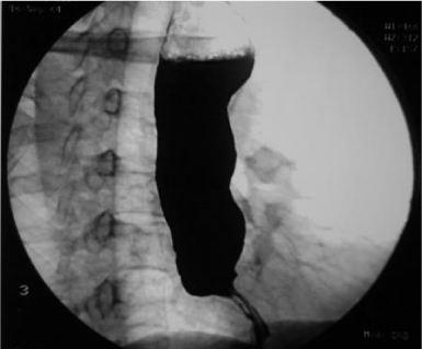
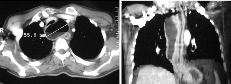
We present the case of a 41-year-old male patient, with an unremarkable medical history. The chief complaint started 3 months before admission, and it was characterized by occasional dysphagia to solids that progressed to dysphagia to liquids. He also presented with hyporexia, sitophobia, asthenia, and weight loss. The initial workup suggested the presence of esophageal stenosis. Therefore, it was decided to perform an Esophagogastroduodenoscopy (EGD) that showed proximal esophageal dilation with distal stenosis (Figure 1). After the EGD, a panendoscopy was performed, and revealed an abscess on the distal 2/3 of the esophagus, that required endoscopic drainage. Furthermore, a biopsy was taken from the site of the stenosis. The histopathologic report revealed an esophageal adenocarcinoma. A new CT scan showed the presence of a cystic structure of 117 x 57 x 59 mm in size, located in the posterior mediastinum, close to the esophageal wall. The cyst was composed by its own walls and they did not communicate with adjacent structures. The CT scan also revealed multiple hepatic lesions with solid characteristics (Figure 2).
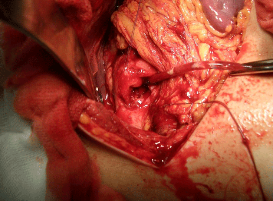
Altogether, these results led to the diagnosis of an esophageal duplication cyst, accompanied by stage IV esophageal cancer that was causing esophageal stenosis. Due to the diagnosis, the patient was not eligible for resective surgery, however; the persistence of symptoms led to the performance of palliative surgery. An exploratory laparotomy with an anterior gastrotomy, retrograde dilation of the distal part of the esophagus and the esophagogastric junction, with placement of endoprosthesis (Figure 3) was carried out. The patient had a favorable evolution and tolerated food at the 4th day after surgery and was discharged afterwards.
Discussion
The diagnosis of an esophageal duplication cyst is incidental in approximately 37% of the cases [12]. In adults, the most frequent symptoms are dysphagia and chest pain. Also, it has been described that the most common location for esophageal duplication cysts is at the posterior mediastinum. It is important to consider that the cyst topography is of greater importance than its volume, due to the risk of compression to adjacent and vital structures [3]. Cysts located in the superior mediastinum may produce more compression symptoms than those located in the middle and lower areas. However, dysphagia only occurs if the esophageal lumen is being considerably compressed by the cyst [13]. In the case presented here, the cyst was located in the posterior mediastinum, with right predominance. It was firmly attached to the esophagus, which could explain the patient’s dysphagia.
Imaging studies like CT, UGI, TTE, and MRI can help to rule out malignancy and evaluate the topographic relationships of the lesion, in order to plan the most appropriate surgical strategy [3,7,8]. Furthermore, esophageal endoscopy allows the evaluation of the esophageal epithelia status, the presence of vessels, and the transesophageal aspiration of the cyst, which has been described as a method for both diagnosis and treatment of the disease [14]. However, it has been reported that puncture and aspiration of the cyst through endoscopy does not provide any additional information for diagnosis and increases the risk of cyst infection [15]. Definitive diagnosis can only be established by the pathology evaluation of the surgical piece. Because of the high risk of neoplastic transformation of the epithelia, the treatment of choice for esophageal duplication cysts is the complete resection of the lesion. This intervention is not inherently complex, but when the cyst becomes symptomatic, it can become dangerous. Therefore, any mediastinal lesion radiologically-compatible with an esophageal duplication cyst, should be resected.
During the surgical procedure, the esophageal duplication cysts should be carefully handled and removed, with preservation of the esophageal muscles and vagus nerve. Then, the integrity of the esophageal mucosa should be verified by insufflating a nasogastric tube. After enucleating the cyst, it is of great importance to adequately approximate the muscular ends of the esophagus, to avoid the appearance of a pseudodiverticulum and postoperative dysphagia. Some authors use the video-assisted thoracoscopy to treat these type of lesion, as it has shown to be as effective and safe as open surgery in the treatment of esophageal duplication cysts. It has been described that the use of video-assisted thoracoscopy leads to a better and faster recovery, when compared to open surgery [9]. These lesions tend to be poorly vascularized and resection should not be complicated. In the presented case, the lesion presented a firm adherence to the esophagus with an accompanying finding of adenocarcinoma located at the distal portion of the esophagus. These were determining factors with the decision to not resect the cyst and to only alleviate the esophageal obstruction with dilation and endoprosthesis.
Conclusion
The diagnosis of an esophageal duplication cyst in the adult is rare. These have a higher predominance in males and they mainly remain asymptomatic [3]. The esophageal duplication cysts may be diagnosed as an incidental finding, however; depending on their location and size, these may be associated with typical symptoms of dysphagia, chest compression, and weight loss. Once diagnosed, even when they are asymptomatic, esophageal cysts should be completely resected.
Other surgical options including esophagectomy, myocutaneous flaps and esophageal replacement procedures, is complex, and carries significant morbidity and mortality but may be indicated as needed [16].
In this case, the diagnosis of an esophageal duplication cyst was an incidental finding when treating an adult with dysphagia secondary to an esophagogastric adenocarcinoma. The present case is extremely rare because the diagnosis of the esophageal adenocarcinoma led to the discovery of the duplication cysts. The prevalence of two intrinsic esophageal diseases is not common and they should always be treated to avoid complications, or in this case; disease progression.
- Kim SK, Lim HK, Lee SK, Park CK. Completely isolated enteric duplication cyst: case report. Abdom Imaging. 2003; 28: 12-14. https://bit.ly/2LCOpaj
- Arbona JL, Fazzi J, Mayoral J. Congenital esophageal cysts: case report and review of literature. Am J Gastroenterol. 1984; 79: 177-182. https://bit.ly/2y9kJZP
- Yeung C, MacDonald B, Gilbert S. Esophageal duplication cyst. Shackelford’s Surgery of the Alimentary Tract. 2 Vol Set. 2019; 1: 490-495. https://bit.ly/2Mbpt9A
- Ikuo Watanobe, Yuzuru Ito, Eigo Akimoto, Yuuki Sekine, Yurie Haruyama, Kota Amemiya, et al. Laparoscopic resection of an intra-abdominal esophageal duplication cyst: a case report and literature review. Case Reports in Surgery. 2015; 1-8. https://bit.ly/2M9xzj2
- Zheng J, Jing H. Adenocarcinoma arising from a gastric duplication cyst. Surgical Oncology. 2012; 21: e97-101. https://bit.ly/2K3Ghwf
- Han IS, Kim GH, Lee SJ, Lee BE, I H, Kim YD. A case of hemorrhage of an esophageal duplication cyst improved by endoscopic drainage. Korean J Gastroenterol. 2019; 69: 363-367. https://bit.ly/2LEuUhA
- Takahashi K, Al-Janabi NJ. Computed tomography and magnetic resonance imaging of mediastinal tumors. J Magn Reson Imaging. 2010; 32: 1325-1339. https://bit.ly/2JYK9il
- Liu R, Adler DG. Duplication cysts: diagnosis, management, and the role of endoscopic ultrasound. Endosc Ultrasound. 2014; 3: 152-160. https://bit.ly/2SzP110
- Herbella FAM, Tedesco P, Muthusamy R, Patti MG. Thoracoscopic resection of esophageal duplication cysts. Dis Esophagus. 2006; 19: 132-134. https://bit.ly/2May9wK
- Noguchi T, Hashimoto T, Takeno S, Wada S, Tohara K, Uchida Y. Laparoscopic resection of esophageal duplication cyst in an adult. Dis Esophagus. 2003; 16: 148-150. https://bit.ly/30POYBe
- Aldrink JH, Kenney BD. Laparoscopic excision of an esophageal duplication cyst. Surg Laparosc Endosc Percutan Tech. 2011; 21: 280-283. https://bit.ly/2Z88DMv
- Cioffi U, Bonavina L, De Simone M, Santambrogio L, Pavoni G, Testori A, et al. Presentation and surgical management of bronchogenic and esophageal duplication cysts in adults. Chest. 1998; 113: 1492-1496. https://bit.ly/32NkIJ7
- Banner K, Helft S, Kadam J, Miah A, Kaushik N. An ususual cause of dysphagia in a young woman: esophageal duplication cyst. Gastrointest Endosc. 2008; 68: 793- 795. https://bit.ly/2SAo9hK
- Wiechowska-Kizlowska A, Wunsch E, Majewski M, Milkiewicz P. Esophageal duplication cysts: endosonographic findings in asymptomatic patients. World J Gastroenterol. 2012; 18: 1270-1272. https://bit.ly/2SDJ0Rg
- Diehl DL, Cheruvattath R, Facktor MA, et al. Infection after endoscopic ultrasound-guided aspiration of mediastinal cysts. Interact Cardiovasc Thorac Surg. 2010; 10: 338- 340. https://bit.ly/32MZuLg
- Gambardella C, Allaria A, Siciliano G, Mauriello C, Patrone R, Avenia N, et al. Recurrent esophageal stricture from previous caustic ingestion treated with 40-year self-dilation: case report and review of literature. BMC Gastroenterol. 2018; 18: 68. https://bit.ly/2JLcHgm


Sign up for Article Alerts