Two Times Hepatic Trauma Treatment: Balloon Tamponade and Hepatectomy-an Ideal Scenario?
Fernandes R1,3,6*, Vasconcelos H2,6, Cano R2,6, Steinbruck K3,6, Delai DB4, Souza JRN4, Duque I4, Maciel L5, Enne M2,6, D Oliveira M6, Capelli R6 and Basilio L2,6
1Professor of Surgery Department, Antonio Pedro University Hospital, Fluminense Federal University, Niterói, Brazil2Hepatobiliary Surgery, Ipanema Federal Hospital, Health Ministry, Rio de Janeiro, Brazil
3Hepatobiliary Surgery, Bonsucesso Federal Hospital, Health Ministry, Rio de Janeiro, Brazil
4Antonio Pedro University Hospital, Fluminense Federal University, Niterói, Brazil
5Nacional Cancer Institute, Rio de Janeiro, Brazil
6 Equipe Multidisciplinar Hepatobiliar, Brazil
*Address for Correspondence: Reinaldo Fernandes, Professor of Surgery Department, Antonio Pedro University Hospital, Fluminense Federal University, Niteroi, Brazil, Tel: +55-212-629-9025; ORCID iD: https://orcid.org/0000-0001-8759-2552; E-mail: rei.fernandes.jr@gmail.com
Submitted: 23 November 2019; Approved: 13 December 2019; Published: 16 December 2019
Citation this article: Fernandes R, Vasconcelos H, Cano R, Steinbruck K, Delai DB, et al. Two Times Hepatic Trauma Treatment: Balloon Tamponade and Hepatectomy-an Ideal Scenario? Open J Surg. 2019;3(2): 040-043.
Copyright: © 2019 Fernandes R, et al. This is an open access article distributed under the Creative Commons Attribution License, which permits unrestricted use, distribution, and reproduction in any medium, provided the original work is properly cited
Keywords: Penetrating liver trauma; Hepatic venous injury; Balloon tamponade; Balloon catheter tamponade; Damage control; Hepatectomy
Download Fulltext PDF
Introduction
Complex hepatic injuries are associated in the majority of cases with high mortality because intraoperative hemorrhage or postoperative sepsis. The mechanism of trauma varies since blunt trauma until penetrating, with white gun or gunshot wound.
The liver is the most frequently injured intraperitoneal organ, consisting of a relatively fragile parenchyma contained within the Glisson capsule, which is thin and does not afford it great protection. Hence, the parenchyma and its vasculature are very susceptible to blunt and penetrating trauma. The vasculature consists of wide-bore, thin-walled vessels with a high blood flow, and injury is usually associated with significant blood loss. Independent of the trauma`s grade, liver injury is still a major challenge even for the most experience surgeon [1].
We presented a case of penetrating liver injury that was successfully managed in two times, first, the damage control surgery at an emergency hospital and second, with a new procedure at a terciary and specialized center.
Case Presentation
A 20-year-old woman arrived at an emergency hospital brought by friends after being stabbed in the epigastrium region, left hemithorax below the eighth rib and right lumbar region. On arrival, the patient found to be lucid, complaining of abdominal pain with tachydyspnea (the respiratory rate 36 breaths per minute), the pulse 135 beats per minute and the systolic blood pressure 80mmHg. The physical examination revealed mucous membranes pale, hyperthympanism and abolished vesicular murmur in the left hemithorax, then, thoracic drainage was performed immediately at the emergency room. Two peripheric vascular access was done with 2 liters saline solution’s infusion with no response in blood pressure. Abdominal ultrassonography (FAST) showed moderate amount of free liquid with peritoneal irritation, then, she was transferred to the operating room with no further imaging examination.
Exploratory laparotomy with xifopubian incision revealed massive haemoperitoneum and right retroperitoneal haematoma. Through the epigastrium lesion, the knife tract traversed the transition of the segments IV and V/VIII of the liver but without exit in the posterior view, through the right lumbar lesion reached the upper pole of right kidney.
During the cavity inventary, Pringle maneuver (clamping of porta hepatis) was performed but the liver haemorrhage did not stopped and the patient was hemodinamically instable despite the transfusion of the only three red blood cells concentrates available, two units of fresh frozen plasma and noradrenaline. Because of that, the team decided to perform damage control surgery, and utilized the balloon tamponade fashioned with one inch Penrose drain and a 20G Foley catheter. This device was made putting the Foley catheter in the Penrose drain with ligation of the port, doing a balloon who was inflated with saline solution. When ready, was introduced through the deep liver lesion and was inflated; the bleeding stopped and the other port was exteriorized by abdominal wall for postoperative control. The superior right kidney lesion was sutured and the cavity was drained with two drains, in the right subfrenic region and the Morrison space.
Two days later, at Intensive Care Unit, the patient was extubated, without amines and satisfatory diuresis although hematuria; the thoracic drainage was functioning.
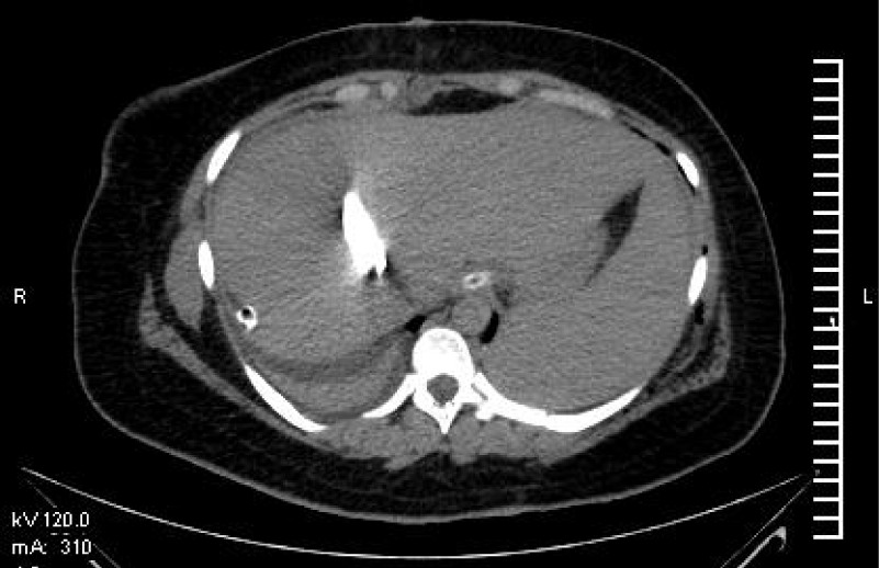
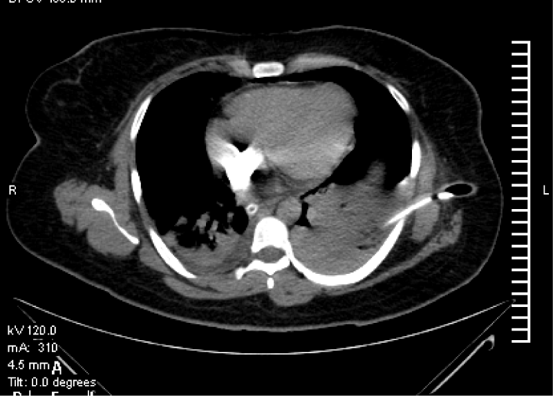
In the third day, the patient was submitted to a contrasted Computadorized Tomography (CT) of thorax and abdômen (Figures 1,2) besides contrast through the device and showed an intrahepatic emplacement with his distal port into the inferior vena cava and right atrium, with a probably right hepatic vein lesion.
After this images-CT scans-showed a high probability of vascular lesion associated with liver parenchyma-grade V AAST-OIS and considering that it had no technical conditions for the second approach, a contact with the Hepatobiliary Unit of Bonsucesso Hospital was made and the patient was transferred.
The preoperative planning made by the hepatobiliar surgery team was to perform right hepatectomy without removing the Foley catheter. An extension called Rio Branco was made joining the previous midline incision. There was a small amount of bile near of the Foley balloon who was inserted in IV/V/VIII liver segments transition. All the major vessels and the liver pedicle were isolated until the parenchyma section that was performed using ultrassonic aspirator SONOCA® after viewed by intraoperative ultrasound, that the distal port’s device was into the right atrium, arriving from the right hepatic vein (Figure 3).
The balloon was our guide during the parenchyma section, when it was finished, the right hepatic vein was isolated (Figure 3-yellow loop); we pulled the balloon, clamped the vein, sectioned and closed, a right hepatectomy was performed. The left remnant liver was fixed in the anterior peritoneum and the abdominal cavity was drained with tubular drain.
In the fifth postoperative day, in the Intensive Care Unit (ICU), the patient presented fever with no thoracoabdominal complications on CT scan but an echocardiogram showed a left atrium’s vegetation with no other anatomic or physiological cardiac alterations and positive S. aureus blood culture enforcing the initiation of venous antibiotic therapy for 3 weeks. On 3rd and 10th day, the thoracic and abdominal drain, respectively, were removed. hospital discharge was obtained 4 days after the end of clinical treatment.
Three months later, a new thoracoabdominal CT scan and echocardiogram were performed without any change and the patient returned for her professional life.
Discussion
Liver trauma corresponds approximately 5% of all trauma admissions [2,3]. The liver is the most common solid organ injured in blunt trauma, up to 40% in some series, and patients with hepatic injury usually have other concomitant injuries and the mortality depends on the degree of injury, becoming often fatal in grade VI. Minor injuries are the majority with 80% to 90% being grades I or II. Liver injury is the primary cause of death in severe abdominal trauma and has a 10% to 15% mortality rate [4].
In the first surgery, when was used the balloon, the patient was still instable after some hemoconcentrates and the team tried to do the fastest treatment; the technique of balloon tamponade using a Foley catheter and a penrose drain, described for us, in management of liver trauma, was first described by Morimoto et al in 1987, for a penetrating gunshot wound transfixing the liver and has subsequently been adopted by others [5]. Over the years some authors (Table 1) have applied a variety of tamponade devices for deep hepatic wounds and the use of catheter for many different situations for emergency control of hemorrhage besides reported the best result in liver trauma with stop bleeding in 83% of the all cases, with Blakemore balloon [6-10]. In summary, balloon catheter tamponade is a valuable tool for damage control of exsanguinating haemorrhage, it can be used in multiple anatomic regions and for variable patterns of injury, and can provide intrahepatic hemostasis through the compression exercised by the insufflated balloon [7,8]. The greatest advantage in relation to perihepatic package is the fact that, in some patients, is possible to desinsulflate without another procedure.
Perhaps one of the emergent treatment is the value of endovascular control of hepatic arterial bleeding. Asensio, et al. [11] showed a mortality rate of 12% in patients with grades IV and V liver injuries who underwent early post-operative angioembolisation, compared to 36% in those who did not. Sclafani, et al. [12] used interventional radiology to control intraperitoneal hepatic artery haemorrhage, as well as late complications of hepatic trauma including arteriovenous fistulas, pseudoaneurysms and intra-abdominal collections. The rate of nonsurgical treatment arrives, in some series, up to 85% of all such injuries with an overall success rate that exceeds 80%, thanks to adjunctive transarterial embolization [13].
The infectious event occurred with this patient, S. aureus infective endocarditis, in our opinion, was related with the presence of balloon catheter over one week and the initial cause of trauma, a white gun, who reached the vein after penetrating the entire abdominal wall.
The tamponade balloon in the hepatic transfixing lesions is a non-expensive, fast and easy procedure to perform. In the emergency rooms of countries that have a high index of abdominal trauma with gunshot wounds and they are not always endowed with all the surgical medical requirements to treat these severe and difficult hepatic lesions, which is the scenario of Brazil - in particular of the Rio de Janeiro state - the tamponade balloon showed to be a good option for surgical treatment [14,15].
Conclusion
Liver trauma is a challenging scenario, mostly in IV-V grade AAST-OIS and the surgeon must have the ability to throw hands of the damage control techniques and tools like tamponade balloon. It was only one case but it represents how difficult it is to have a survival in these cases and maybe in two times be the best way.
- Badger SA, Barclay R, Campbell P, Mole DJ, Diamond T. Management of liver trauma. World J Surg. 2009; 33: 2522-2537. PubMed: https://www.ncbi.nlm.nih.gov/pubmed/19760312
- Beitner MM, Suh N, Dowling R, Miller JA. Penetrating liver injury managed with a combination of balloon tamponade and venous stenting. A case report and literature review. Injury. 2012; 43: 119-122. PubMed: https://www.ncbi.nlm.nih.gov/pubmed/21917256
- Fabian TC, Bee TK. Liver and biliary tract. 6th ed. Feliciano DV, Mattox KL, Moore EK, editors. Trauma. McGraw-Hill; 2008. https://bit.ly/2Ph3HlJ
- Morimoto RY, Birolini D, Junqueira AR Jr, Poggetti R, Horita LT. Balloon tamponade for transfixing lesions of the liver. Surg Gynecol Obstet. 1987; 164: 87-88. PubMed: https://www.ncbi.nlm.nih.gov/pubmed/3798317
- Fraga GP, Zago TM, Pereira BM, Calderan TRA, Silveira HJV. Use of sengstaken-blakemore intrahepatic balloon: An alternative for liver-penetrating injuries. World J Surg. 2012; 36: 2119-2124. PubMed: https://www.ncbi.nlm.nih.gov/pubmed/22562452
- Demetriades D. Balloon tamponade for bleeding control in penetrating liver injuries. J Trauma. 1998; 44: 538-539. PubMed: https://www.ncbi.nlm.nih.gov/pubmed/9529186
- Ball CG, Wyrzykowski AD, Nicholas JM, Rozycki GS, Feliciano DV. A decade’s experience with balloon catheter tamponade for the emergency control of hemorrhage. J Trauma. 2011; 70: 330-333. PubMed: https://www.ncbi.nlm.nih.gov/pubmed/21307730
- Smaniotto B, Bahten LC, Nogueira Filho DC, Tano AL, Thomaz Junior L, Fayad O. Hepatic trauma: Analysis of the treatment with intrahepatic balloon in a university hospital of Curitiba. Rev Col Bras Cir. 2009; 36: 217-222. PubMed: https://www.ncbi.nlm.nih.gov/pubmed/20076901
- Poggetti RS, Moore EE, Moore FA, Mitchell MB, Read RA. Balloon tamponade for bilobar transfixing hepatic gunshot wounds. J Trauma. 1992; 33: 69-697. PubMed: https://www.ncbi.nlm.nih.gov/pubmed/1464918
- Doklestic K, Djukic V, Ivancevic N, Gregoric P, Loncar Z, Stefanovic B, et al. Severe blunt hepatic trauma in polytrauma patient - management and outcome. Srp Arh Celok Lek. 2015; 143: 416-422. PubMed: https://www.ncbi.nlm.nih.gov/pubmed/26506751
- Asensio JA, Demetriades D, Chahwan S, Gomez H, Hanpeter D, Velmahos G, et al. Approach to the management of complex hepatic injuries. J Trauma. 2000; 48: 66-69. PubMed: https://www.ncbi.nlm.nih.gov/pubmed/10647567
- Sclafani SJ, Shaftan GW, McAuley J, Nayaranaswamy T, Mitchell WG, Gordon DH, et al. Interventional radiology in the management of hepatic trauma. J Trauma. 1984; 24: 256-262. PubMed: https://www.ncbi.nlm.nih.gov/pubmed/6708146
- Carrillo EH, Spain DA, Wohltmann CD, Schmieg RE, Boaz PW, Miller FB, et al. Interventional techniques are useful adjuncts in nonoperative management of hepatic injuries. J Trauma. 1999; 46: 619-624. PubMed: https://www.ncbi.nlm.nih.gov/pubmed/10217224
- Pellegrino A, Taronna I, Vento G, Mathison L, Del Medico P, Smitter J, et al. Balloon tamponade in penetrating liver lesions. Technical note. Minerva Chir. 1999; 54: 363-366. PubMed: https://www.ncbi.nlm.nih.gov/pubmed/10443119
- Kodadek LM, Efron DT, Haut ER. Intrahepatic balloon tamponade for penetrating liver injury: Rarely needed but highly effective. World J Surg. 2019; 43: 486-489. PubMed: https://www.ncbi.nlm.nih.gov/pubmed/30280221


Sign up for Article Alerts