Post-Renal Acute Kidney Injury after Ureteral Obstruction Due to Signet Ring Cell Adenocarcinoma of the Bladder
Alex Pereira Ramos1*, Raimundo Marcial de Brito Neto1 and Fernando de Almeida Werneck2
1Student of Medicine, University of Vassouras, Brazil
2Pediatric Oncohematologist at Servidores do Estado (RJ) Hospital, Brazil
*Address for Correspondence: Alex Pereira Ramos, Student of Medicine, University of Vassouras, Brazil, Tel: +557-399-1616171; E-mail: alexramosmed@gmail.com
Submitted: 28 March 2019; Approved: 16 May 2019; Published: 16 May 2019
Citation this article: Ramos AP, de Brito Neto RM, de Almeida Werneck F. Post-Renal Acute Kidney Injury after Ureteral Obstruction Due to Signet Ring Cell Adenocarcinoma of the Bladder. Am J Urol Res. 2019;4(1): 013-017.
Copyright: © 2019 Ramos AP, et al. This is an open access article distributed under the Creative Commons Attribution License, which permits unrestricted use, distribution, and reproduction in any medium, provided the original work is properly cited
Download Fulltext PDF
Introduction
Bladder Cancer (BC) goes for approximately 95% of urothelial cell carcinomas or transitional cell carcinomas, making the disease the most common cancer of the urinary tract [1,2]. BC is more common in males than in females (1: 2.44) [2-4], although the diagnosis in females usually occurs in more advanced stages. In Brazil, the estimative for each year of the biennium 2018-2019 is approximately 9,500 new cases in the South, where it occupies the seventh place of more incidental types of cancer (excluding non-melanoma skin tumors), the region with the highest incidence in the country [3].
Smoking continues to be the most important risk factor in the carcinogenesis [1-5], which explains the epidemiological behavior of the disease over the years in the world in general. Other factors are also present in the prevalence of the disease, such as occupational factors [workers in the plastic industry, rubber; painters; hairdressers; exposure to pesticides; truck drivers (exposure to diesel exhaust gas)] [4,6].
The morbidity of BC occurs commonly in the older age patients, the diagnosis is frequently made around the age of 70 [7]. Young patients (younger than 55 years) are responsible for only 8% of the cases [8].
The clinical presentation of the disease is quite variable [7], and the onset of polaciria and dysuria may occur. Hematuria is the most common sign in bladder cancer, expressing macroscopically or microscopically, in large or small quantities, leading to a change in urine color to rosy tones or a characteristic red color [4,7,9].
Although there is no standardized method of screening, most patients are diagnosed in an initial staging, non-invasive forms of the disease, which allows for a less aggressive therapeutic approach to the patient10. In patients at more advanced stages, when palliative care is necessary [10], complications are expected, such as ureteral and urethral obstruction, leading to acute renal injury after renal [1].
Clinical reasoning that raises the suspicion of cancer in the early stages is one of the main ways of acting for a greater survival of the diagnosed patients [1]. Epidemiologically, South America has one of the lowest mortality rates in the world, although some countries still have a very high and/or non-decreasing number of cases, such as Cuba and Brazil [1], which is a common milestone in developing countries.
In this study, a young patient with late diagnosis of bladder cancer is presented, rapidly evolving to post-renal acute kidney injury due to left ureter obstruction. During the course of the disease, the tumor partially occluded the urethral ostium of the bladder, which also led to a dysfunctional function of the right kidney, vertiginously deteriorating the renal function of the patient. The aim of this study was to elucidate a case of late diagnosis bladder cancer, drawing attention to the possible consequences of delayed effective therapy.
Methodology
This is a case report carried out at the University Hospital of Vassouras in May 2018. Its beginning occurred after a case discussion in the ward and after searching the literature on the subject through the Google Scholar, PubMed and Scielo, under the search descriptors “bladder cancer adenocarcinoma”, “bladder cancer”, “urinary bladder neoplasms”. In this search, articles were accepted from any year of publication, as long as they were at indexed journals.
A field request was made in the teaching coordination of the University Hospital of Vassouras, and it was approved. The Patient’s Free and Informed Consent Form was also collected, leaving a copy of the document with the patient. Data collection was performed between May and June.
Case Report
A 53-year-old male patient, truck driver, married, father of three, resident of the city of Rio De Janeiro (RJ), born in São Gonçalo (RJ). He started with hematuria complaint in December 2017, associated with left flank pain of intensity 7/10, abrupt onset during his work. He used ibuprofen in this episode, with relative and momentary improvement of pain. The condition remained constant for two weeks, worsening hematuria on January 4, 2018, when the patient went to emergency care in a hospital in the city of Rio de Janeiro. There, he was given a prescription for PACO® 500 mg / 300 mg (1 tablet every 8 hours).
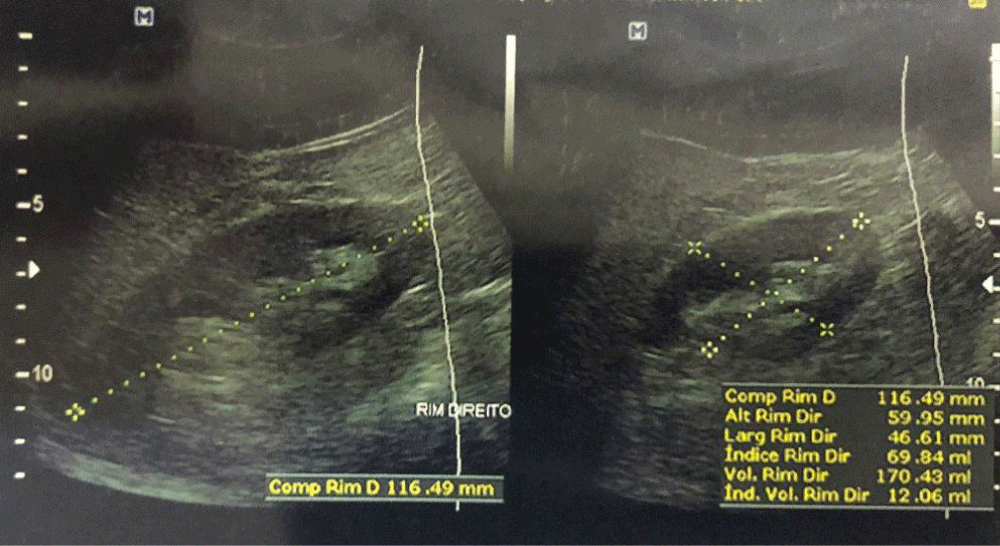
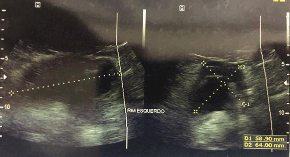
With the persistence of hematuria, he sought care on February 18, 2018. On this occasion, it was asked for serum laboratory tests (Table 1), prostatic USG and urinary tract (Figures 1 and 2) and EAS. In table 1, the hemogram and the leukogram are only with the primordial data and/or with the ones with alteration.
In continuation of the diagnostic search, an Abdominal and Pelvic Ultrasonography was requested and performed on March 7, 2018. The images obtained by the examination demonstrated an initial impairment in the left kidney (Figure 1), which presented with normal size (111 x 64 x 58 mm), regular surface, homogeneous texture and with marked dilatation of the pielocalicial system.
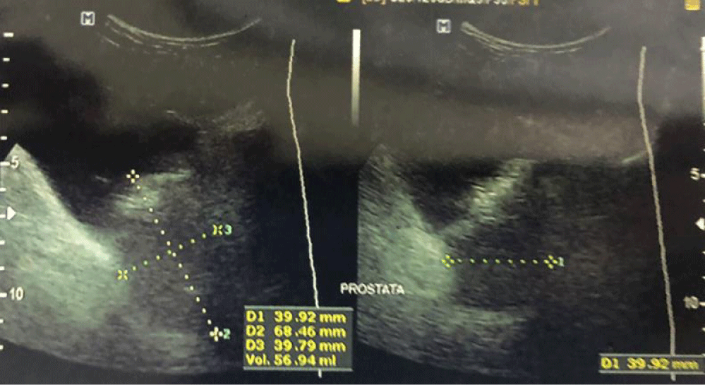
In the ultrasound images of the prostate, the examining physician calculated the size of the gland erroneously, as can be seen in figure 3. In the report, a diagnosis of prostatic disease was suggested, and another examination was indicated to confirm the hypothesis. Thus, a transurethral resection of the prostate was performed to collect samples of prostate cells.
As expected, the sample came inconclusive for neoplasia on May 17th 2018 leading to a new diagnostic search for the patient’s complaints. Cystoscopy with a biopsy was then scheduled for August 2018, eight months after the first symptom of the disease reported by the patient.
However, at the end of May 2018, the patient was admitted to the university hospital emergency with complaint of oliguria, weakness for a week and inappetence two days ago. Laboratory tests (Table 2) show a worsening of the renal function of the patient and the electrolytes, and emergency dialysis was performed.
After a detailed anamnesis done after clinical stabilization of the patient, the diagnosis of bladder cancer was suggested and a cystoscopy with biopsy was scheduled three days later. A tomography of the abdomen and pelvis was also requested.
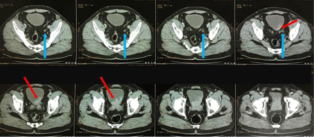
As expected, the tomography image was suggestive of bladder mass, later confirmed as bladder adenocarcinoma. The biopsy report was bladder adenocarcinoma, mucinous pattern, with vacuolated cells suggesting “signet ring” cells, infiltrative in muscle layer.
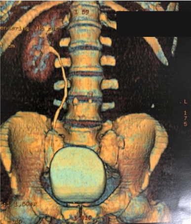
The tomography image shows a vegetative solid, poorly defined expansive lesion with irregular contour and contrast enhancement, located on the pelvic floor and the postero-lateral wall of the bladder, as seen in figure 4.
It was possible to notice in the image that the tumor infiltrated the area of left ureter implantation in the bladder, which caused a dilation of the left ureter and, consequently, hydronephrosis. This explains the poor renal function of the patient, with elevated creatinine and urea.
In another tomographic section and with contrast, it is possible to see that the left kidney no longer shows filtration, and its function is completely compromised. The size of the organ is larger than the contralateral one, with loss of the tomographic density of the parenchyma of the organ, as seen in figure 5.
After the patient’s clinical condition stabilization and J-J stent implantation, the patient was referred to the National Cancer Institute (INCA) in Rio de Janeiro, his home city, for the necessary treatment and follow-up.
Discussion
There are two broad categories of bladder tumor: urothelial and non-urothelial. Although urothelial types are more common, about 90% of the cases, adenocarcinomas enter in non-urothelial type. Primary urinary Bladder Adenocarcinoma (PBA) corresponds to less than 2% of the total cases of bladder cancers [11,12]. Signet Ring Cell Variant of Mucinous Adenocarcinoma (SRCC) of the urinary bladder is even rarer, which may correspond to only 2% to 43% of PBA [13]. Unlike other bladder tumors, such as urothelial tumors, PBA tends to be diagnosed in later stages because of its more aggressive and rapidly invasive character [13]. The majority of patients are over 70 years of age, and the presentation in younger patients is more rare [14].
In this case, the patient is a young patient of 53 years with no clear risk factors for the pathology. Risk factors such as smoking and occupational exposure are known in the carcinogenesis of bladder tumors, although these factors are more commonly associated with the urothelial subtype [5,9,14]. As a truck driver, the patient could fit as a risk group.
Chronic urinary infections and parasitic infections in the bladder are known causes of bladder tumor of the non-urothelial subtype [6,9]. Such factors were not recognized in the patient of the case report.
Obstructions, metastatic invasion and metabolic changes are known consequences in the late diagnosis of cancers [13,14]. In bladder cancer, the diagnosis is generally given in early stages of the disease, since hematuria appears early in the cancer, common in all subtypes of the disease. Some possible complications in the late diagnosis of bladder cancer are ureter obstructions, metastatic invasion of the pelvic floor, obstruction of intestinal transit, among others.
The patient in this case report had an obstruction of the left ureter, leading to ipsilateral hydronephrosis, and, consequently, loss of renal function. Although catheter placement has been performed, the return of renal function from the left kidney is unlikely, and is therefore another serious consequence of late diagnosis bladder cancer. Laboratory abnormalities of urea, creatinine, potassium and blood counts can be explained by the pathophysiology of the disease.
The loss of renal function due to obstruction most likely caused increased urea, creatinine and potassium, which led to uremic symptoms in the patient. Thus, urgent dialysis was justified. Some events may explain the delay in the patient’s diagnosis, such as the dubious presentation of the onset of hematuria.
Although hematuria is the most important sign of bladder cancer, even more at later ages, it is usually painless. The presentation of a hematuria with pain is more sensitive to other types of diagnosis, such as urinary lithiasis [10]. Once the initial medical care has been done at another service, it is impossible to determine the actual source of the pain. Hypothetically, hematuria of bladder cancer may have started along with the hematuria of a possible urinary lithiasis.
Anyway, the persistence of macroscopic hematuria after urinary lithiasis is uncommon, and a further cause of hematuria should be sought. Hematuria due to the concomitance of prostate cancer is not uncommon, although other obstructive and irritating prostate symptoms are expected to occur if the bladder is invasive, which the patient did not have. The PSA dosage was also inconclusive to prostate cancer.
Another factor that did not help in the prognosis and evolution of the disease was the misinterpretation of the pelvic ultrasound, which ratifies the need for a critical analysis of the physician when he has reports and images of complementary tests.
Conclusion
Adenocarcinoma is a rare type of bladder cancer, with the subtype signet ring cell adenocarcinoma even rarer. Despite having the same initial clinical presentation symptoms as the other types, it usually presents with a more aggressive and invasive disease, resistant to the standard treatments. It can be seen from this case report that the depletion of the patient’s health can occur very fast from the beginning of the symptomatology, and the physicians in general should be attentive to the differential diagnosis of hematuria. Also critical analysis of physicians when receiving complementary examinations from other co-workers should always be careful, so as to avoid misunderstanding. Any delay in the treatment of the cancer patient, not only in bladder cancer, can be fatal.
- Antoni S, Ferlay J, Soerjomataram I, Znaor A, Jemal A, Bray F. Bladder cancer incidence and mortality: a global overview and recent trend. Eur Urol. 2017; 71: 96-108. https://bit.ly/2VHUbge
- Instituto Nacional de Câncer José Alencar Gomes da Silva. Estimativa da incidência e mortalidade por câncer no Brasil. 2017; 128: 48-49.
- Khan R, Ibrahim H, Tulpule S, Iroka N. Bladder cancer in a young patient: undiscovered risk factors. Oncol Lett. 2016; 11: 3202-3204. https://bit.ly/2vZSn30
- Wein AJ, Kavoussi LR, Novick AC. Campbell-Walsh Urology. Vol 3. 10th ed. Philadelphia, PA: Elsevier Saunders. 2012; 2309-2506.
- Nomikos M, Pappas A, Kopaka ME, Tzoulakis S, Volonakis I, Stavrakakis G, et al. Urothelial carcinoma of the urinary bladder in young adults: presentation, clinical behavior and outcome. Adv Urol. 2011; https://bit.ly/2VpAx3T
- Ng M, Freeman MK, Fleming TD, Robinson M, Dwyer-Lindgren L, Thomson B, et al. Smoking prevalence and cigarette consumption in 187 countries, 1980-2012. JAMA. 2014; 311: 183-192. https://bit.ly/2Yvk5AT
- SeN V, Bozkurt O, Demir O, Esen DA, Mungan U, Guven A, et al. Clinical behavior of bladder urothelial carcinoma in young patients: a single center experience. Scientifica. 2016. https://bit.ly/2W3Wkm1
- Telli O, Sarici H, Ozgur BC, Doluoglu OG, Sunay MM, Bozkurt S, et al. Urothelial cancer of bladder in young versus older adults: clinical and pathological characteristics and outcomes. Kaohsiung J Med Sci. 2014; 30: 466-470. https://bit.ly/2WOeOUK
- Hardt J, Gillitzer R, Schneider S, Fischbeck S, Thüroff JW. Coping styles as predictors of survival time in bladder cancer. Health. 2010; 2: 429-434. https://bit.ly/2VsSTAX
- Lopes M, Nascimento LC, Zago MMF. Paradox of life among survivors of bladder cancer and treatments. Rev Esc Enferm USP. 2016; 50: 224-231. https://bit.ly/2VGGsGv
- Vasudevan G, Bishnu A, Singh BMK, Nayak DM, Jain P. Bladder adenocarcinoma: a persisting diagnostic dilemma. J Clin Diagn Res. 2017; 11: ER01-ER04. https://bit.ly/2LNbs32
- Niedworok C, Panitz M, Szarvas T, Reis H, Reis AC, Szendröi A, et al. Urachal carcinoma of the bladder: impact of clinical and immunohistochemical parameters on prognosis. J Urol. 2016; 195: 1690-1696. https://bit.ly/2Ed5d2z
- Holmang S, Borghede G, Johansson SL. Primary signet ring cell carcinoma of the bladder: a report on 10 cases. Scand J Urol Nephrol.1997; 31: 145-148. https://bit.ly/2HuU65S
- Shringarpure SS, Thachil JV, Raja T, Mani R. A case of signet ring cell adenocarcinoma of the bladder with spontaneous urinary extravasation. Indian J Urol. 2011; 27: 401-403. https://bit.ly/2LNm4PE

Sign up for Article Alerts