OCT Dilemma in Juxtaluminal Cavity: Ruptured Plaque versus Intraplaque Hemorrhage?
Ioannis A. Stathopoulos1*, Khady Fall1 and Akiko Maehara2
1Columbia Presbyterian Hospital, NY
2Cardiovascular Research Foundation, NY, Columbia University Medical Center, NY
*Address for Correspondence: Ioannis A. Stathopoulos, Columbia Presbyterian Hospital, NY, 30-10 38th Street, 2nd Floor, Astoria, New York 11103, Tel: +718 278-7376; Fax: +718 865 9192; E-mail: jastathopoulos@hotmail.com
Submitted: 27 December 2016; Approved: 16 March 2017; Published: 21 March 2017
Citation this article: Stathopoulos IA, Fall K, Maehara A. OCT Dilemma in Juxtaluminal Cavity: Ruptured Plaque versus Intraplaque Hemorrhage. Adv J Vasc Med. 2017;2(1): 017-019.
Copyright: © 2017 Stathopoulos IA, et al. This is an open access article distributed under the Creative Commons Attribution License, which permits unrestricted use, distribution, and reproduction in any medium, provided the original work is properly cited
Download Fulltext PDF
We present the first reported finding, during optical coherence tomography evaluation of a right coronary artery, of a juxtaluminal cavity filled with signal poor tissue without attenuation. We discuss the possible explanations according to previous descriptions of OCT findings..
Introduction
Advances in high-resolution imaging modalities, such as OCT, have provided new insights into evolving thrombotic lesions, especially intraluminal. But, we still have not validated all findings and our understanding of this new modality is increasing with its more widespread use. We present the first reported finding, during optical coherence tomography evaluation of a right coronary artery, of a juxtaluminal cavity filled with signal poor tissue without attenuation. We discuss the possible explanations according to previous descriptions of OCT findings.
Case report
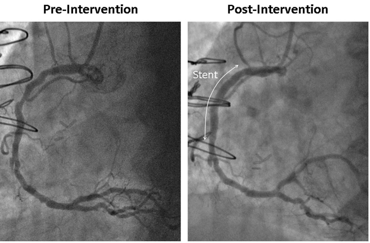
A 58-year-old lady, active smoker, with a history of hypertension, hypothyroidism, known coronary artery disease with chronically occluded left main artery, status post coronary bypass grafting with Saphenous Vein Graft (SVG) to Left Anterior Descending artery (LAD) with several Percutaneous Coronary Interventions (PCI) of the LAD through the SVG, was admitted with unstable angina and underwent coronary angiography that revealed patent SVG to LAD and worsened – compared to previous angiography - stenoses of the proximal and mid Right Coronary Artery (RCA). The left circumflex artery had mild diffuse disease. IVUS interrogation of the lesion was not performed in our case but instead, the RCA was interrogated by frequency domain Optical Coherence Tomography (OCT) imaging (C7 Dragonfly imaging catheter, St. Jude Medical). Because the patient reported intolerance to aspirin with possible allergy, she was first desensitized and subsequently underwent PCI of the RCA with a 3.5 x 30 mm Integrity stent (Medtronic) (Figure 1). Patient had an uneventful hospital stay and she has been well since then.
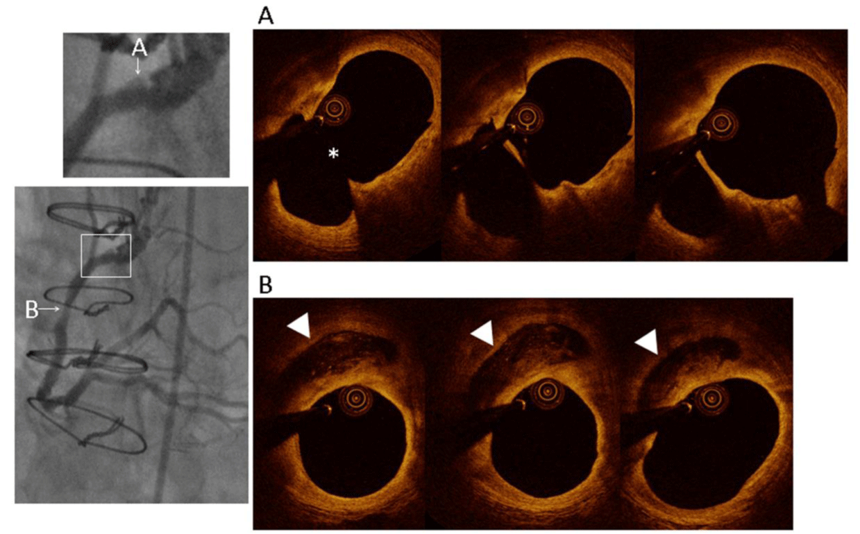
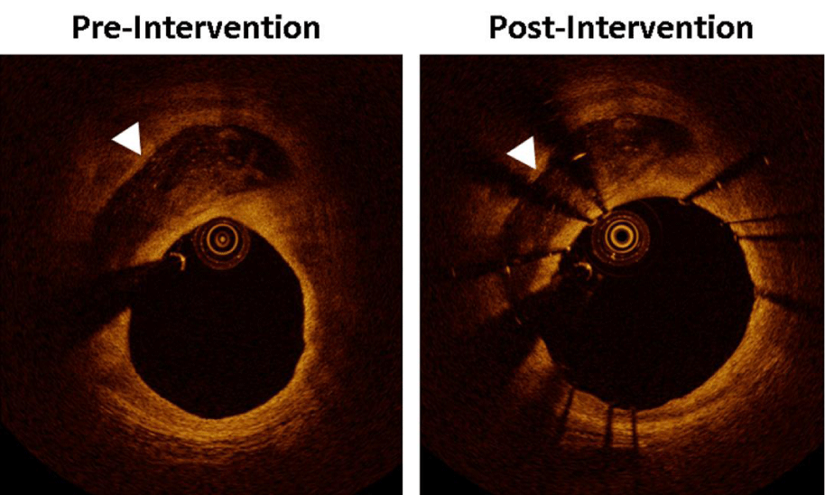
OCT evaluation revealed the following interesting findings: 1) a plaque rupture that correlated with the proximal RCA angiographic finding of ulceration (Figure 2, lesion A), 2) a stenotic area with minimum lumen area 3.2 mm2 (not shown) and 3) a cavity filled with signal poor tissue without attenuation (Figure 2, lesion B). Figure 3 shows the OCT findings at the lesion B location, at matched slice, with the cavity structure before and after the stent placement.
Discussion
To our knowledge, this is the first time that such a juxtaluminal finding is reported by OCT. In our opinion, this cavity probably represents a site of previous plaque rupture, filled with an organized thrombus that manifests features in the range of organized thrombus as described previously by Nakano, et al. [1]. Nevertheless, we could not definitively identify the entry site, probably due to the wire artefact obscuring the presumed rupture entry point or the entry point had been already sealed.
Other investigators have also reported lack of definitive evidence of plaque rupture [2], as plaque rupture may be obscured by organized thrombi or other structures. An organized thrombus is shown by OCT as a signal poor area with or without clear border and, it has been proposed that, a signal-poor tissue represents proteoglycan-rich tissue.
Although this cavity structure may represent intraplaque hemorrhage, it is less likely because the shape of the cavity did not change after the stent placement which was post-dilated with high pressure balloon catheter (18 atmospheres, NC Sprinter 3.5 x 15 mm, Medtronic) achieving adequate size and strength to “squeeze” softer structures like intraplaque hemorrhage.
Conclusion
Advances in high-resolution imaging modalities, such as OCT, have provided new insights into evolving thrombotic lesions, especially intraluminal. But, we still have not validated all findings and our understanding of this new modality is increasing with its more widespread use. OCT can provide us with information that can help us with the decision for the need to proceed to treatment with stent placement, since this can stem not only from the absolute measurement of the lumen area but also, as in our case, from the morphological observations that define a more vulnerable lesion with increased chance for future complications.
- Nakano M, Vorpahl M, Otsuka F, Taniwaki M, Yazdani SK, Finn AV, et al. Ex vivo assessment of vascular response to coronary stents by optical frequency domain imaging. JACC Cardiovasc Imaging. 2012; 5: 71-82. https://goo.gl/HhtrbI
- Kang SJ, Nakano M, Virmani R, Song HG, Ahn JM, Kim WJ, et al. OCT findings in patients with recanalization of organized thrombi in coronary arteries. JACC Cardiovasc Imaging. 2012; 5: 725-732. https://goo.gl/qflNPU


Sign up for Article Alerts