Primary Squamous Cell Carcinoma of the Lacrimal Duct: About Two Cases and Review of the Literature?
Omar Iziki*, Chaker K, Rouadi S, Abada R, Roubal M, and Mahtar M
Department of Otorhinolaryngology, Head and Neck Surgery, King Hassan II University, Ibn Rochd Hospital, Casablanca, Morocco
*Address for Correspondence: O.Iziki, Department of Otorhinolaryngology, Head and Neck Surgery, King Hassan II University, Ibn Rochd Hospital, Casablanca, Morocco. Tel: +212-061-395-3183; ORCid: https://orcid.org/0000-0002-0697-9374; E-mail: omar.iziki@gmail.com
Submitted: 14 September 2019; Approved: 12 December 2019; Published: 14 December 2019
Citation this article: Iziki O, Chaker K, Rouadi S, Abada R, Roubal M, et al. Primary Squamous Cell Carcinoma of the Lacrimal Duct: About Two Cases and Review of the Literature. Int J Case Rep Short Rev. 2019;5(12): 066-069.
Copyright: © 2019 Iziki O, et al. This is an open access article distributed under the Creative Commons Attribution License, which permits unrestricted use, distribution, and reproduction in any medium, provided the original work is properly cited
Download Fulltext PDF
Primary squamous cell carcinoma is an unusual finding in clinical practice. The symptoms are non-specifics, usually mistaken as dacryocystitis, thus the diagnosis is often delated. The treatment of primary malignant lacrimal sac duct tumors depends essentially on surgery from the resection to the reconstruction, followed by radiation and/or chemotherapy. We report, two cases of primary squamous cell carcinoma of the lacrimal duct presented in our department.
The objective of this study is to describe - from our clinical cases and from literature review- the clinical, radiological and histological features of the squamous cell carcinoma and to discuss its therapeutic management.
Introduction
Lacrimal sac tumors are malignant in 55% of cases most are epithelial tumors represented on the squamous cell carcinoma [1], they are easily mistaken for more benign processes such as idiopathic nasolacrimal duct blockage or chronic dacryocystitis. The management of these tumors still inconsistent with high level of recurrence and mortality rate goes between 38 and 44% [2,3].
Case 1
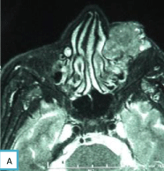
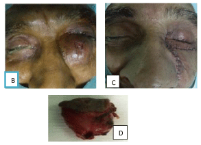
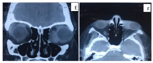
A 64-year-old man, chronic smoker, presented to our department for a 6-month history of a left medial canthal mass. The preoperative Computed Tomography (CT) and Magnetic Resonance Imaging (MRI) showed an irregular shaped tissular lesion involving the left lacrimal sac, lower eyelid and sinonasal tract. Surgical biopsy confirmed the diagnosis of squamous cell carcinoma. The patient underwent only surgical treatment consisting of wide excision of the tumor with health margins. The evolution was good without recurrence after more than 5years (Figure 1 and Figure 2).
Case 2
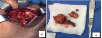
A 48-year-old women, none smoker, presented to our department for one-year history of dacryocystitis, the preoperative CT and MRI, showed an irregular lesion involving the left larcimal sac. Surgical biopsy confirmed the diagnosis. The patient underwent only surgical treatment consisting of wide excision of the tumor, drilling the canalicular portion of the upper and lower eyelids and the medial wall of the orbit and reconstruction of the conjunctive using oral mucosa flap after being sure that the margins were clean. Six months after surgery, she presented for a mass in the surgical site, fixation of the globe, with negative light perception, the biopsy confirmed the local recurrence of the squamous cell carcinoma. She underwent exenteration of her eye with radiation therapy. The evolution was good without recurrence after 5years (Figure 3 and Figure 4).
Discussion
Malignancy of Lacrimal sac and nasolacrimal duct are extremely rare and their frequency is poorly known [4-6], 300 cases have been published in the literature [7], most as isolated case studies [6]. Usually it’s considered there is no predominance in terms of ethnic origin or gender [6]. Early symptoms including chronic unilateral tearing, purulent secretions, inner canthal swelling, pain and rarely epistaxis, are nonspecific and can usually be mistaken for symptoms of dacryocystitis thus the diagnosis is often delayed and made often during Dacryocystorhinostomy (DCR) [1]. Early diagnosis and treatment are curative, can preserve the visual function without recurrence [8].
The radiological exploration should include the couple CT and MRI with and without injection of contrast material, with axial and coronal views [6]. MRI is the modality of choice for the study of the local extension into the soft tissues nasal, paranasal cavity, the orbit and for the post-treatment follow-up. MRI shows an intermediate-signal lesion on T1-weighted sequences and a hypo-intense signal on T2-weighted images compared to the inflammatory dacryocystitis which presents a hyper intense signal on T2-weighted images. It takes up the contrast agent moderately and sometimes heterogeneously after gadolinium injection [9]. When the radiological appearance is suspicious of a tumor, a biopsy of the mass is appropriate. The CT has the advantage of providing an accurate analysis of the relationship of the tumor with bony walls.
The treatment is based essentially on surgery which depends for the most part on the histological type, patient’s general health, the tumor extension. Even though the diagnosis was confirmed by biopsy it is recommended to extemporaneously analyze the tumor in all cases. Wide resection with complete resection of the lacrimal drainage ducts is the choice treatment. It is recommended not to perform osteotomy before the final histological result if the diagnosis cannot be made during extemporaneous examination [10]. A combined external-endonasal approach is indicated if extension to the nasal cavity or the ethmoid [10]. Bony erosion of the nasolacrimal canal does not modify the prognosis, but it may influence the size of the bony resection which could be a medial maxillectomy, an ethmoidectomy. Exenteration is indicated for tumor having larger diameter ≥ 30 mm with / or an invasion of the intraconal space of the orbit may necessitate orbital exenteration [11]. In our team, invasion of the orbital fatty tissue with a limited tumor extension is not an absolute indication for exenteration [6]. Because of the low number of cases reported in the literature orbital exenteration do not provide a gain in terms of local control and long-term survival is difficult to assess when orbital invasion is limited; it is justified in cases of major invasion of the orbital cone or recurrence after adjuvant radiotherapy [6]. Posterior ethmoidectomy is indicated for tumors with more posterior extension. Frequently, tumors erode through the bone of the posterior lacrimal crest on the medial orbital floor to involve the periorbita, necessitating a careful dissection [12]. The disfigurement is major and requires repair solutions after radiotherapy when the disease has been confirmed cured [6]. Our team prefers local reliable flaps such as a nasofrontal flap or a Mustarde flap.
Radiotherapy can be exclusive when surgery is contra-indicated or refused by the patient or the tumor is deep-rooted [1]. Valenzuela, et al. recommend adjuvant radiotherapy is indicated for cases with narrow margins or incomplete or for an aggressive histologic Subtype [13]. Radiation therapy has a critical role in postoperative management [1]. The risk of immediate toxicity of radiotherapy is low and is limited to conjunctival or cutaneous hyperemia. The risks of reduction in visual acuity may occur with doses between 50 and 70 Gy delivered to the optic nerve or the retina. Xerophthalmia, radiation retinopathy, glaucoma, cataract, or keratitis with corneal ulcerations are Delayed toxicity [1,14,15].
The mortality rate was 43.7% in a series of 71 patients despite orbital exenteration and other radical surgical measures [1]. In another study, the mortality rate was 37.5% for all patients and 50% in the subset with large and invasive lesions [6]. Ni et al., the recurrence rate is related to initial tumor excision, with 12.5% recurrence for wide excision versus 43.7% recurrence in cases of isolated lacrimal sac resection [5]. They also showed that, the prognosis remains poor when exenteration is required, with mortality reaching 50% even in cases of radical surgery with tumor excision via the trans facial approach associated with orbit exenteration followed by radiation treatment [5]. Recurrences are frequent, with a 50% rate for carcinomatous lesions and in 50% of cases recurrence is fatal. Local recurrences are treated on a case-by-case basis with surgery and/or radiotherapy [16].
It is recommended that Imaging surveillance every 3 months for the first year, every 6 months the second year, and annually in the third year and beyond.
Conclusion
In conclusion, it should be that squamous cell carcinoma of the lacrimal ducts are rare and often initially diagnosed as dacryocystitis. The vital, functional, esthetic prognosis depend essentially on the time of diagnosis which influence the management.
- Stefanyszyn MA, Hidayat AA, Pe'er JJ, Flanagan JC. Lacrimal sac tumors. Ophthal Plast Reconstr Surg. 1994; 3: 169-184. PubMed : https://www.ncbi.nlm.nih.gov/pubmed/7947444
- Ni C, D'Amico DJ, Fan CQ, Kuo PK. Tumors of the lacrimal sac: clinicopathological analysis of 82 cases. Int Ophthalmol Clin. 1982; 1: 121-140. PubMed: https://www.ncbi.nlm.nih.gov/pubmed/6277817
- Choussy O, Babin E, Delas B, Bailhache A, François A, Marie JP, et al. Primary malignant tumors of the lacrimal sac and nasolacrimal duct. Ann Otolaryngol Chir Cervicofac. 2007; 6: 309-313. PubMed: https://www.ncbi.nlm.nih.gov/pubmed/17583669
- Parmar DN, Rose GE. Management of lacrimal sac tumours. Eye (Lond). 2003; 17: 599-606. PubMed: https://www.ncbi.nlm.nih.gov/pubmed/12855966
- Hornblass A, Jakobiec FA, Bosniak S, Flanagan J. The diagnosis and management of epithelial tumors of the lacrimal sac. Ophthalmology. 1980; 87: 476-490. PubMed: https://www.ncbi.nlm.nih.gov/pubmed/7413136
- Montalban A, Lietin B, Louvrier C, Russier M, Kemeny JL, Mom T, et al. Gilain Malignant lacrimal sac tumors Eur Ann Otorhinolaryngol Head Neck Dis. 2010; 127: 165-172. PubMed: https://www.ncbi.nlm.nih.gov/pubmed/21036121
- Ishida M, Iwai M, Yoshida K, Kagotani A, Kohzaki H, Arikata M, et al. Primary ductal adenocarcinoma of the lacrimal sac: the first reported case. Int J Clin Exp Pathol. 2013; 6: 1929-1934. PubMed : https://www.ncbi.nlm.nih.gov/pubmed/24040460
- De Stefani A, Lerda W, Usai A, et al. Squamous cell carcinoma of lacrimal drainage system: Case report and literature review. Tumori. 1998; 84: 506-510. PubMed: https://www.ncbi.nlm.nih.gov/pubmed/9825006
- Rahangdale SR, Castillo M, Shockley W. MR in squamous cell carcinoma of the lacrimal sac. AJNR Am J Neuroradiol. 1995; 6: 1262-1264. PubMed: https://www.ncbi.nlm.nih.gov/pubmed/7677021
- Sullivan TJ, Valenzuela AA, Selva D, McNab AA. Combined external-endonasal approach for complete excision of the lacrimal drainage apparatus. Ophthal Plast Reconstr Surg. 2006; 3: 169-172. PubMed: https://www.ncbi.nlm.nih.gov/pubmed/16714923
- Kumar VA, Esmaeli B, Ahmed S, Gogia B, Debnam JM, Ginsberg LE. Imaging features of malignant lacrimal sac and nasolacrimal duct tumors. http://dx.doi.org/10.3174/ajnr.A4882
- El-Sawy T, Frank SJ, Hanna E, Sniegowski M, Lai SY, Nasser QJ, et al. Multidisciplinary management of lacrimal sac/Nasolacrimal duct carcinomas. Ophthal Plast Reconstr Surg. 2013; 29: 454-457. PubMed: https://www.ncbi.nlm.nih.gov/pubmed/24195987
- Valenzuela AA, Selva D, McNab AA, Simon GB, Sullivan TJ. En bloc excision in malignant tumors of the lacrimal drainage apparatus. Ophthal Plast Reconstr Surg. 2006; 22: 356-360. PubMed: https://www.ncbi.nlm.nih.gov/pubmed/16985419
- Nakamura K, Uehara S, Omagari J, Kunitake N, Kimura M, Makino Y, et al. Primary non-Hodgkin’s lymphoma of the lacrimal sac: a case report and a review of the literature. Cancer. 1997; 11: 2151-2155. PubMed : https://www.ncbi.nlm.nih.gov/pubmed/9392338
- Lumbroso Le Rouic L, Decaudin D. Orbitopalpebral or adnexal lymphoma. EMC Ophthalmology. Elsevier Masson. 2007; 21: 110-120. http://bit.ly/2sEWBiv
- Choussy O, Babin E, Delas B, Bailhache A, Francois A, Marie JP, et al. Primary malignant tumors of the lacrimal sac and nasolacrimal duct. Ann Otolaryngol Chir Cervicofac. 2007; 6: 309-313. PubMed: https://www.ncbi.nlm.nih.gov/pubmed/17583669


Sign up for Article Alerts