Anesthetic Management in a Child with Morquios Syndrome - A Case Report?
Garima Daga*and Mohit Tyagi
Department of Anesthesiology, Lady Harding Medical College and Hospital New Delhi, India
*Address for Correspondence: Garima Daga, Department of Anesthesiology, Lady Harding Medical College and Hospital New Delhi, India, Tel: +783-867-2612; ORCid: 0000-0002-4334-3885; E-mail: [email protected]
Submitted: 27 September 2020; Approved: 03 October 2020; Published: 05 October 2020
Citation this article: Daga G, Tyagi M. Anesthetic Management in a Child with Morquios Syndrome - A Case Report. Int J Case Rep Short Rev. 2020; 6(10): 063-068. https://dx.doi.org/10.37871/ijcrsr.id86
Copyright: © 2020 Daga G, et al. This is an open access article distributed under the Creative Commons Attribution License, which permits unrestricted use, distribution, and reproduction in any medium, provided the original work is properly cited
Keywords: Pediatric anesthesia; Difficult airway; Morquios syndrome; Unstable neck; Mucopolysaccharidoses; Metabolic syndrome
Download Fulltext PDF
Morquios syndrome is a progressive lysosomal storage disorder with AR inheritance. It is a type IV Mucopolysaccharidoses occurring due to deficiency of enzymes: N-acetyl-galatosamine 6 sulphatase (type IVa) and Beta galactosidase (type IVb). The Incidence of MPS type IVA is very rare up to 1:216,400. We are presenting a case of MS and its anesthetic implications. There are various important factors to be considered before administration of general anesthesia, regional anesthesia and even during transportation of such patients. Very few case reports are available for the same. We describe the anesthetic management of a 12-year-old male child diagnosed with Morquios Syndrome type IVa presented for osteotomy in view of genu valgum. CT scan of spine revealed anterior displacement of atlas causing Cranio-Vertebral (CV) junction narrowing and Odontoid hypoplasia was reported.
The decided anesthetic plan was general anesthesia combined with caudal block, with minimal neck movements during transport and positioning. Child had uneventful surgery and remained stable intraoperatively and postoperatively.
Abbreviations
MS: Morquios Syndrome; AR: Autosomal Recessive; MPS: Mucopolysaccharidoses; CV: Cranio-Vertebral; CT: Computed Tomography; MRI: Magnetic Resonance Imaging; GA: General Anesthesia; ABG: Arterial Blood Gases; GAGs: Glycosaminoglycans; LMA: laryngeal mask airway
Introduction
Morquio’s Syndrome (MS) or Mucopolysaccharidosis (MPS) is a progressive lysosomal storage disorder with autosomal recessive inheritance. Deficiency of the N-acetylgalactosamine-6-sulfate sulfatase enzyme (type IVa) and beta galactosidase (type IVb) results in accumulation of the glycosaminoglycans (GAGs), keratan sulphate, and chondroitin-6-sulfate principally in cartilage and the extracellular matrix of connective tissue [1].
Typically, a child with MS presents with marked skeletal changes and disturbances of linear growth. The abnormalities of chondrogenesis and subsequent osteogenesis are largely responsible for the typical phenotype with characteristic features such as short stature, kyphoscoliosis and rib deformities causing prominent barrel-shaped chest, frontal bossing, genu valgum (‘‘knock knees’’), and epiphyseal changes with joint laxity [2,3]. The latter two abnormalities frequently necessitate orthopedic interventions to achieve limb realignment.
Late onset of aortic regurgitation, hepatosplenomegaly and hearing loss have also been associated with this defect. Of note, mental retardation is not usually present [4,5].
In this report, we discuss the anaesthetic management of a patient with MS and review the literature, giving special consideration to the issues relevant to the anaesthesiologist such as restriction of chest excursion and thereby limited pulmonary function, abnormalities of the aortic and mitral valves [1,2], and the frequent occurrence of spinal deformities such as hypoplastic odontoid process that leads to atlanto-axial joint instability, that mandates extreme caution with airway management [6].
Case Presentation
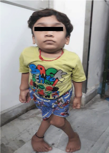
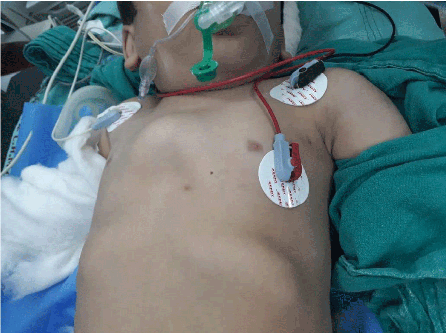
The patient (figure 1) was a 12-year-old male child (20 kg of weight and 106 cm height) who presented for bilateral osteotomies in view of genu valgum (knock knees). He had already undergone growth modulation by bilateral femoral manipulation with figure of 8 application under general anesthesia one year back. The patient was born out of a consanguineous marriage, was low weight at birth, had history of delayed cry and delayed physical growth. As the child started walking at 3 years of age, mother noted bilateral outwards deviation of both the legs and touching of both the knees while walking. This was not associated with any history of traumatic injury or febrile episodes. His facial features were notable for a flattened nose, prominent maxilla and mandible and large tongue. He had an extremely short neck. A Mallampati Class III was assigned to his airway based on our ability to visualize only the base of uvula. However, there was no documented history of obstructive sleep apnea [7]. Higher mental functions were normal. Further physical examination revealed pectus carinatum, laxity of the wrists, ankle, and finger joints and a protuberant abdomen. Bilateral lower extremity weakness was present. Neurological examination showed no sensory deficits. Mucopolysaccharide spot urine test (toluidine blue) was positive. Rest of the laboratory studies, including prothrombin time/partial thromboplastin time, were within the normal range.
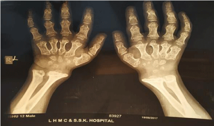
X- ray bilateral hands (figure 2) showed tapering of proximal portions of metacarpals with small irregular carpal bones and that of bilateral feet reported small tarsal bones and widened metaphysis of metatarsals.
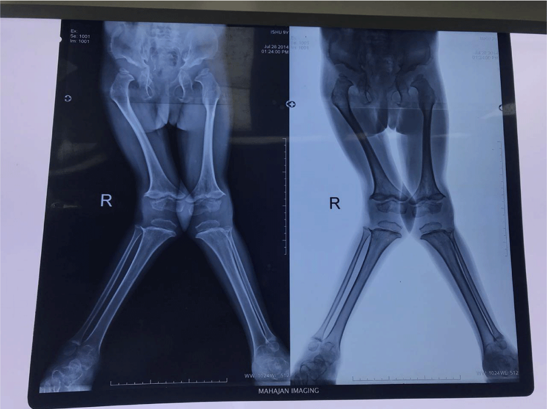
All these findings were consistent with the associated osteogenesis defects (dysostosis multiplex with MS ) [8]. Digital skiagram of bilateral pelvis and hip in AP view (figure 3) reported widened deep acetabular cavity with small sacro-sciatic notch resulting in triangular pelvic activity. There was evidence of small bilateral femoral capital epiphysis with crenated appearance of epiphysis of greater tuberosity.
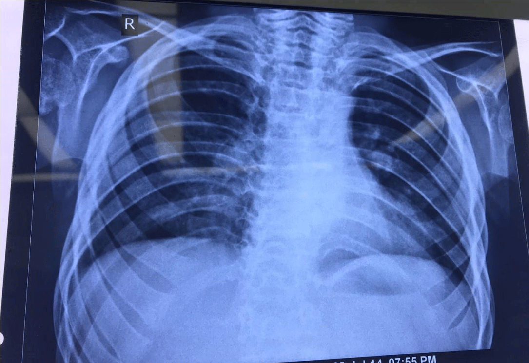
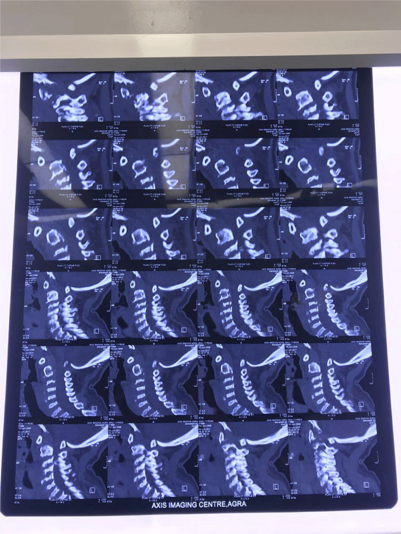
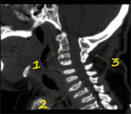
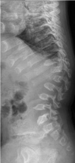
Chest X-ray PA view (figure 4) revealed no abnormalities. Plain CT cervical spine from C1 to T1 vertebrae level (figure 5) revealed anterior displacement of atlas vertebra causing narrowing at craniovertebral junction leading to compression of cervical cord with hyperintense signal in cord suggestive of compressive myelopathy. Hypoplasia of odontoid process was reported. The visualized vertebrae were decreased in height with anterior beaking (platyspondyly) and normal intervertebral disc spaces (figure 6). The atlanto-axial distance did not change on flexion-extension scans and thus there was no evidence of atlanto-axial instability (figure 7).
Magnetic Resonance Imaging (MRI) of the cervical spine with cranio-vertebral junction was performed and it reported findings consistent with those of CT scan cervical spine. 2D echocardiography was also normal. Arterial blood gases were also within normal limits.
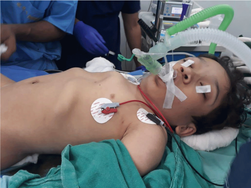
After a thorough pre anesthetic check-up and an adequate fasting of 8 hours, patient was carefully transferred onto the operating room table where the routine noninvasive monitors were applied (EKG, noninvasive blood pressure monitor, pulse oximeter, end tidal CO2 monitoring, precordial stethoscope). Difficult airway cart was kept ready in the OR. An intravenous catheter was placed and intravenous dose of 0.08 mg of glycopyrrolate as pre-medication was administered. After preoxygenation with 100% oxygen, anaesthesia was induced via inhalation with 4% sevoflurane along with injection fentanyl 40 micrograms and injection propofol 50 mg. Slight airway obstruction occurred promptly as anaesthetic-induced relaxation of laryngeal structures took place as identified by stridor. This was easily relieved with the two-handed jaw-thrust maneuver. As the patient became more deeply anaesthetized, LMA proseal 1.5 number was inserted with minimal movements at atlanto-occipital junction while practicing manual in-line stabilization of spine (figure 8). To aid for analgesia, regional anesthesia via caudal block was decided and the patient was carefully positioned in left lateral position with always someone maintaining neck stabilization. Under all aseptic precautions, caudal anesthesia was administered using 22 gauge caudal needle and 10 ml 0.25% bupivacaine with 400 micrograms morphine was administered. Anesthesia was maintained with 2% sevoflurane in a 50:50 oxygen: nitrous oxide mixture. Surgery lasted for 1.5 hours and patient had stable intraoperative vitals. LMA proseal was removed after the patient was fully wake and airway reflexes completely recovered. Patient had a smooth emergence and recovery implying good pain relief and was shifted to post anesthesia care unit where all vital parameters monitored closely. His postoperative course was uneventful and he was discharged on third postoperative day from pediatric surgery ward.
The absence of enzymes N-acetyl galactosamine and beta-galactosidase in Morquios Syndrome (MS) type IVa and type IVb respectively, leads to accumulation of glycosaminoglycans mainly chondroitin sulphate and keratan sulphate. They accumulate largely in mucous membranes, soft tissues, cartilages and bones causing characteristic skeletal dysplasias. It is also reflected by excess excretion of these GAGs in urine. Characteristic features are dwarfism, knock knees, epiphyseal changes with joint laxity, osteoporosis, coarse facies with prominent maxillae, broad mouth, short anteverted nose, widely spaced teeth with defective enamel and corneal opacities, and the typical barrel chest due to spinal curvature and rib deformities [9,10].
Excessive accumulation of GAGs in mucosal membrane lined tonsils, adenoids and tongue leads to their hypertrophy that results in an airway prone to obstruction [11]. Bag and mask ventilation may be difficult and the chances of airway obstruction are increased during induction. Difficult mask ventilation is common and may need ‘two-person technique’ to achieve. Chances of bleeding also increases during intubation due to hypertrophied tonsils and bulgy soft tissue. Trachea-bronchial secretions are usually copious in amount, and administering anti-sialagogue is advised as pre medication. Narrowed airway along with atlanto-axial instability together present a challenging situation for any anesthesiologist. It is known that airway difficulties increase with age in these patients, most likely secondary to continued infiltration of the soft tissues with glycosaminoglycans [12]. Due to the anticipated smaller caliber airway, smaller endotracheal tubes must be kept available.
Spinal involvement with characteristic features like odontoid hypoplasia, platyspondyly and large intervertebral discs as found in this patient, contribute to the frequent occurrence of spinal stenosis in such patients [13]. Complete spinal imaging should be a part of comprehensive care in patients with MS. In the situations where cord imaging might not be available, e.g., emergency procedures, it would be wise to assume the presence of above-mentioned spinal stenosis (as well as atlantoaxial instability). Also, avoiding precipitous fall of mean arterial pressure intra and post-operatively can prevent grave consequences like spinal cord infarction/ischemia [14]. It is therefore important to establish intraoperative neuromonitoring baseline assessments prior to turning patients to the prone position following induction of anesthesia and to monitor cardiac output during prone positioning as in spinal surgeries.
Another area that requires great attention is while transporting and positioning of these patients. It requires knowledge of the stability of the individual patient’s cervical spine. If in doubt we must assume the possibility of an unstable cervical spine due to a high prevalence and known risk of cervical subluxation. Thus, all measures may be taken to stabilize cervical spine while transporting MS patients inside the operating room, on the table and while shifting the patient to the recovery unit.
Pulmonary involvement in patients with MS is multifactorial. Kyphoscoliosis and restricted chest excursion together, leads to restrictive pattern of pulmonary disease and increased chances of recurrent infections. Alveolar capacity is decreased in severe cases. Ventilation-perfusion mismatch is common and these patients may have hypoxemia with hypercarbia type of presentation in the arterial blood gas report. In long standing cases, pulmonary hypertension and cor pulmonale may be associated [15]. Our patient had normal ABG. Thorough preoperative assessment for identifying these issues and their optimization is imperative.
The most common associated cardiac lesion in MS type IV is aortic insufficiency [16], although it occurs late in the course of disease. Deposits of GAGs in the myocardium of the affected individuals, decrease their myocardial compliance making them susceptible to peri-operative systolic/diastolic dysfunction [11,17]. Rarely, GAGs accumulation in the coronary arteries increase the chances of MI. Judicious use of fluids, avoiding tachycardia and hypotension are important strategies to be implemented while managing these cases.
Regardless of the operation to be performed, the anaesthesia care provider must have a thorough understanding of the disease process and its implications in the perioperative period. While deciding a plan of anesthesia for such cases, the pros and cons of general as well as regional anesthesia must be carefully weighed out.
Administering regional anesthesia with or without general anesthesia, decreases the requirement of anesthetic drugs and avoids multi pharmacy. It provides good peri-operative analgesia to the patient that is a pre-requisite for early ambulation and recovery. Also, avoids unnecessary airway manipulations. There are a few studies, in which spinal cord infarction was reported after administering epidural infusions [13]. The possibility that an epidural infusion increased neuraxial pressures and thereby decreased the local perfusion pressures of the spinal cord was considered. Along with this, the GAGs deposition in the intra-vertebral foramina may have been a contributing factor.
Keeping in mind these reasons, caudal block might just be a better and a safer alternative in patients with MS. The chances of increase in local neuraxial pressures are considerably decreased as the sacral canal has a larger capacity and loss of some drug volume occurs through wider sacral foraminas. Also, the point of entry of needle is further away from termination of dural sac in caudal approach as compared to thoracic or lumbar epidural. Better hemodynamic control can be achieved with caudal approach as compared to lumbar/thoracic epidural and subarachnoid block. The latter may mask the features of spinal cord ischemia and contributing further in the injury to spinal cord, if any.
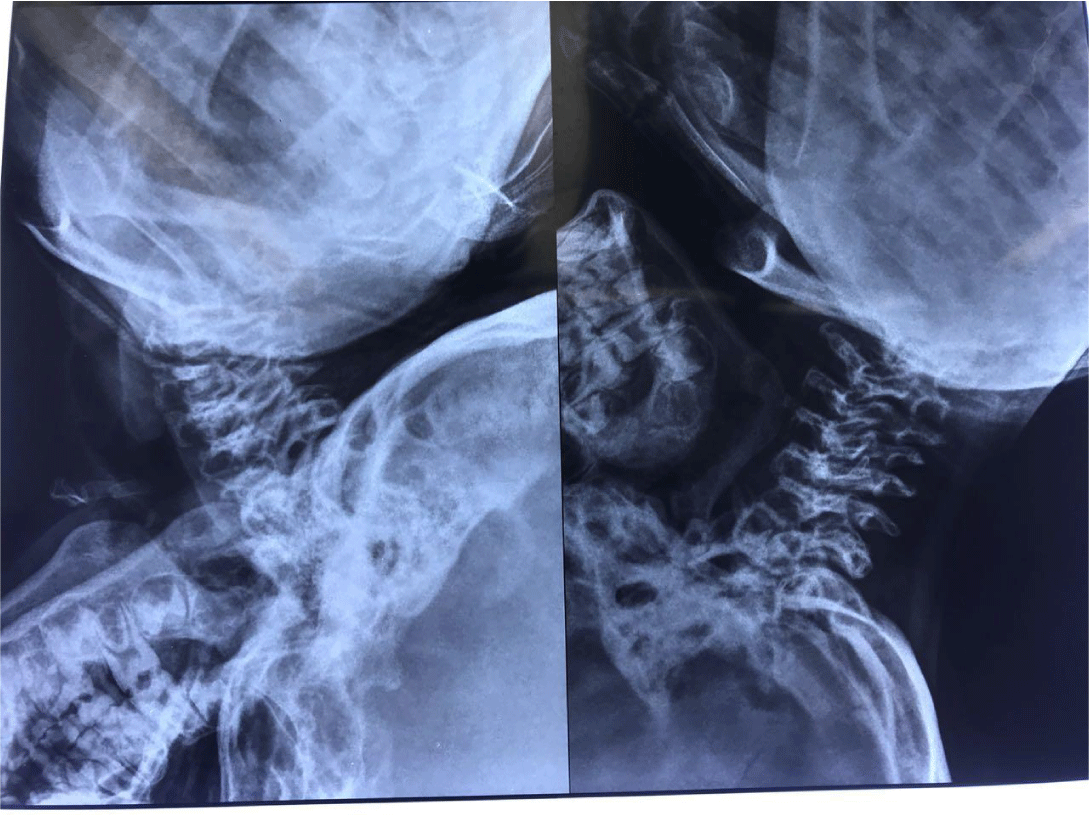
General anesthesia is the gold standard while managing pediatric cases. It allays anxiety, provides better hemodynamic control and decreases the risk of direct injury to spinal cord. However, patients of MS who present with an unstable cervical spine and extremely short neck where head flexion and extension are further limited by the pectus carinatum (figure 9) and kyphotic spine (figure 10), the anesthesiologist must be prepared for difficult scenarios. The airway should be examined by experienced personnel and, if deemed prone to obstruction, preoperative sedation should be minimized or avoided. For anticipated difficult airway, alternative plans, equipment (i.e. laryngeal mask airways, fiberoptic bronchoscope, emergency surgical airway), and personnel (ENT surgeons in situations of can’t intubate can’t ventilate) must be available and prepared for immediate action. One of the safe approaches for induction of anaesthesia is not to abolish spontaneous ventilation by the patient until control is established either by tracheal intubation or adequate positive pressure ventilation via a face mask/supraglottic airway device. Avoiding drugs like narcotics should be considered to avoid postoperative respiratory depression, and postoperative mechanical ventilation should be considered in high risk patients
We preferred LMA over endotracheal intubation in our patient as he had no history of obstructive sleep apnea or snoring that advocates need for postoperative elective mechanical ventilation. It avoids excessive neck movement and need for lower cervical flexion as needed while intubation. LMA proseal was chosen as it displaces soft tissue in a definitive and rigid manner. Also, the previous surgical history under LMA was uneventful. In patients with documented with atlanto-axial instability, it is prudent to always keep the neck always in a neutral position and permit very less movements before, during and after surgery. Manual in-line stabilization is one method to achieve it, while intubating or inserting any supra-glottic airway device. Some use cervical collar or a halo cast that itself may be a hindrance to intubation.
Conclusion
To summarise, Morquio’s syndrome is an inherited multisystem disorder affecting organ systems of interest to the anaesthesiologist. Patients with MS undergo multiple surgical procedures in their lifetime and as the disease progresses with age, majority of which are orthopaedic, heart valve, and ear-nose-throat related. With a thorough understanding of the disease process and potential consequences, it is possible to evaluate and manage these patients with a high margin of safety in the perioperative period. Airway management is challenging and general anaesthesia is a high-risk procedure involving the risk of death. Anticipation of difficulties and preparation are of paramount importance. Airway tools necessary to manage difficult airway should be readily available. Caudal anesthesia can prove to be a much safer alternative with lesser risk of injury to spinal cord in these patients. Thus, having complete knowledge and being well versed with the patient before taking in the operating room can go a long way and result in better outcomes.
Acknowledgement
I express my gratitude to the patient for granting his permission for the presentation of this report and to Dr. Mohit Kumar Tyagi MD for review of and suggestions about the case report.
- Yasuda E, Fushimi K, Suzuki Y, Shimizu K, Takami T, Zustin J, et al. Pathogenesis of morquio a syndrome: An autopsied case reveals systemic storage disorder. Mol Genet Metab. 2013; 109: 301-311. DOI: 10.1016/j.ymgme.2013.04.009
- Tomatsu S, Montano AM, Oikawa H, Smith M, Barrera L, Chinen Y, et al. Mucopolysaccharidosis type IVA (Morquio A disease): Clinical review and current treatment. Curr Pharm Biotechnol. 2011; 12: 931-945. DOI: 10.2174/138920111795542615
- Tomatsu S, Mackenzie WG, Theroux MC, Mason RW, Thacker MM, Shaffer TH, et al. Current and emerging treatments and surgical interventions for Morquio A syndrome: A review. Res Rep Endocr Disord. 2012; 2012: 65-77. DOI: 10.2147/RRED.S37278
- Jones KL. Smith’s recognizable patterns of human malformation, 5th ed. Philadelphia: WB Saunders. 1997: 466-467.
- Jones AE, Croley TF. Morquio syndrome and anesthesia. Anesthesiology. 1979; 51: 261-262.
- Theroux MC, Nerker T, Ditro C, Mackenzie WG. Anesthetic care and perioperative complications of children with morquio syndrome. Paediatr Anaesth. 2012; 22: 901-907. DOI: 10.1111/j.1460-9592.2012.03904.x
- Mallampati SR, Gatt SP, Gugino LD, Desai SP, Waraksa B, Freiberger D, et al. A clinical sign to predict difficult tracheal intubation: A prospective study. Can Anaesth Soc J. 1985; 32: 429. DOI: 10.1007/BF03011357
- White KK, Jester A, Bache CE, Harmatz PR, Shediac R, Thacker MM, et al. Orthopedic management of the extremities in patients with morquio a syndrome. J Child Orthop. 2014; 8: 295-304. DOI: 10.1007/s11832-014-0601-4
- Morquio L. On a form of familial bone dystrophy. Arch Med Children.1929; 32: 129-140.
- Holzgreve W, Grobe H, von Figura K, Kresse H, Beck H, Mattei JF. Morquio syndrome: Clinical findings in 11 patients with MPS IVA and two patients with MPSIVB. Hum Genet. 1981; 57: 360-365.
- Baines D, Keneally J. Anaesthetic implications of the mucopolysaccharidoses: A fifteen-year experience in a children’s hospital. Anaesth Intens Care. 1983; 11: 198-202.
- Kempthorne PM, Brown TCK. Anaesthesia and the mucopolysaccharidoses. A survey of techniques and problems. Anaesth Intens Care. 1983; 11: 203-207. DOI: 10.1177/0310057X8301100304
- Drummond JC, Krane EJ, Tomatsu S, Theroux MC, Lee RR. Paraplegia after epidural-general anesthesia in a morquio patient with moderate thoracic spinal stenosis. Can J Anaesth. 2015; 62: 45-49.
- Tong CKW, Chen JCH, Cochrane DD. Spinal cord infarction remote from maximal compression in a patient with Morquio syndrome. J NeurosurgPediatr. 2012; 9: 608-612. DOI: 10.3171/2012.2.PEDS11522.
- Smith RM. Anesthesia for Infants and Children, 6th ed. St Louis: CV Mosby Co. 1996: 620-622. https://bit.ly/3jxBrIf
- Jones KL. Smith’s recognizable patterns of human malformation, 5th ed. Philadelphia: WB Saunders. 1997: 466-467.
- King DH, Jones RM, Barnett MB. Anaesthetic considerations in the mucopolysaccharidoses. Anaesthesia. 1984; 39: 126-131.

Sign up for Article Alerts