Role of BNP as a Screening Tool to Identify Asymptomatic Cardiac Disease in Chronic Type 2 DM Patients?
SM. Rajendran1*, R. Senthinathan2, EN. Vedhashri3, S. Abinaya3 and Jayashri4
1Profesor of Diabetology and Internal Medicine, Bharat University, Chennai, Tamil Nadu, India
2Professor of Orofasciomaxillary surgery, Bharat University, Chennai, Tamilnadu, India
3Resident, Saroja Hospital, Chennai, Tamil Nadu, India
4Nursing Instructor, Saroja Hospital, Chennai, Tamil Nadu, India
*Address for Correspondence: SM. Rajendran, Professor of Diabetology and Internal Medicine, Bharat University, Chennai, Tamil Nadu, India, E-mail: drshanmugamrajendran@gmail.com
Submitted: 17 March 2017; Approved: 01 July 2017; Published: 12 July 2017
Citation this article: Rajendran SM, Senthinathan R, Vedhashri EN, Abinaya S, Jayashri. Role of BNP as a Screening Tool to Identify Asymptomatic Cardiac Disease in Chronic Type 2 DM Patients. Int J Clin Endocrinol. 2017;1(1): 020-024.
Copyright: © 2017 Rajendran SM, et al. This is an open access article distributed under the Creative Commons Attribution License, which permits unrestricted use, distribution, and reproduction in any medium, provided the original work is properly cited
Download Fulltext PDF
Aim: To study the value of BNP as a screening tool to identify silent ischemia and diastolic dysfunction in asymptomatic type II diabetic patients.
Objectives: The objective of the study is how far BNP value will be useful in early detection of LV dysfunction and ischemia without subjecting the patient to treadmill test and ECHO, as both are even though specific but not sensitive. Our effort is to identify a simple blood test which is highly sensitive in identifying them.
Study population This study was conducted in the in the Department of Medicine and Department of cardiology Sree Balaji Medical College, Chennai, Tamil Nadu during the period of August 2010 to August 2011. Total number of patients included in this study was 77 out of which 22 were males and 55 were females’ patients ranging from 30 years to 70 years.
Study Design
This study is a prospective study and is aimed to assess the prognostic role of serum BNP level in asymptomatic type 2 Diabetic patients for silent myocardial infarction.
Exclusion criteria
1. Subjects with the presence of pathological Q waves, LV hypertrophy on voltage criteria or ST/ T wave abnormalities in ECG.
2. History of Congestive Heart Failure.
3. History of End Stage Renal Failure.
4. History of smoking.
5. History of consumption of alcohol.
6. Subject with uncontrolled hypertension.
7. Clinically significant abnormal laboratory results at screening.
8. Patient who is not willing to participate in the study.
9. Participating in a clinical research trial within 30 days of our study.
10. Individuals who are cognitively impaired and/ or who are unable to give informed consent.
11. Any other health or mental condition that in investigator’s opinion may adversely affect the subject’s ability to complete the study or its measures or that may pose significant risk to the subject.
Description of the Study
The subjects are invited to the study center from local population who are diabetic and willing to participate in the trial. Each subject will be informed both orally and in writing about the study prior to inclusion in the study and only the subjects who have given written consent were included in the study. The selection of the subjects was taken by the inclusion and exclusion criteria. The selected subjects were given a screening number and underwent a screening procedure to find out eligible candidates. The screening procedure includes obtaining subject’s demographic data and medical history, physical examination, baseline symptomatology, laboratory investigations like fasting blood glucose, HbA1c, Lipid profile, urea, creatinine, liver function test and ECG.
The selected subjects were given subject number. They will be asked to come on the next day for the next set of investigation like BNP, TMT and resting ECHO.
BNP assay
The test measures BNP by immunoassay and utilizes a fluorescence detection system. The assay is linear between 20-1300 pg/ ml, with a lower limit of detection of 20 pg/ ml. Samples. 1300 pg/ ml cannot be re-assayed after dilution. Collect whole blood in 4 ml lavender top tube- a minimum of 2 ml of blood is required. Moderately hemolyzed specimens are acceptable. Samples greater than 4 hours old are not acceptable for testing. The rapid BNP will be available only on STAT basis with a turnaround time of less than 60 minutes. No panic value levels have been set [1].
Resting echocardiography
Subjects are examined in the left lateral decubitus position using a standard commercial ultrasound machine using a 2.5 MHz phased array probe. Three apical views (apical 4 chamber, 2 chamber, and long axis views) are acquired using standard harmonic imaging. Mitral and pulmonary flow velocities were recorded by using conventional pulsed wave Doppler ECHO. Left ventricular diameters and wall thickness are measured from 2 dimensional targeted M-mode echocardiography. Left ventricular mass is determined by Devereux’s formula [2]. Left ventricular hypertrophy is defined as LV mass index (g/ m2) greater than 131 g/ m2 in men and greater than 100g/ m2 in women [3]. Resting LV end diastolic, end systolic volumes and ejection fraction are computed using a modified Simpson’s biplane method. Regional wall motion analysis is scored as normal, mildly hypokinetic, severely hypokinetic and akinetic by two people who blinded to the patient’s clinical data. Infarction was identified by resting wall motion abnormalities.
Stress testing
Exercise echocardiography is performed in patients who have fulfilled the inclusion and exclusion criteria. Blood pressure and cardiac status by 12 lead electrocardiograms are monitored during the exercise test. Regional wall motion is compared before and after stress, and patients with ischemia are identified by inducible wall motion abnormalities.
Results
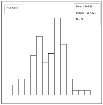
Of the 77 patients 28 had diastolic dysfunction and 23 had ischemia (inducible wall motion abnormalities and peak exercise), with 17 having both LVD and ischemia. (Figure 1) (Table 1-3).
Echocardiography
Among 77 patients 28 had subclinical left ventricular diastolic dysfunction identified by a 2D ECHO. These patients NT pro BNP were compared. NT pro BNP > 600 could predict diastolic dysfunction at sensitivity of 64% and negative predictive value of 73%, p = 0.000. Suggestive BNP could be used effectively as screening tool to identify diastolic dysfunction. (Figure 1) (Table 4).
Treadmill
Among the 77 patients 23 patients had developed silent ischemic changes on performing Exercise tolerance test. When BNP > 600 of this patients were observed was found BNP could predict silent ischemia at sensitivity of 50%, negative predictive value 61%, p = 0.000.
Both treadmill and echocardiography77 enrolled in the study, 17 of them had features of both diastolic dysfunction and silent ischemic changes. When compared with BNP cut off value it had sensitivity and negative predictive value was 100%.
Discussion
Both echocardiography and BNP have clinically been used for screening and monitoring of LV dysfunction, particularly in those with high possibilities of development of heart failure. The present study has demonstrated that echocardiographic screening of asymptomatic diabetic subjects with apparently normal cardiac function may identify significant numbers of patients with asymptomatic ischemia, LVH and subclinical LV dysfunction. Measuring BNP is simple and rapid and has been suggested as a screening test for LV dysfunction in patients with diabetes mellitus [4], BNP levels in the present study were significantly different between patients with and without subclinical LV dysfunction.
Screening for LVD and CAD
Conventional echocardiography is routinely used for screening for LVD in clinical settings. The current study demonstrated that BNP was significantly increased in older female patients, confirming previous findings [5]. However, although BNP was higher in patients with LVD than those without, consistent with previous findings [6], an abnormal BNP level was found in 22 patients, suggesting usefulness of BNP as a screening tool for LVD [7,8].
Coronary artery disease is commonly silent in patients with DM and screening with stress ECHO may be of value in identifying patients with occult CAD. However, although the coronary natriuretic peptode system is involved in the pathobiology of intimal plaque formation in a human being and plasma BNP concentration has been shown to be markedly increased in patients with CAD, even without concomitant LV dysfunction [9], the present study suggests that BNP levels can be used for CAD screening. Our findings are different from the previous work, showing that BNP is elevated even in uncomplicated ischemic heart disease without increased LV wall stress or myocardial dysfunction [10].
Subclinical diabetic heart disease
Recent application of new sensitive techniques such as myocardial velocities, strain and strain rate has enabled non invasive detection of the abnormal LV function in diabetic hearts at an early stage [11,12]. However, age and sex are important factors associated with LV dysfunction [13] 2 and have not individually been taken into account in the determination of cardiac functional status in these studies. By application of recently described age and sex adjusted normal myocardial velocity ranges to define the prevalence of subclinical diabetic heart disease in patients without known heart disease, the present study demonstrated that 36% patients had significant subclinical LV dysfunction in patients with diabetes but without LVA or CAD.
BNP in diabetic
Brain natriuretic peptide has been found to be elevated in patients with heart failure and asymptomatic or minimally symptomatic LV dysfunction [14] and reliably predicts the presence of overt LV dysfunction by echocardiography [15]. However, BNP measurement seems to have usefulness in identifying patients with LVH and seems to have value in detection of CAD in diabetic patients without known heart disease.
Importantly, the balance of evidence suggests that BNP is uninfluenced by diabetes itself- BNP release has no relationship with acute hyperinsulinemic conditions [16], does not rise in response to acute hyperglycemia in type I patients [17] and diabetes does not influence the expression of BNP Messenger RNA (mRNA) in the ventricular myocardium [18]. However, some data do suggest that BNP release is reduced in diabetes, after ligation of coronary artery for 1 week, the ventricular BNP mRNA increased less in diabetic than non diabetic rats [19]. The association of obesity and type 2 diabetes may be important, as obesity is associated with low BNP [20,21]. Overall these data suggest that diabetes may inhibit or at least have no effect on LV BNP synthesis.
Since the study population group is very small it cannot be generalized for entire population. So, further studies are required in larger groups to identify whether NT pro BNP can be used as a screening tool at primary health care center level to identify asymptomatic cardiac disease among diabetic patients. The currently cost NT pro BNP is high, when we use it in a larger scale we hope the cost might get reduced.
Summary
Adults with diabetes have a two to four fold greater risk for dying from cardiovascular diseases compared to those without diabetes [22]. The poor prognosis of these patients has been explained by a greater incidence of heart failure and the adverse impact of diabetes on heart failure. Many patients with LV dysfunction remain undiagnosed and untreated until advanced disease causes disability. This delay could be avoided if screening techniques could be used to identify LV dysfunction in its preclinical phase.
Currently standard echocardiography and stress echocardiographic testing are used to screen for LVH and CAD. The objective of this study is how BNP value will be useful in early detection of LVD and ischemia without subjecting the patient to TMT and ECHO as both are even though specific not sensitive. Our effort is to identify a simple blood test which is highly sensitive in identifying them.
This study was conducted in the department of medicine and department of cardiology at Sree Balaji Medical College, Chennai. Tamil Nadu, India during the period of August 2010 to August 2011. Total number of patients was 77 who were included after the screening procedure out of which 22 were males and 55 were females ranging from the ages 30-70 years.
Among 77 patients 28 had subclinical left ventricular diastolic dysfunction identified by a 2D ECHO. These patients NT pro BNP were compared. NT pro BNP > 600 could predict diastolic dysfunction at sensitivity of 64% and negative predictive value of 73%, p = 0.000. Suggestive BNP could be used effectively as screening tool to identify diastolic dysfunction.
Among 77 patients 23 had developed silent ischemic changes on performing TMT. When BNP > 600 of this patients were observed it was found that BNP could predict silent ischemia at sensitivity of 50%, negative predictive value of 61%, p = 0.000.
77 enrolled in the study 17 of them had features of both diastolic dysfunction and silent ischemic changes. When compared BNP cut off value (> 600), it has sensitivity and negative predictive value of 100%.
Conclusion
This studies a clear correlation between the occurrence of an increase in diastolic dysfunction and ischemia in diastolic dysfunction and ischemia with an increase in BNP concentration. Furthermore, the results suggest that NT-pro BNP value above 600 is a good indicator for referring a patient for ECHO and exercise tolerance test. We conclude that a single measurement of NT pro BNP at diabetic OPD can provide important information about the cardiac status of asymptomatic diabetes patients who might require a cardiac evaluation. Early diagnosis of cardiovascular complications with the lab test would help preventing and reducing morbidity in diabetic patients.
- Devereux RB, Reichek N. Echocardiographic determination of left ventricular mass in man. Anatomic validation of the method. Circulation. 1977; 55: 613-8. https://goo.gl/vFHKp2
- Levy D, Savage DD, Garrison RJ, Anderson KM, Kannel WB, et al. Echocardiographic criteria for left ventricular hypertrophy: the Framingham herat study. Am J Cardiol. 1987; 59: 956-60. https://goo.gl/XMpFCH
- Dawson A, Struthers AD. Screening for treatable left ventricular abnormalities in diabetic patients. Expert Opin Biol Ther. 2003; 3: 107-12. https://goo.gl/5jT9GK
- Redfield MM, Rodeheffer RJ, Jacobsen SJ, Mahoney DW, Bailey KR, Burnett JC Jr. Plasma brain natriuretic peptide concentration: impact of age and gender. J Am Coll Cardiol. 2002; 40: 976-82. https://goo.gl/NE8RFP
- Almeida P, Azevedo A, Rodrigues R, Dias P, Friões F, Vazquez B, et al. B-Type natriuretic peptide and left ventricular hypertrophy in hypertensive patients. Rev Port Cardiol. 2003; 22: 327-36. https://goo.gl/EyxCjT
- Vasan RS, Benjamin EJ, Larson MG, Leip EP, Wang TJ, Wilson PW, Et al. Plasma natriuretic peptides for community screening for left ventricular hypertrophy and systolic dysfunction: the Framingham heart study. JAMA. 2002; 288: 1252-9. https://goo.gl/R6GrkH
- Nakamura M, Tanaka F, Yonezawa S, Satou K, Nagano M, Hiramori K. The limited value of plasma B-type natriuretic peptide for screening for left ventricular hypertrophy among hypertensive patients. Am J Hypertens. 2003; 16: 1025-9. https://goo.gl/8x5kCP
- Goetze JP, Christoffersen C, Perko M, Arendrup H, Rehfeld JF, Kastrup J, et al. Increased cardiac BNP expression associated with myocardial ischemia. FASEB J 2003; 17: 1105-7. https://goo.gl/qSuysC
- Arad M, Elazar E, Shotan A, Klein R, Rabinowitz B. Brain and atrial natriuretic peptides in patient with ischemic heart disease with and without heart failure. Cardiology. 1996; 87: 12-7. https://goo.gl/fZ2tSP
- Andersen NH, Poulsen SH, Eiskjaer H, Poulsen PL, Mogensen CE. Decreased left ventricular longitudinal contraction in normotensive and normoalbuminuric patients with type II diabetes mellitus: A Doppler tissue tracking and strain rate echocardiography study. Clin sci (LOND) 2003; 105: 59-66. https://goo.gl/pTRRro
- Vinereanu D, Nicolaides E, Tweddel AC, Mädler CF, Holst B, Boden LE, Et al. Subclinical left ventricular dysfunction in asymptomatic patients with type II diabetes mellitus, related to serum lipids and glycated haemoglobin. Clin Sci (Lond). 2003; 105: 591-9. https://goo.gl/yZS1nu
- Chen HH1, Burnett JC Jr. The natriuretic peptides in heart failure: diagnostic and therapeutic potentials. Proc Assoc Am Physicians. 1999; 111: 406-16. https://goo.gl/nYM6jw
- Tsutamoto T, Wada A, Maeda K, Hisanaga T, Mabuchi N, Hayashi M, et al. Plasma brain natriuretic peptide level as a biochemical marker of morbidity and mortality in patients with asymptomatic or minimally symptomatic left ventricular dysfunction. Comparison with plasma angiotensin II and endothelin I. Eur Heart J. 1999; 20: 1799-807. https://goo.gl/QuCMeU
- Maisel AS, Koon J, Krishnaswamy P, Kazenegra R, Clopton P, Gardetto N, et al. Utility of B-natriuretic peptide as a rapid, point-of-care test for screening patients undergoing echocardiography to determine left ventricular dysfunction. Am Heart J. 2001; 141: 367-74.
- Tanabe A, Naruse M, Wasada T, et al. Acute hyperglycemia causes elevation in plasma atrial natriuretic peptide concentrations in Type I diabetes mellitus. Diabet Med. 2000; 17: 512-7. https://goo.gl/R5WzKc
- McKenna K, Smith D, Tormey W, Thompson CJ. Acute hyperglycaemia causes elevation in plasma atrial natriuretic peptide concentrations in type I diabetes mellitus. Diabet Med. 2000; 17: 512-7. https://goo.gl/YkSsGc
- Christoffersen C, Goetze JP, Bartels ED, Larsen MO, Ribel U, Rehfeld JF, et al. Chamber- dependent expression of brain natriuretic peptide and its mRNA in normal and diabetic pig heart. Hypertension. 2002; 40: 54-60. https://goo.gl/96psvP
- Inoue M, Kanda T, Arai M, Suga T, Suzuki T, Kobayashi I, et al. Impaired expression of brain natriuretic peptide gene in diabetic rats with myocardial infarction. Exp Clin Endocrinol Diabetes. 1998; 106: 484-8. https://goo.gl/jDDe6j
- Wang TJ, Larson MG, Levy D, Benjamin EJ, Leip EP, Wilson PW, et al. Impact of obesity on plasma natriuretic peptide levels. Circulation. 2004; 109: 594-600. https://goo.gl/mgVcyR
- Mehra MR, Uber PA, Park MH, Scott RL, Ventura HO, Harris BC, et al. Obesity and suppressed B-type natriuretic peptide levels in heart failure. J Am Coll Cardiol. 2004; 43: 1590-5. https://goo.gl/UTawni
- Self reported heart disease and stroke among adults with and without diabetes- United states, 1999-2001. MMWR Morb mortal Wkly rep. 2003; 52:1065-70
- Fang ZY, Leano R, Marwick TH. Relationship between longitudinal and radial contractility in sub clinical diabetic hard disease. Clin Sci (Lond). 2004; 106: 53-60. https://goo.gl/9zu5yV


Sign up for Article Alerts