Location of Anterior Pituitary Gland Tissue is interrelated with Sella Turcica Morphology in Human Fetuses - Review and Perspective ?
Inger Kjær*
Faculty of Health and Medical Sciences, Institute of Odontology, University of Copenhagen, 20 Noerre Alle, DK2200 Copenhagen N, Denmark
*Address for Correspondence: Inger Kjær Professor emer. dr. odont et dr. med., Faculty of Health and Medical Sciences, Institute of Odontology, University of Copenhagen, 20 Noerre Alle, DK2200 Copenhagen N, Denmark, E-mail: ik@sund.ku.dk
Submitted: 29 November 2017; Approved: 23 December 2017; Published: 31 December 2017
Citation this article: Kjær I. Location of Anterior Pituitary Gland Tissue is interrelated with Sella Turcica Morphology in Human Fetuses - Review and Perspective. Int J Clin Endocrinol. 2017;1(2): 059-062.
Copyright: © 2017 Kjær I. This is an open access article distributed under the Creative Commons Attribution License, which permits unrestricted use, distribution, and reproduction in any medium, provided the original work is properly cited
Download Fulltext PDF
This mini review is based on immunohistochemical studies on the human pituitary gland during the last 25 years. The normal development of the pituitary gland and its supporting sella turcica is presented. Furthermore focus is on pathological development, demonstrating ectopic adeno-pituitary gland tissue and abnormal sella turcica morphology. It is highlighted how the sella turcica morphology can predict abnormal formation and ectopic locations of the anterior pituitary gland tissue.
Introduction
Normal and pathological development of pituitary gland tissue and sella turcica morphology in human fetuses have received sporadic attention in the literature. The pituitary gland (or hypophysis) is composed of two main lobes, the adenohypophysis (anterior lobe) and the neurohypophysis (posterior lobe). It is well known from human and animal studies that the anterior lobe of the human pituitary gland originates from the surface - ectodermally derived pharyngeal epithelium immediately anterior to the buccopharyngeal membrane, while the posterior lobe of the pituitary gland has a neuro-ectodermal origin. The neurohypophysis is connected through the infundibulum cerebri (neural stalk) to the hypothalamus. [1-4]. Accordingly, the different components of the pituitary gland have different embryological origins.
Development of the pituitary gland starts at the age of 3 weeks of gestation (GA). After the gland has developed, and the invaginated adenohypophysis has lost its connection through Rathke’s pouch with the pharyngeal tissue, the gland-supporting cartilaginous cranial base appears [5-7]. Earlier studies state that a small portion of the adenohypophysis normally persists in the wall of the pharynx as a pharyngeal hypophysis [8-11].
The pituitary gland often evades examination in routine fetal autopsies. The reason for this may be that the gland is difficult to access and that a throughout examination should include the adjacent cranial base.
Since 1995, systematic studies on mid-axial tissue blocks from human fetuses, focusing on pituitary gland tissue and sella turcica in human normal and pathological fetuses, have been published by Inger Kjær and co-workers. In this short overview of the publications by Kjær et al, the development of the normal pituitary gland and the development of the sella turcica, will be introduced. Thereafter several pathological conditions in the development of this key region in the early brain and cranial base development will be highlighted.
Normal development
Pituitary gland
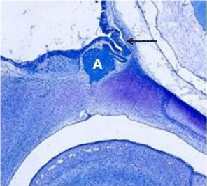
At the initial stage of pituitary gland formation, there is a pituitary gland placode in the pharyngeal region, consisting of an adhesion between the infundibulum cerebri (offshoot from the brain) and the pharyngeal ectoderm [12-16]. Due to migration of the neural crest cells in connection with craniofacial development, this early placodal region is drawn cranially, resulting in appearance of a pocket, named Rathke’s pouch (Figure 1, 2) [14,16]. Immunohistochemical NGFR (Nerve Growth Factor Receptor) reaction has been observed in the neuro-pituitary lobe at the border-line against the anterior lobe of the gland (pars intermedia) from the age of 16 weeks GA.
Sella Turcica
The gland develops before the formation of the sella turcica. This timing in normal development is important to consider in pathological cases. The sella turcica appears in the initial stages in cartilage and later as a bony structure, supporting the mature gland. Sella turcica has an anterior wall and a posterior wall, connected by the floor in the sella. The posterior wall is the first structure to develop in the sella. This posterior wall develops from the paranotocordal mesoderm [16-18]. The anterior wall and the floor of the sella develop shortly after from the neural crest cells, ectomesenchymally derived.
Pathological Development
Abnormal development of the tissues in the pituitary gland placode and/or abnormal development in the surrounding tissues (neural crest cells or the mesenchyme in the cranial part of the notochord), could cause pathological development in the region. The abnormal gland development might result in absence of the sella or in sella malformations, such as abnormal posterior wall formation or malformation in the floor or in the anterior wall [19].
Absence of sella Turcica
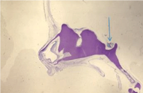
In certain craniofacial malformations it has been observed that the entire anterior pituitary gland tissue persist in the pharyngeal epithelium (Figure 3) [20].
Posterior wall deficiency in sella Turcica
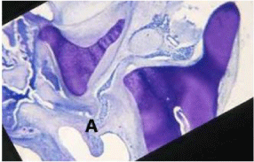
In anencephaly there seems to be a malformation in the infundibulum cerebri in the pituitary gland placode, resulting in absence of the neuropituitary gland tissue and ectopic posteriorly located adeno-pituitary gland tissue in a malformed (not concave) sella turcica [21]. Also in trisomy 18 and in Meckel Syndrome malformation of the posterior wall, extending to the floor are associated with posteriorly located ectopic adeno-pituitary gland tissue (Figure 4) [22,23].
Anterior wall deficiency in sella turcica
In holoprosencephaly and other craniofacial abnormalities caused by abnormal neural crest cells, the anterior wall is malformed, often combined with a canal in the floor [16,19]. In this case, most parts of the gland tissue are located subpharyngially (Figure 5). In Down syndrome and in combined cleft Lip and palate conditions [24] malformations in the floor of the sella turcica is often combined with anterior wall deficiency.
Absence or malformed floor in sella Turcica
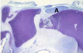
In different types of encephaloceles, the floor can be missing partly and the adeno-pituitary gland tissue located subpharyngeally (Figure 6) [25,26]. The subpharyngeal gland tissue reacts positively on TSH (Thyroid Stimulating Hormone) immunohistochemically staining.
Conclusion and Perspectives
This short review demonstrates how sella turcica can predict abnormal formation and ectopic locations of the anterior pituitary gland tissue. Former studies also demonstrates that immunochemical analyzes can reveal early functions of the gland. Pituitary gland tissue and sella turcica have been investigated in the few cases of more rare conditions, such as human Triploidy [27], Fragile X Syndrome [28] and Fetal Hypohydrotic Ectodermal Hypoplasia [29].
Perspectives
The observations of the early prenatal interrelationship between the formation of the anterior lobe of the pituitary gland and the morphology of the supporting sella might be important for postnatal clinical diagnostics. The shape of the sella turcica has been described radiographically in postnatal Encephaloceles [30], Down Syndrome [31], and postnatal Combined Cleft Lip and Palate [32]. In all cases, the postnatal morphology of the sella was identical to prenatal morphologies, but associated postnatal analyzes of the pituitary gland in these cases are still lacking. In newer studies radiographic investigations on sella turcica morphology are combined with neuro-radiographic imaging of the brain [33,34]. Studies on sella turcica and the pituitary gland are important, not only for analyzes in endocrinology, but also for analyzes in other medical disciplines, such as craniofacial surgery [34] and orthodontics [35].
Acknowledgement
These studies have been constantly scientifically supported by former chief pathologists Birgit Fischer Hansen, MD dr. med. and Jean Keeling, MD. Sincerely thanks are forwarded to these esteemed colleagues.
- Enemar A. The development of the hypophysial vascular system in the bank-vole (clethryionomys glareolus) Acta Anat. 1957; 31: 76-111. https://goo.gl/zH891U
- Kuhlenbeck H. The Central Nervous System of Vertebrates. Vol. 5, Part I: Derivatives of the Prosencephalon, Diencephalon and Telencephalon. Basel Karger. 1977; 5. https://goo.gl/ry321b
- Dorland w. Dorland’s illustrated medical dictionary. 27th ed.W.B. Saunders Company, Philadelphia. 1988. https://goo.gl/axQkt9
- Williams PL, Warwick R, Dyson M, Bannister L (eds): “Gray’s Anatomy” Edinburgh: Churchill Livingston. 1989. https://goo.gl/Y91VES
- Sperber GH. “Craniofacial Embryology”. Dental Practitioner Handbook No. 15, London; Wright. 1989. https://goo.gl/hJYKDH
- O’Rahilly R. “The Embryonic Human Brain –an atlas of developmental stages” 2nd ed. New York, Wiley-Liss. 1999. https://goo.gl/q1VEp5
- Kjaer I, Keeling JW, Hansen BF. The Prenatal Human Cranium-normal and pathological development. Copenhagen, Munksgaard. 1999. https://goo.gl/KPkjdg
- Boyd JD. Observations on the human pharyngeal hypophysis. J. Endocrinol 1956; 14: 66-77. https://goo.gl/JDCL1a
- McGrath P. Aspects of the human pharyngeal hypophysis in normal and anencephalic fetuses and neonates and their possible significance in the mechanism of its control. J anat. 1978; 127: 65-81. https://goo.gl/QV55p9
- Hamilton WJ. Boyd JD, Mossman HW. “Human Embryology” 4th ed. Cambridge, England: W. Heffer and Sons Ltd. 1972. https://goo.gl/omUvAu
- Sadler TW (ed). “Longman’s Medical Embryology” 6th ed. Baltimore, Williams & Wilkins. 1990. https://goo.gl/XUSfQw
- Kjaer I, Hansen BF. The adenohypophysis and the cranial base in early human development. J Craniofac Genet Dev Biol. 1995; 15: 157-161. https://goo.gl/tFv12K
- Kjaer I. Neuro-osteology. Crit Rev Oral Biol Med. 1998; 9: 224-244. https://goo.gl/tXHWrV
- Kjaer I, Nolting D, Hansen BF. 75-NGFR Expression in the human prenatal pituitary gland. Pediatric neurology. 2004; 5: 345-348. https://goo.gl/XfV1WD
- Kjaer I, Hansen BF. The prenatal pituitary gland - hidden and forgotten. Pediatric Neurology. 2000; 22: 155-156. https://goo.gl/9GJK1W
- Kjaer I. Etiology based dental and craniofacial diagnostics. Wiley. 2017. https://goo.gl/vSepvM
- Kjaer I, Becktor KB, Nolting D, Hansen BF. The association between prenatal sella turcica morphology and notochordal remnants in the dorsum sellae. J Craniofac Genet Dev Biol. 1997; 17: 105-111. https://goo.gl/rfNW7W
- Kjær I, Mygind H, Hansen BF. Notochordal remnants in human iniencephaly suggest disturbed dorso-ventral axis signaling. Am J Med Genet. 1999; 84: 425-432.
- Kjaer I. Orthodontics and foetal pathology: a personal view on craniofacial patterning. Eur J Orthod. 2010; 32: 140-147. https://goo.gl/EygMqe
- Kjaer I, Reintoft I, Poulsen H, Nolting D, Prause JU, Jensen OA, Hansen BF. A new craniofacial disorder involving hypertelorism and malformations of external nose, palate and pituitary gland. J Craniofac Genet Dev Biol. 1997; 17: 23-34. https://goo.gl/htVd5v
- Kjaer I, Hansen BF. Human fetal pituitary gland in holoprosencephaly and anencephaly. J Craniofac Genet Dev Biol. 1995; 15: 222-229. https://goo.gl/kWNhLA
- Kjaer I, Keeling JW, Reintoft I, Hjalgrim H, Nolting D, Hansen BF. Pituitary gland and sella turcica in human trisomy 18 fetuses. Am J Med Genet. 1998; 76: 87-92. https://goo.gl/rekjWU
- Kjaer KW, Hansen BF, Keeling JW, Nolting D, Kjaer I. Malformation of cranial base structures and pituitary gland in prenatal Meckel syndrome. APMIS 1999; 107: 937-944. https://goo.gl/8c5V3o
- Kjaer I, Keeling JW, Reintoft I, Nolting D, Hansen BF. Pituitary gland and sella turcica in human trisomy 21 fetuses related to axial skeletal development. Am J Med Genet. 1998; 80: 494-500. https://goo.gl/2ybPNB
- Kjaer I, Hansen BF, Keeling JW. Axial skeleton and pituitary gland in human fetuses with spina bifida and cranial encephalocele. Pediatr Pathol.1996; 16: 909-926. https://goo.gl/mCZjEE
- Kjaer I, Hansen BF, Reintoft I, Keeling JW. Pituitary gland and axial skeleton malformations in human fetuses with spina bifida. Eur J Pediatr Surg. 1999; 9: 354-358. https://goo.gl/AbcEeM
- Kjaer I, Keeling JW, Smith NM, Hansen BF. Pattern of malformations in the axial skeleton in human triploid fetuses. Am J Med Genet. 1997; 72: 216-221. https://goo.gl/9dJxi9
- Hjalgrim H, Hansen BF, Brøndum-Nielsen K, Nolting D, Kjaer I. Aspects of skeletal development in Fragile X syndrome fetuses. Am J Med Genet. 2000; 95: 123-129. https://goo.gl/Jjkok5
- Nordgarden H, Reintoft J, Nolting D, Hansen BF, Kjaer I. Craniofacial tissues including tooth buds in fetal hypohidrotic ectodermal dysplasia. Oral Dis. 2001; 7: 163-170. https://goo.gl/t5FYa8
- Kjær I, Wagner Aa, Madsen P, Blichfeldt S, Rasmussen K, Russell B. The sella turcica in children with lumbosacral myelomeningocele. Eur J Orthod. 1998; 20: 443-448. https://goo.gl/JRoZ9r
- Russell BG, Kjær I. Postnatal structure of the sella turcica in Down syndrome. Am J Med Genet. 1999; 87: 183-188. https://goo.gl/YLkzVK
- Nielsen BW, Molsted K, Kjær I. Maxillary and sella turcica morophology in newborns with cleft lip and palate. Cleft Palate Craniofac J. 2005; 42: 610-617. https://goo.gl/C82Kxn
- Kjaer I. Sella turcica morphology and the pituitary gland-a new contribution to craniofacial diagnostics based on histology and neuroradiology. Eur J Orthod. 2015; 37: 28-36. https://goo.gl/R6RwJz
- Cho CH, Barkhoudarian G, Hsu L, Bi WL, Zamani AA, Laws ER. Magnetic resonance imaging validation of pituitary gland compression and distortion by typical sellar pathology. J Neurosurg. 2013; 119: 1461-1466. https://goo.gl/NXGZ9f
- Sathyanarayana HP, Kailasam V, Chitharanjan AB. Sella turcica-Its importance in orthodontics and craniofacial morphology. Dent Res J (Isfahan). 2013; 10: 571–575. https://goo.gl/GbysDt


Sign up for Article Alerts