The Determination of the Portal Vein Variations in Subjects aged between 18-80 Years
Ayşe Gül Kabakcı1*, Mahmut Öksüzler2, Sema Özandaç Polat1, Musa İnal3 and Ahmet Hilmi Yücel1
1Cukurova University, Faculty of Medicine, Department of Anatomy, Turkey
2Adana Medline Hospital, Department of Radiology, Turkey
3Adana Medline Hospital, Department of General Surgery, Turkey
*Address for Correspondence: Ayşe Gül KABAKCI, Çukurova University, Faculty of Medicine, Department of Anatomy, 01330 Adana-Turkey, Tel: +90-322-338-606-034-89; Fax: +90-322-338-6572; ORCID ID: orcid.org/0000-0001-7144-8759; E-mail: [email protected]
Submitted: 03 March 2020; Approved: 11 April 2020; Published: 13 April 2020
Citation this article: Kabakcı AG, Öksüzler M, Polat SÖ, İnal M, Yücel AH. The Determination of the Portal Vein Variations in Subjects aged between 18-80 Years. Int J Hepatol Gastroenterol. 2020; 6(1): 005-009.
Copyright: © 2020 Kabakcı AG, et al. This is an open access article distributed under the Creative Commons Attribution License, which permits unrestricted use, distribution, and reproduction in any medium, provided the original work is properly cited
Keywords: Liver; Portal vein types; Portal vein variation
Download Fulltext PDF
Aim: The aim of this research is to determine the frequency of the portal ven variations, and to show a new Typeportal vein variation (Type IV) with multislice abdominal computed tomography.
Material and Method: This study was carried out from the 340 healthy adult subjects (161 Males; 179 females) aged 18-80 years. All CT scans were obtained using a 64x2-slice multidetector CT (Siemens Somatom Definition AS, Siemens Healthcare). The axial and coronal image were used to evaluate the portal vein variations. The SPSS 21.0 program was used for statistical analysis of the measurement results. In all statistical analyses; p value under 0.05 was considered statistically significant. Furthermore, Chi Square Test was chosen.
Type1: Typeof bifurcation branch of normal main portal vein. The portal vein separates into two branches as left and right in the liver hilum; It is defined as the division of the right portal vein into the anterior and posterior major branches, the V-VIII segmental branches, and the left portal vein into the I-IV segmental branches.
Type2: Typeof trifurcation in the main portal vein. The portal vein divides into three parts as right anterior portal vein, right posterior portal vein and left portal vein.
Type3: The right posterior portal vein branch is the first branch from the main portal vein.
The left portal vein and right anterior portal vein are separated as a common branch, and the right posterior portal vein is separated as a other branch.
Type4: It was observed that the right posterior portal vein and the left portal vein were directly separated and the right anterior portal vein was branched from the left portal vein.
Results: Type4 portal vein variation were seen both females (n=3) and males (n=4). There were no significant different in four Types portal vein variations (p=0.138). The most frequent portal vein variation was Type1 in females and males followed by Type2.
Conclusion: The determine of the Portal vein variations may be important for decrease in risk for complications during surgical prosedure. Also, the knowledge of the presence of variation in the venous system and its prognosis in the preoperative period may reduce the complications thay may occur during interventional radiological prosedures and abdominal surgery operations and increase in the success of the operation.
Introduction
The portal vein is formed by the convergence of the superior mesenteric and splenic veins posterior to the neck of pancreas at the level of L2 vertebra [1,2]. It is approximately 8 cm long and lies anterior to the inferior vena cava, posterior to the neck of the pancreas and obliquely to the right. It ascends behind the first part of the duedonum, common bile duct and gastroduedonal artery. At this point it is directly anterior to the inferior vena cava. It enters the right border of the lesser omentum, ascends anterior to the epiploic foramen to reach the right end of the porta hepatis, and then divides into right and left main branches. In the lesser omentum it lies posterior to both the common bile duct and hepatic artery. The right branch of the portal vein usually receives the cystic vein and then enters the right lobe of the liver. The left branch receives only obliterated umblical vein via the ligamentum teres which connects to its vertical portion. Varaitons usually involve the right portal vein: absence of a right portal vein with the resulting portal trifurcation in the form of left portal vein in the form of left portal vein, right medial and right lateral portal veins, is present in 10-15% of livers. Occasionally, right medial vein arises from the left portal vein, a variant which is important to remember during left sided liver resection. The portal trifurcation has implications for split liver and live donor liver transplantation, where its presence might be considered as a relative contra-indication. On rare occasions, the portal bifurcation is absent, in which case the main portal vein enters the liver giving off the right segmental branches and then turns left to supply the left lobe of the liver [1,3]. Variations of the Main Portal Vein (MPV) and Right Portal Vein (RPV) branching at the hepatic hilum are quite frequent. Identification and reporting of such variations are necessary prior to interventions such as liver transplantation, as some are contraindications to living donor lobectomy or they at least require different techniques of anastomosis.
The portal vein carries about 70% of the blood to the liver [4]. Knowledge of normal anatomical variants in branching patterns are necessary pre-operatively for portal vein embolization, liver transplantation, hepatic tumor resection, and placement of trans jugular intrahepatic portosystemic shunts and for accurate tumor localization [2,5]. The aim of this study was to evaluate and estimate the prevalence of variations of portal vein branching by Multi detector computed tomography.
Material and Methods
This study was carried out from the 340 healthy adult subjects (161 males; 179 females) aged 18-80 years. This study was approved by the Cukurova University, Clinical Researches Ethics Committee, with Decision No:2020/95-37. All the test procedures were performed after ethics committee approval. This study was a retrospective observational study which done in Department of Radiology in and subjects who admitted to the hospital for various reasons between January 2014 and December 2019 were participated. All CT scans were obtained using a 64x2-slice multidetector CT (Siemens Somatom Definition AS, Siemens Healthcare). The axial and coronal image were used to evaluate the portal vein variations.
The patients having large hepatic masses, distorted intrahepatic portal venous anatomy and poor opacification of the vessels, choledochlithiasis or urinary calculus were excluded. The data were divided into both two groups according to gender (healthy adult female and male subjects), and into seven groups according to ages (Group I, 18-20 years; Group II, 21-30 years; Group III, 31-40 years; Group IV, 41-50 years; Group V, 51-60 years; Group 6, 61-70 years; and Group 7, 71-80 years).
The SPSS 21.0 program was used for statistical analysis of the measurement results. In all statistical analyses; p value under 0.05 was considered statistically significant. Furthermore, Chi Square Test was chosen.
These parameters were as follows:
Type1: Typeof bifurcation branch of normal main portal vein. The portal vein separates into two branches as left and right in the liver hilum; It is defined as the division of the right portal vein into the anterior and posterior major branches, the V-VIII segmental branches, and the left portal vein into the I-IV segmental branches (Figure 1).
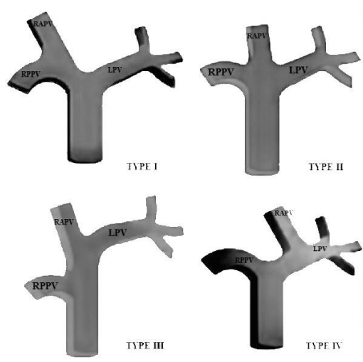
Type2: Typeof trifurcation in the main portal vein. The portal vein divides into three parts as right anterior portal vein, right posterior portal vein and left portal vein (Figure 1).
Type3: The right posterior portal vein branch is the first branch from the main portal vein.
The left portal vein and right anterior portal vein are separated as a common branch, and the right posterior portal vein is separated as a other branch (Figure 1).
Type4: Unlike the three Types of portal system variations existing in the literature, the fourth Typevariation was determined. It was observed that the right posterior portal vein and the left portal vein were directly separated and the right anterior portal vein was branched from the left portal vein (Figure 1).
Inclusion Criteria
1. Adult patients aged above 18 years
2. Patients undergoing Contrast enhanced CT abdomen for indications other than Suspected liver pathology.
Exclusion Criteria
1. Patients with hepatic lesions as it can distort the intrahepatic portal venous anatomy.
2. Patients with history major abdominal surgery.
3. Cases with insufficient portal venous opacification.
4. Cases with motion artifacts resulting in poor quality images.
5. Patients with allergy to contrast media.
Results
The portal vein variations of 340 healthy adult subjects (161 Males; 179 females) aged 18-80 years were shown in Tables 1-2. All four Types portal vein variations were seen both females (n = 3) and males (n = 4). There were no significant different in four Types portal vein variations (p = 0.138). The most frequent portal vein variation was Type1 in females and males followed Type2. The least seen Typewas 4. This Typewas defined firstly in this study (Table 1). The distribution of portal vein variations were shown in table 2. In evaluation of the distribution of the portal vein variations, there were no a significant difference. There was only Type1 portal vein variation in the age of 18-20 years, however there was no seen Type2, 3 and 4 portal vein variations in the age of 18-20 years. In the age of the 21-30 years, Type2 and Type4 portal vein variation was no found in Group 1, and 2 and 4. The most frequent seen portal vein variation was Type1 (46), followed by Type2 (12), Type3 (1), Type4 (1) in Group 3. In the age of 41-50 years, Type1, 2 and 3 portal vein variations were also noted. Additionally, the most frequent seen portal vein variation was Type1 (52), followed by Type2 (10), Type3 (2). There were Type1 (64), Type2 (4), Type3 (3) and Type4 (2) portal vein variations in Group 5, respectively. In Group 6, the most frequent seen portal vein variation was Type1 (63), followed by Type3 (4), Type2 (3), Type4 (3), respectively. In the age of 71-80 years, the least seen portal vein variation was Type4 (1), followed by Type3 (2), Type2 (5), Type1 (29), respectively.
Discussion and Conclusion
Embryologically, the PV is formed between 4th and 10th week. Initially, three paired venous systems are present: umbilical veins of chorionic origin, vitelline veins from the yolk sac and cardinal veins from the body of the embryo. Selective involution of the vitelline veins results in the formation of PV during the second month of gestation [6,7]. Portal Vein, which is formed dorsally by the superior mesenteric vein and spleen vein to the neck of the pancreas, is located at the level of the second lumbar vertebra. In the hilum of liver, the main Portal Vein is divided into larger right and left Portal Vein branches. The Right Portal Vein then divides into the anterior and posterior truncus. The Right Anterior Portal Vein provides the V and VIII Segments, The Right Posterior Portal Vein provides VI and VII Segments and then it turns medially towards the ligamentum teres giving branches to provide Segments II, III and IV and the caudate lobe [8,9]. Standard anatomy in branching pattern is seen in only 65% of the cases. Most common anatomic variation of MPV is trifurcation variation followed by Right Posterior Portal Vein as a first branch of Main Portal Vein [10].
This study revealed the prevalence of Portal Vein variations by age and gender. All four Types portal vein variations of 340 healthy adult subjects (161 Males; 179 females) aged 18-80 years were seen both females (n = 3) and males (n = 4). There were no significant different in four Types portal vein variations (p = 0.138). There was no significant difference between 7 age groups in terms of portal variations (p = 0.160). In a study in which Koç et al. examined the cross-sectional CT images of 1384 patients (721 men and 663 women) in 2007, A statistically significant difference in the prevalences of PV variations was not detected between male and female patients (p = 0.582). In addition, normal intrahepatic Portal venous branching was observed in 1005 of 1384 patients, while portal venous variation was found in 379 subjects. In Koç et al’s study performed with multislice CT images, portal vein branching was found to be 78.5% of Type1, followed by 11.1% of Type2 and 9.7% of Type3 [11].
In a study conducted by Sarı et al. in 2014, CT images of 48 healthy individuals [(female, 11 - 22.9%); (male, 37 - 77.1%)] who applied as donors for liver transplantation were examined. They detected normal main portal vein bifurcation in 25 (52%) cases (Type1). The most detected main portal vein variation was the trifurcation (Type2) Typebranching in the main portal vein seen in 7 cases (14.6%) and the variation (Type3: the right posterior portal vein branch) was the first branch coming out of the main portal vein in four cases (8.4%). Also, Sarı, et al. [12] found that there was no significant difference between the frequency of portal venous variation and the relationship between gender (p = 0.34). In a study of Yanmaz, et al. retrospectively evaluated the multislice CT of 750 patients images, 363 of whom were male and 387 subjects were female. As a result of evaluations, the most frequent seen Typewas Type1 variation in 616 patients (82.1%), followed by Type2 variation in 72 patients (9.6%), Type3 variation in 53 patients (7.4%), right portal vein trifurcation in 8 patients (0.9%), and quadrification in 1 patient (0.1%). They detected the variation of the main portal vein in 142 patients (19.1%) in total [13]. In another study performed by Kamel et al. to determine liver portal venous variations in 40 donors (26 males and 14 females) aged between 18-57 years, it was found that 8 patients (20%) had no right portal vein, 6 patients (15%) trifurcation, 1 patient (2.5%) had quadrifurcation and 1 patient (2.5%) the right posterior portal vein emerged from the main portal vein [14].
Sureka, et al. performed with 967 Indian patients, and reported that the most seen frequent Typewas Type1 (normal anatomy, 773 subjects, 79.94%), followed by Type2 (trifurcation, 66 patients, 6.83%); Type3 (right posterior vein as first branch of main Portal Vein, 48 subjects, 4.96%); Type4 (origin of right anterior portal vein from left portal vein, 26 patients, 2.69%) and Type5 (total ramification, 13 patients, 1.34%). Also, 19 cases showed other Types of variations [15]. In an another study performed with 507 patients using ultrasound method, trifurcation Typewas found in 55 (10.8%), right posterior segmental branch Typearising from the main portal vein in 24 patients (4.7%), right anterior segmental branch Typeoriginating from the left portal vein in 22 patients (4.3%) and absence of the horizontal segment of the left portal vein in one patients (0.2%) [15].
Akgül, et al. studied on 585 cases (280 females, 47.9%) and 305 males, 52.1%. According to these findings, 504 subjects had Type1 (normal portal vein anatomy) whereas, 72 patients (12.3%) showed trifurcation (the Main Portal Vein entered the porta hepatis and divided into the Right Anterior Portal Vein, Right Posterior Portal Vein and Left Posterior Vein branches). Moreover, the right anterior portal vein originated from the left portal vein in 5 patients (0.9%). The left portal vein originated from the right anterior portal vein in 2 patients (0.3%). Additionally, the right posterior portal vein arose from the main portal vein ın 2 patients (0.3%) [15].
In a study performed with 69 American patients by Soyer et al., normal portal vein anatomy (Type1) was observed in 60 patients. Also, variations in intrahepatic portal anatomy were found in four patients (6%), and trifurcation of the portal vein in three patients (4%), and left main portal vein originating from the right anterior portal branch in one patient (2%) [17]. In another study conducted by Covey et al. in 200 healthy American population, the most frequent seen Typewas normal portal vein Type1 (130 subjects), followed by trifurcation in 18 subjects, Type 3 (Right Posterior Portal Vein as first branch of Main Portal Vein) in 26 subjects, Type5 (Segment VI branch as separate branch of Right Portal Vein) in 12 subjects, undefined different Typein 12 subjects, and Type4 in 2 subjects (Segment VII branch as separate branch of Right Portal Vein) [18].
As a result of the examination carried out by Atasoy and Özyürek in 200 patients, they found that there were Type1 (Main portal vein dividing into left portal vein and right portal vein, right portal vein then dividing into right anterior portal vein and right posterior portal vein) in 131 patients, Type 2 (Main portal vein trifurcating into left portal vein, right anterior portal vein, and right posterior portal vein) in 19 patients, Type 3 (Main Portal Vein dividing into Right Posterior Portal Vein and a common Right Anterior Portal Vein –Left Posterior Vein trunk) in 47 patients and other Types of variations in 3 patients [18].
In a conclusion, in the studies of the main portal vein variation Types there were no difference between gender, whereas, in these studies various methodologies such as CT, US, and cadaver were used. The observations presented in studies showed that the main portal vein had more than 3 branches or less than 3. These findings obtained healthy subjects may provide to occur the reference data and also showed the portal vein variations were seen in healthy subjects as well. Additionally, they helped to define the segmental and subsegmental location of intrahepatic tumors, and to assess the feasibility of tumor resection and planning the operative approach. The knowledge of portal vein variations may be important for portal vein embolization, anatomic resection, and transplantation for general surgeon due to normal variant portal vein anatomy increasingly important, and the complexity of hepatic interventions.
- Standring S. Gray’s Anatomy. Kırkıncı baskı. Spain: Elsevier; sayfa. 2008. p. 1166-1167. https://bit.ly/2UZV6GZ
- Kumar V, Sundar J. Evaluation of variations of portal vein branching by multidetector computed tomography. J Evid Based Med Healthc. 2019; 6: 154-159. https://bit.ly/2Rtjv5t
- Yücel AH. Dere Anatomi Atlası ve Ders Kitabı, 7. Baskı, Akademisyen Kitabevi, Adana, 2018.
- Snell RS. Topografik Klinik Anatomi. Çev-ed. Ankara, Türkiye: Palme Yayıncılık, 2015; 194-198.
- Varotti G, Gondolesi GE, Gondolesi GE, Goldman J, Wayne M, Florman SS, Schwartz ME, Miller CM, Sukru E. Anatomic variations in right liver living donors. Journal of the American College of Surgeons. 2004; 198: 577-582. https://bit.ly/34x5EAp
- Hu GH, Shen LG, Yang J, Mei JH, Zhu YF. Insight into congenital absence of the portal vein: is it rare? World J Gastroenterol. 2008; 14: 5969-5979.
- Walsh G, , Williams MP. Congenital anomalies of the portal venous system: CT appearances with embryological considerations. Clin Radiol 1995; 50: 174-176. PubMed: https://www.ncbi.nlm.nih.gov/pubmed/7889709
- Burroughs AK. The hepatic artery, portal venous system and portal hypertension: The hepatic veins and liver in circulatory failure. In: Dooley JS, Lok ASF, Burroughs AK, Heathcote EJ, editors. Sherlock's diseases of the liver and biliary system. 12th ed. New York, NY: Wiley Blackwell Publishing Ltd; 2011. p. 152-209. https://bit.ly/2V37TIY
- Covey AM, Brody LA, Getrajdman GI, Sofocleous CT, Brown KT. Incidence, patterns, and clinical relevance of variant portal vein anatomy. AJR Am J Roentgenol. 2004; 183: 1055-1064. PubMed: https://www.ncbi.nlm.nih.gov/pubmed/15385304
- Sureka B, P Yashwant, Bansal K, Rajesh S, Agrawal N, Arora A. Portal vein variations in 1000 patients: Surgical and radiological importance. Br J Radiol. 2015; 88: 20150326. PubMed: https://www.ncbi.nlm.nih.gov/pubmed/26283261
- Koç Z, Oğuzkurt L, Ulusan Ş. Portal vein variations: Clinical implications an frequencies in routine abdominal multidetector CT. Diagn Interv Radiol. 2007; 13: 75-80. PubMed: https://www.ncbi.nlm.nih.gov/pubmed/17562511
- Sarı ZŞ, Yılmaz FG, Kervancıoğlu S, Kervancıoğlu R. The evaluation of potential donors’ hepatic vascular anatomy in liver transplantation with multislice computed tomography. Eur J Ther. 2014; 20: 245-253. https://bit.ly/2xdk5Od
- Yanmaz R, Karazincir S. Evaluation of portal vein anatomic variations with multidetector computed tomography. Mustafa Kemal Üniv Tıp Derg. 2017; 8(29): 19-26.
- Kamel IR, Kruskal JB, Pomfret EA, Keogan MT, Warmbrand G, Raptopoulos V. Impact of multidetector CT on donor selection and surgical planning before living adult right lobe liver transplantation. AJR Am J Roentgenol. 2001; 176: 193-200. PubMed: https://www.ncbi.nlm.nih.gov/pubmed/11133565
- Atri M, Bret PM, Fraser-Hill MA. Intrahepatic portal venous variations: Prevalence with US. Radiology. 1992; 184: 157-158. PubMed: https://www.ncbi.nlm.nih.gov/pubmed/1609075
- Akgül E, Inal M, Soyupak S, Binokay F, Aksungur E, Oğuz M. Portal venous variations. Acta Radiologica. 2002;43:315-9.
- Soyer P, Bluemke DA, Choti MA, Fishman EK. Variations in the intrahepatic portions of the hepatic and portal veins: findings on helical CT scans during arterial portography. AJR Am J Roentgenol. 1995; 164: 103-108. PubMed: https://www.ncbi.nlm.nih.gov/pubmed/7998521
- Atasoy C, Ozyürek E. Prevalence and types of main and right portal vein branching variations on MDCT. AJR Am J Roentgenol. 2006; 187: 676-681. PubMed: https://www.ncbi.nlm.nih.gov/pubmed/16928929
Sign up for Article Alerts