Bilateral Deep Brain Stimulation of the Subthalamic Nuclei in Parkinson’s disease Patients with Camptocormic Posture?
Fadi Almahariq1, Marina Raguz1*, Vladimira Vuletic2, Darko Oreskovic1, Ivica Franciskovic1, Petar Marcinkovic1 and Darko Chudy1
1Department of Neurosurgery, University Hospital Dubrava, Zagreb, Croatia
2Department of Neurology, University Hospital Center Rijeka, Rijeka, Croatia
*Address for Correspondence: Marina Raguz, Department of Neurosurgery, University Hospital Dubrava, Avenija Gojka Suska 6 Zagreb HR-10000, Croatia, Tel: +385 1 290 2476; Fax: +385 1 290 2934; E-mail: [email protected]
Submitted: 05 August 2017; Approved: 17 August 2017; Published: 18 August 2017
Citation this article: Almahariq F, Raguz M, Vuletic V, Oreskovic D, Franciskovic I, et al. Bilateral Deep Brain Stimulation of the Subthalamic Nuclei in Parkinson’s disease Patients with Camptocormic Posture. Scientific Journal of Neurology & Neurosurgery. 2017;3(2): 037-040.
Copyright: © 2017 Raguz M, et al. This is an open access article distributed under the Creative Commons Attribution License, which permits unrestricted use, distribution, and reproduction in any medium, provided the original work is properly cited
Keywords: Camptocormia; Deep Brain Stimulation (DBS); Parkinson’s disease (PD); Subthalamic Nucleus (STN); Globus Pallidus Internus (GPI).
Download Fulltext PDF
Background: Camptocormia is a disabling syndrome characterized by forward flexion that can be an idiopathic or associated with numerous diseases like movement disorders. Posture improvement could be expected after bilateral deep brain stimulation (DBS) of the Globus Pallidus Internus (GPI) or Subthalamic Nucleus (STN) in Parkinson’s disease (PD) patients with camptocormia. The aim of this study was to determine the efficacy of bilateral STN DBS in alleviating the degree of camptocormia in PD patients.
Methods: Two patients (67 year old female and 66 year old male) suffering from PD in the last ten years were subjected to bilateral STN DBS procedure. The positions of electrodes were verified with a postoperative magnetic resonance imaging. The results were objectivized by measuring thoracolumbar flexion angle before and after operation and utilizing all recommended scales for the international survey of DBS.
Results: The degree of forward flexion of the spine substantially decreased and the quality of life, motor symptoms and functioning was improved in both patients.
Conclusions: STN DBS should be considered as a potential treatment option for PD patients with camptocormia. Further analysis is needed to conclude which PD patients are suitable candidates for bilateral STN or GPi stimulation in the treatment of camptocormia.
Introduction
Camptocormia, also known as ‘’Bent Spine Syndrome’’ (BSS), is one of the most disabling features of Parkinson’s disease (PD). It manifests as a marked forward flexion of the thoracolumbar spine that worsens during walking or standing with a tendency to worsen with fatigue [1,2], but disappears in the recumbent position [3-5]. Camptocormia occurs in approximatelly 10% of PD patients [2], progressively over a year or more, and the trunk flexion is usually associated with scoliosis in these patients. Long standing camptocormia often results in low back pain from sagittal misalignment and potentially increases the risk of falling due to axial instability. These debilitating symptoms are further complicated by degenerative or osteoporotic changes of the spine, which are evidenced to be common in patients with PD. Magnetic Resonance Imaging (MRI) described paraspinal muscle signal abnormalities and fatty replacement in camptocormia patients [5,6], while spinal erector electromyogram shows motor unit reductions [6]. Camptocormia can be an idiopathic condition, but can also be associated with numerous diseases like spinal or metabolic disorders, neuromuscular and movement disorders [2,4]. Camptocormia in PD patients did not show significant improvement with levodopa treatment [4,7]. Conservative treatment for camptocormia, such as psychotherapy, physiotherapy, drugs and botulinum toxin injections are limited and usually futile [8]. Surgical treatment includes bilateral pallidal (GPi) or bilateral Subthalamic Nucleus (STN) DBS [8]. Several reports showed successful bilateral pallidal and bilateral subthalamic nucleus stimulation leading to alleviation of camptocormia in PD patients [7,9-13], and patients without PD [14]. Reports of success after STN or GPi stimulation support the important role of the basal ganglia in axial posture control [15]. However, outcome results are inconsistent, especially STN DBS data. The aim of this study was to determine the efficacy of bilateral STN DBS in alleviating the degree of camptocormia in two PD patients by measuring the thoracolumbar angle preoperatively and on pre-defined postoperative follow up.
Patients and Methods
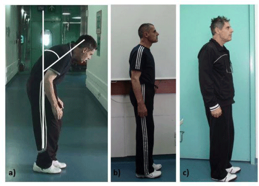
Two patients suffering from PD associated with camptocormia underwent operative treatment. The Thoracolumbar Flexion Angle (TLA) was assessed before and after the surgery according to recommended scales for the international survey of DBS; Functional Independence Measure (FIM) instrument, Unified Parkinson Disease Rating Scale – UPDRS, especially motor score and Activities Of Daily Living (ADL) and levodopa dosage. TLA was measured from the lateral view of the patient and it is the angle between the thoracolumbar spine and a vertical line [Figure 1]. Both patients (67 year old female and 66 year old male) were diagnosed with an advanced PD and severe camptocormia at least ten years prior to operation. It started with unilateral tremor, rigidity and dyskinesia and was characterized by bilateral spreading. Over the last 3 years camptocormia progressed. Pharmacotherapy showed poor effect on PD and camptocormia management. Therefore, patient’s life quality was significantly decreased. Preoperatively, patients UPDRS III score, TLA degrees and UPDRS ADL score were recorded [Table 1].
Both patients underwent bilateral stereotactic Subthalamic Nucleus Deep Brain Stimulation (STN DBS) surgery. In a local anesthesia, a Leksell frame was fixed on the patient’s head and Multi-Slice Computed Tomography (MSCT) scan was performed (slides thickness 0,75 mm). Afterwards, the MSCT scan was merged with a preoperative MRI T2 sequences (slides thickness 1,5 mm) using Medtronic Stealth Station TREON plus Surgical Navigation. Target point (STN bilaterally) was defined alongside with the entry point, whereupon Medtronic electrodes (lead model 3389) were implanted bilaterally in the STN. The target point was determined according to the anterior (AC) and posterior (PC) commissures as follows: for the first patient 16.5 millimeters (mm) posterior from the AC, 14.5 mm lateral on the left and 13.3 mm on the right from the AC-PC line and 4 mm ventral from the AC-PC line, while AC-PC line was 27 mm in length; for the second patient 16 mm posterior from the AC, 13.5 mm lateral on the left and 13 mm on the right, while the AC-PC line was 26 mm in length. The entry point was approximately 2.5 centimeters (cm) from the midsagittal line and 1 cm anterior from the coronal suture. The ACTIVA PC programmable pulse generator was implanted subcutaneously in the left pectoral region. Postoperative MSCT and MRI were performed to confirm the exact location of electrodes placement in the STN. The programming parameters for the first patient were as follows: right electrode: 9(-) 2.8 V, 130 Hz, 90 µs; left electrode: 1(-) 2.7 V, 130Hz, 90µs. Stimulation parameters for the second patient were: left electrode: 8(-) 1.8V, 130 Hz, 90 µs; right electrode: 0(-) 1.8V, 130 Hz, 90µs. In both patients the case of the neurostimulator was set as an anode and the stimulation parameters mentioned above were reached gradually during the first few postoperative days. The follow up period alongside with postoperative measurement of the TLA were done 1 month, 3 months, 6 months for both patients, and one year after operation for female patient.
Results
PD symptoms and camptocormia showed rapid improvement after the initial programming, one week postoperatively. The degree of thoracolumbar flexion substantially decreased in both patients. In female patient TLA was decreasing over time; 20° after the first month, 16° after 3 months, 13° after 6 postoperative months and finally 10° a year after the surgery. UPDRS III score improved from 52 to 16, and the UPDRS ADL score improved from 39 to 10. FIM score improved from 70 to 116 postoperatively. Levodopa daily dose was reduced from 1000 mg preoperatively to 645 mg postoperatively [Table 1]. In male patient TLA was decreasing over time; 15° after the first month, 13° after 3 months and 10° after 6 months. UPDRS III score improved from 44 to 20 postoperatively, UPDRS ADL score improved from 30 to 10, FIM improved to 118 postoperatively. Levodopa daily dose was reduced from 1860 mg preoperatively to 1000 mg postoperatively [Table 1]. Quality of life increased significantly in both patients, pain and sleep problems included. There were no postoperative complications and the positions of implanted electrodes were within STN in both patients. The stimulation parameters were slightly changed on the follow-ups if the patients experienced dyskinesia or other adverse effects.
Discussion
DBS is widely accepted form of therapy for PD patients, especially in patients within pharmacy resistant PD [15]. Unlike several papers reporting beneficial effect of DBS on motor symptoms and dyskinesia in PD patients, there are only few reports showing the effect of DBS in PD patients with camptocormia [1,11,13,16-18]. Bilateral STN DBS showed significant improvement of camptocormia with a substantial improvement of the TLA of around 78 ± 9.1% [12], which is consistent with our result. Like in other studies, our patients showed major improvement after the DBS procedure. Their results in UPRDS III ‘’off medication’’ are more than 50% improved after 6 and 12 months, respectively. A substantial improvement was observed in their UPDRS III ‘’on medication’’ with a decline from 39 and 30 preoperatively to 10 postoperatively. TLA in both patients notably reduced to only 10° for about 6 and 12 months after the operation, respectively. Reduced TLA implicate posture improvement for more than 85% and increased life quality in our patients. It should be mentioned that in several studies [1,10] beneficial effect of STN DBS didn’t occur within all patients included. The efficacy of DBS in PD patients with camptocormia seems to depend whether camptocormia is caused by predominant basal ganglia dysfunction or myopathy [18]. Camptocormia caused by myopathy is more frequent and doesn’t respond to DBS, unlike camptocormia caused by basal ganglia dysfunction [18]. Patients who do not respond to DBS treatment should be screened for other camptocormia causes [18]. Also, the efficacy of STN DBS depends on the duration of camptocormia. Patients with shorter duration of camptocormia experience a beneficial DBS effect [19]. Reports of posture improvement in PD patients with bilateral STN or GPi DBS surgical procedure confirm that basal ganglia has a great role in posture and gait control [20,21]. Besides STN, the GPi can be considered an alternative target for PD patients with camptocormia, because it’s assumed that most STN efferent neurons send axons that simultaneously innervate the GPi [20,21]. There are studies confirming that bilateral GPi DBS can alleviate the severity of camptocormia in PD patients [13,17].
Conclusion
Although results may vary from excellent improvement to no improvement at all, STN DBS should be considered as a potential treatment option for PD patients with camptocormia. Majority reports of bilateral STN and GPi DBS in PD patients with camptocormia speak in favor of successful control of axial posture in these patients. Further and bigger studies are needed to conclude which PD patients are candidates for bilateral STN or GPi stimulation and what kind of screening should they undergo to determine if they are appropriate candidates for STN or GPi DBS.
- Asahi T, Taguchi Y, Hayashi N, Hamada H, Dougu N, Takashima S, et al. Bilateral subthalamic deep brain stimulation for camptocormia associated with Parkinson’s disease. Stereotactic and functional neurosurgery. 2011; 89: 173-177. https://goo.gl/rivDqr
- Yoritaka A, Shimo Y, Takanashi M, Fukae J, Hatano T, Nakahara T, et al. Motor and non-motor symptoms of 1453 patients with Parkinson's disease: prevalence and risks. Parkinsonism & related disorders. 2013; 19: 725-731. https://goo.gl/irURYm
- Djaldetti R, Mosberg-Galili R, Sroka H, Merims D, Melamed E. Camptocormia (bent spine) in patients with Parkinson's disease - characterization and possible pathogenesis of an unusual phenomenon. Movement Disorders. 1999; 14: 443-447. https://goo.gl/Zk9m53
- Lenoir T, Guedj N, Boulu P, Guigui P, Benoist M. Camptocormia: the bent spine syndrome, an update. European Spine Journal. 2010; 19: 1229-1237. https://goo.gl/yUwRor
- Wrede A, Margraf NG, Goebel HH, Deuschl G, Schulz-Schaeffer WJ. Myofibrillar disorganization characterizes myopathy of camptocormia in Parkinson’s disease. Acta neuropathologica. 2012; 123: 419-432. https://goo.gl/PYh5py
- Djaldetti R, Melamed E, Lepoutre AC, Devos D, Blanchard-Dauphin A, Pardessus V et al. A specific clinical pattern of camptocormia in Parkinson's disease. Commentary. Journal of neurology, neurosurgery and psychiatry. 2006; 77: 1229-34. https://goo.gl/SfFmcV
- Yamada K, Goto S, Matsuzaki K, Tamura T, Murase N, Shimazu H, et al. Alleviation of camptocormia by bilateral subthalamic nucleus stimulation in a patient with Parkinson's disease. Parkinsonism & related disorders. 2006; 12: 372-375. https://goo.gl/avTNY9
- Finsterer J, Strobl W. Presentation, etiology, diagnosis, and management of camptocormia. European neurology. 2010; 64: 1-8. https://goo.gl/AV8bBn
- Hellmann MA, Djaldetti R, Israel Z, Melamed E. Effect of deep brain subthalamic stimulation on camptocormia and postural abnormalities in idiopathic Parkinson's disease. Movement disorders. 2006; 21: 2008-2010. https://goo.gl/XtKFzo
- Umemura A, Oka Y, Ohkita K, Yamawaki T, Yamada K. Effect of subthalamic deep brain stimulation on postural abnormality in Parkinson disease: clinical article. Journal of neurosurgery. 2010; 112: 1283-1288. https://goo.gl/fFhstx
- Lyons M, Boucher O, Patel N, Birch B, Evidente V. Long-term benefit of bilateral subthalamic deep brain stimulation on camptocormia in Parkinson's disease. Turkish neurosurgery. 2011; 22: 489-492. https://goo.gl/nNqFXA
- Sako W, Nishio M, Maruo T, Shimazu H, Matsuzaki K, Tamura T et al. Subthalamic nucleus deep brain stimulation for camptocormia associated with Parkinson's disease. Movement Disorders. 2009; 24: 1076-1079. https://goo.gl/2PM7Xn
- Micheli F, Cersósimo MG, Piedimonte F. Camptocormia in a patient with Parkinson disease: beneficial effects of pallidal deep brain stimulation: case report. Journal of neurosurgery. 2005; 103: 1081-1083. https://goo.gl/3FvYCw
- Nandi D, Parkin S, Scott R, Winter JL, Joint C, Gregory R et al. Camptocormia treated with bilateral pallidal stimulation: case report. Journal of neurosurgery. 2002; 97: 461-466. https://goo.gl/gC7itN
- Lyons MK. Deep brain stimulation: current and future clinical applications. In Mayo Clinic Proceedings. 2011. Elsevier. https://goo.gl/qAdr1X
- Chieng LO, Madhavan K, Wang MY. Deep brain stimulation as a treatment for Parkinson’s disease related camptocormia. Journal of Clinical Neuroscience. 2015; 22: 1555-1561. https://goo.gl/3At8Mv
- Thani NB, Bala A, Kimber TE, Lind CR. High-frequency pallidal stimulation for camptocormia in Parkinson disease: case report. Neurosurgery. 2011; 68: E1501-1505. https://goo.gl/ANjH74
- Allert N, Miskiewicz A, Coenen V. Deep brain stimulation for Parkinson's disease and camptocormia? A case report. Fortschritte der Neurologie-Psychiatrie. 2011; 79: 97-101. https://goo.gl/MpkaCW
- Schulz-Schaeffer WJ, Margraf NG, Munser S, Wrede A, Buhmann C, Deuschl G, et al. Effect of neurostimulation on camptocormia in Parkinson's disease depends on symptom duration. Movement Disorders. 2015; 30: 368-372. https://goo.gl/jKcCVr
- Lanciego JL, Luquin N, Obeso JA. Functional neuroanatomy of the basal ganglia. Cold Spring Harb Perspect Med. 2012; 2: a009621. https://goo.gl/hCoL2v
- Takakusaki K. Functional neuroanatomy for posture and gait control. Journal of movement disorders. 2017; 10: 1. https://goo.gl/QM3kdC
Sign up for Article Alerts