Germinoma in Corpus Callosum with Severe Restrictive Anorexia?
Enrique Caro Osorio1, Roberto J. Alcazar Felix2, Jose A. Figueroa Sanchez1, Alvaro Barbosa Quintana1, Jose C. Herrera Castro1 and Hector R. Martinez1*
1Neurology and Neurosurgery Institute, Hospital Zambrano Hellion, TecSalud. Tecnologico de Monterrey. San Pedro Garza Garcia, Nuevo Leon, Mexico
2AANS Medical Student Chapter Tec de Monterrey, Escuela de Medicina y Ciencias de la Salud TecSalud del Tecnologico de Monterrey. Monterrey, Nuevo Leon, Mexico
*Address for Correspondence: Hector R. Martinez, Batallon de San Patricio 112 Colonia Residencial San Agustin, San Pedro Garza Garcia, Nuevo Leon, Mexico, 66278, Fax number & Tel: +528-188-880-676; E-mail: [email protected]
Submitted: 17 June 2017; Approved: 13 July 2017; Published: 16 July 2017
Citation this article: Osorio EC, Alcazar-Felix RJ, Figueroa-Sanchez JA, Quintana AB, Martinez HR, et al. Germinoma in Corpus Callosum with Severe Restrictive Anorexia. Sci J Neurol Neurosurg. 2018;4(1): 005-007.
Copyright: © 2017 Martinez HR, et al. This is an open access article distributed under the Creative Commons Attribution License, which permits unrestricted use, distribution, and reproduction in any medium, provided the original work is properly cited
Keywords: Germinoma; Corpus callosum; Unspecified eating disorder
Download Fulltext PDF
Intracranial germ cell tumors are a rare group of neoplasms constituting 1% to 2% of primary intracranial tumors in North America and Europe. Germinomas of the corpus callosum are exceedingly rare, accounting for only 0.7% of all intracranial germ cell tumors. We report a case of germinoma in the corpus callosum of a 17-year-old woman with a 2-year history of personality change, anorexia, amnesia, hypersomnia, and depression. Magnetic Resonance Imaging (MRI) showed a well-circumscribed, heterogeneous mass measuring 2.9 × 5 × 3.1 cm, with multiple cystic areas and heterogeneous enhancement with gadolinium. It arose in the corpus callosum and extended to the fornix and frontal lobes. There was mild perilesional edema but no evidence of hypothalamus or hippocampus involvement. No spinal drop metastases were visualized on MRI. Cerebrospinal fluid and serum levels of alpha-fetoprotein, beta-human chorionic gonadotropin, carcinoembryonic antigen, and Placental Alkaline Phosphatase (PLAP) were all normal. Immuno histologic staining of tumor cells was positive for OCT3/4, PLAP, and CD117 and negative for CD30 and GPC3. Radiotherapy led to a substantial decrease in tumor size. This is a case of germinoma arising in the corpus callosum that presented clinically with an eating disorder manifested as restrictive anorexia.
Introduction
Primary intracranial Germ Cell Tumors (GCTs) are an uncommon heterogeneous group of neoplasms affecting mainly children and adolescents. GCTs account for approximately 5% of all intracranial tumors seen in patients younger than 20 years of age. GCTs in the Central Nervous System (CNS) are broadly divided into germinomas and nongerminomas [1-4]. Germinomas usually arise from midline CNS structures and may be the only such lesions that originate from primordial germ cells [3-5]. GCTs occur in the pineal and suprasellar regions in 76% to 90% of cases [1,2,6] and are considered ectopic if found in other CNS areas.
Intracranial germinomas localized to the corpus callosum are extremely rare, with a reported incidence of 0.7% of all intracranial germ cell tumors and they are usually associated with other intracranial lesions [3,7]. We report a patient with an ectopic germinoma arising in the corpus callosum who presented with chronic restrictive anorexia.
Case Report
A 17-year-old female presented with a 2-year history of social isolation, repetitive behavior, memory loss, attention deficit, and hypersomnia. She had a psychiatric assessment and was treated with fluoxetine but showed no improvement. Two months later the patient developed anorexia based upon lack of interest in food, clinically significant weight loss (weighing 20.5 kg), impaired psychosocial functioning, electrolyte imbalance and absence of distortion of the body image or fear of being fat. Initially the diagnosis of Anorexia Nervosa was considered but due to the DSM-V criteria, an Unspecified Eating Disorder diagnosis was made.
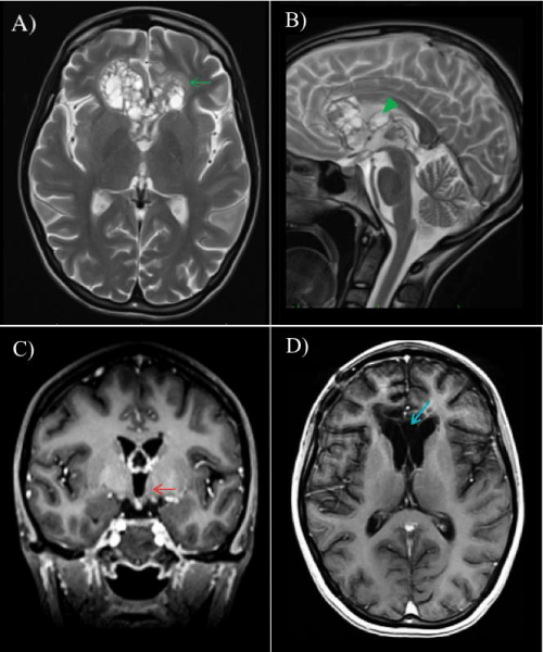
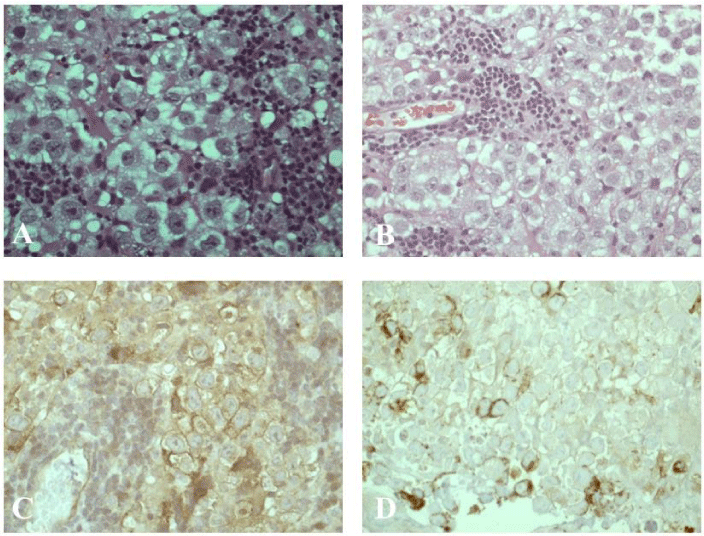
The neurologic exam was normal. Magnetic Resonance Imaging (MRI) of the head revealed an intraaxial lesion in an enlarged corpus callosum at the genu level. It was a heterogeneous mass measuring 2.9 × 5 × 3.1 cm, containing multiple cystic areas, and extending to the fornix and frontal lobes. There was mild perilesional edema but no evidence of hypothalamus or hippocampus involvement (Figure 1). No spinal drop metastases were visualized on MRI. Cerebrospinal Fluid (CSF) and serum levels of Alpha-Fetoprotein (AFP), Beta-Human Chorionic Gonadotropin (bHCG), Carcinoembryonic Antigen (CEA), and Placental Alkaline Phosphatase (PLAP) were all within the normal ranges. After adequate hydration and nutritional support, the mass was biopsied. On microscopy, the specimen, stained with hematoxylin and eosin, was a pure germinoma. Immunohistochemistry stains indicated that the tumor cells were positive for OCT3/4, PLAP, and CD117 (c-kit) but negative for CD30 and GPC3 (Figure 2).
Treatment
The patient was treated with radiotherapy alone, 25.2 Gy of craniospinal radiation, followed by a boost to the primary site of 45 Gy. This led to a substantial decrease in tumor size (Figure 1) and total remission of the anorexia. Two years after treatment, the patient remained asymptomatic. She was eating well and weighed 50 kg. Her psychiatric symptoms had all resolved, and she was taking no antidepressant medications.
Discussion
GCTs in the CNS may be explained by the abnormal incorporation of primordial germ cells into the intraembryonic mesoderm that migrates cranially during the formation of the primitive streak in the third week of embryogenesis [8]. These misplaced germ cells migrate before the formation of the corpus callosum, which occurs in the tenth week of embryogenesis [9]. Because the pineal gland and suprasellar structures are already formed by the seventh week, the abnormal migration of germ cells could explain the higher incidence of GCTs originating in these regions and the lower incidence in the corpus callosum.
The differential diagnosis of corpus callosum masses includes glioblastoma, lymphoma, oligodendroglioma, tumefactive multiple sclerosis, gliomatosis cerebri, and lipoma.
To the best of our knowledge, there are less than 39 cases of GCTs involving the corpus callosum described in the literature and only a few of them presented with symptoms of an eating disorder. GCTs affecting the hypothalamus or hippocampus can present with psychiatric symptoms, including anorexia. Our patient had no evidence on MRI of direct tumor involvement in the hypothalamus or hippocampus. However, the perilesional edema could explain all the symptoms. Kamoshima et al. [10] reported a case of pineal germinoma presenting with an eating disorder. However, 12 years after that tumor was found, MRI follow-up revealed a second mature teratoma in the corpus callosum, though without symptoms. Classical eating disorders have been found to have a positive correlation between aversion ratings and activity in the right medial apical prefrontal cortex [11]. In our patient, changes in appetite and eating behavior could attributed to a tumor mass effect on the hypothalamus or right frontal lobe [11].
Germinoma tumor markers such as AFP, bHCG, CEA, PLAP may help indicate the diagnosis as well as being useful for posttreatment surveillance. However up to 60% of patients may have normal serum and CSF bHCG levels [12] Histologic subtype and tumor size are predictors of GCT prognosis. Survival rates are substantially lower if a germinoma is >4 cm [13]. Complete remission of the tumor after treatment is associated with a better prognosis, although there is a 10% to 17% probability of recurrence within 2 years [14] Recurrence even much later could be due to G0-phase tumor cells that survive treatment and later proliferate to form a new tumor [15]
In conclusion, germinomas in the corpus callosum are exceedingly rare but should be considered in any young patient with a characteristic mixed cystic and solid intraaxial mass. Ectopic germinomas are a possible cause of neuropsychiatric symptoms in young patients. The optimal management of CNS GCTs remains unsettled because of the rarity of these tumors and, hence, the lack of prospective trials. An individualized approach is recommended. When the tumor does not affect directly the hypothalamus or hippocampus it is infrequent to clinically present as an eating disorder.
- Sumida M, Uozumi T, Kiya K, et al. MRI of intracranial germ cell tumors. Neuroradiology. 1995; 37: 32-37. https://goo.gl/dk3JKW
- Liang L, Korogi Y, Sugahara T, Ikushima I, Shigematsu Y, Okuda T, et al. MRI of intracranial germ-cell tumours. Neuroradiology. 2002; 44: 382-388. https://goo.gl/tmELCE
- Yonezawa H, Shinsato Y, Obara S, Hirano H, Kitajima S, Arita K. Germinoma with syncytiotrophoblastic giant cells arising in the corpus callosum. Neurol Med Chir (Tokyo). 2010; 50: 588-591. https://goo.gl/ctBt9h
- Gul Niflioglu G, Unlu M, ozer E, Yuceer N. Childhood intracranial germinoma with granulomatous reaction. Turk Patoloji Dergisi. 2011; 27: 87-90. https://goo.gl/QhCdF1
- Sano K. Pathogenesis of intracranial germ cell tumors reconsidered. J Neurosurg. 1999; 90: 258-264. https://goo.gl/Z8UJ7T
- Jennings MT, Gelman R, Hochberg F. Intracranial germ-cell tumors: natural history and pathogenesis. J Neurosurg. 1985; 63: 155-167. https://goo.gl/Jjz9P9
- Soejima T, Takeshita I, Yamamoto H, Tsukamoto Y, Fukui M, Matsuoka S. Computed tomography of germinomas in basal ganglia and thalamus. Neuroradiology. 1987; 29: 366-370. https://goo.gl/7GHDU3
- Margolin E, Noble J, Kassel E, Croul S, Gilbert J. Germinoma of the corpus callosum. Neuroophthalmology. 2011; 35: 15-21. https://goo.gl/3yNavC
- Wilson JT, Wald SL, Aitken PA, Mastromateo J, Vieco PT. Primary diffuse chiasmatic germinomas: differentiation from optic chiasm gliomas. Pediatr Neurosurg. 1995; 23: 1-5. https://goo.gl/5snFUZ
- Kamoshima Y, Sawamura Y, Iwasaki M, Iwasaki Y, Sugiyama K. Metachronous mature teratoma in the corpus callosum occurring 12 years after a pineal germinoma. J Neurosurg. 2008; 109: 126-129. https://goo.gl/6zAXd9
- Uher R, Treasure J. Brain lesions and eating disorders. J Neurol Neurosurg Psychiatry. 2005; 76: 852-857. https://goo.gl/W9pMUS
- Allen J, Chacko J, Donahue B, Girish Dhall , Cynthia Kretschmar, Regina Jakacki, et al. Diagnostic sensitivity of serum and lumbar CSF bHCG in newly diagnosed CNS germinoma. Pediatr Blood Cancer. 2012; 59: 1180-1182. https://goo.gl/DU4oPH
- Huo LEI, Wang XIA, Allen PK, Wang L, Liao Y, Han Z, et al. Predictors of long-term survival following postoperative radiochemotherapy for pathologically confirmed suprasellar germ cell tumors. Mol Clin Oncol. 2015; 3: 430-434. https://goo.gl/3v2kDs
- Ono N, Isobe I, Uki J, Kurihara H, Shimizu T, Kohno K. Recurrence of primary intracranial germinomas after complete response with radiotherapy: recurrence patterns and therapy. Neurosurgery. 1994; 35: 611-615. https://goo.gl/14DfsT
- Wenger M, Lovblad KO, Markwalder R, Taub E. Late recurrence of pineal germinoma. Surg Neurol. 2002; 57: 34-40. https://goo.gl/ijX3Yp
Sign up for Article Alerts