Developing a Scale to Assess Hypoxia severity in Hospitalized Elderly Patients with Poor Glycemic Control
Yuriy Zamota1, Scott Edele1, Yuliya Zamota2, Ivan Rodriguez2, Bishoy Goubran3 and Marcos A Sanchez Gonzalez3*
1Department of Family Medicine, Larkrin Community Hospital, South Miami, FL, USA
2College of Osteopathic Medicine, New York Institute of Technology, Glen Head, NY, USA
3Division of Clinical and Translational Research, Larkin Community Hospital, FL, USA
*Address for Correspondence: Marcos A Sanchez Gonzalez, Division of Clinical and Translational Research, Larkin Community Hospital, FL, USAMain, Tel: 305-284-7608; Fax: 305-284-7787; E-mail: [email protected]
Submitted: 26 August 2017; Approved: 18 October 2017; Published: 21 October 2017
Citation this article: Zamota Y, Edele S, Zamota Y, Rodriguez I, Goubran B, Developing a Scale to Assess Hypoxia severity in Hospitalized Elderly Patients with Poor Glycemic Control. Sci J Pulm Respir Med. 2017;1(2): 024-027.
Copyright: © 2017 Zamota Y, et al. This is an open access article distributed under the Creative Commons Attribution License, which permits unrestricted use, distribution, and reproduction in any medium, provided the original work is properly cited
Keywords: Type II Diabetes Mellitus; Hypoxia; Hypoxic Scale; Decreased Length of Stay in Hospital; Decreased Cost of Healthcare
Download Fulltext PDF
Tissue hypoxia has been previously described in diabetes, it is usually ascribed to local inflammatory changes and micro vascular complications. Hypoxia manifests clinically by an array of signs and symptoms, leading to detrimental effects including aggravating the diabetic complications and worsening the prognosis. However, a simple bed side scale for detection of hypoxic manifestations in inpatients remains to be developed. The aim of this study was to identify the most common clinical signs of hypoxia patients with uncontrolled type II Diabetes mellitus age 70 or older and formulate an effective clinical tool for recognition of hypoxic states in this population. To that end medical records from admission to discharge were assessed to look for presence of any of the following parameters: BP > 140/90, HR > 100, RR > 20, arrhythmias, ABG, cyanosis, impaired judgment, supplemental oxygenation, presence respiratory treatment as hospital medications, history of cardiovascular disease, history of pulmonary disease, use of cardiovascular medications at home, use of respiratory medications at home, and length of stay in hospital. Results revealed an association between uncontrolled Type II Diabetes Mellitus and hypoxic states in the elderly, and six statistically significant criteria were identified and grouped together to create a hypoxic scale. Practical implementation of this scale can result in lower cost of healthcare by decreasing length of hospital stay through early recognition of hypoxic state, activation of medical care to prevent destabilization of the patient, and exercise higher level of medical care later in hospital course.
Introduction
Type II Diabetes Mellitus (DMII), a chronic metabolic disease affecting the microcirculation, is characterized by insulin resistance and elevated blood glucose levels. Adequate control of DMII is measured using serum levels of Glycated hemoglobin (HgbA1C) with levels of ≥ 7.0% considered indicative of poor glycemic control [1]. Interestingly, diabetic patients with poor glycemic control usually display high levels of glucose outside the cell, but low glucose levels intracellulary [2]. Metabolic anomalies present in DMII may precipitate hypoxia defined here as inadequate oxygen tension at the cellular level. Deficiency of oxygen for aerobic cellular metabolism is compensated by a glucose dependent anaerobic pathway which in turn may trigger clinical manifestations of hypoxia including increased lactate concentration in blood, increased respiratory rates and hemodynamic changes.
Poor glycemic control may precipitate acute symptoms of hypoxia such as tachycardia, arrhythmias, dyspnea, tachypnea, hypertension, anxiety, blurred vision, impaired judgment, euphoria, lethargy/weakness, and tremors [3]. Tissue hypoxiaas well as lower levels of cellular oxygenation have been previously described in decompensated diabetes [4]. Interestingly, hypoxia may occur even in the absence of hypoxemia as a consequence of a chronic pro-inflammatory state with intensive microangiopathy exuded by DM [5]. To note, the dissociation rate constant of hemoglobin from O2 are not typically affected by DMII, and paradoxically normal saturation levels are usually found in these patients [6].
Previous studies have shown that hyperbaric oxygen therapy improves fasting blood glucose in diabetic patients and thus can be used as a therapeutic intervention for DMII [5]. Moreover an improved tissue oxygenation has been shown to aid in prophylaxis against diabetic microangiopathy [7,8] further emphasizing the importance of tackling hypoxia in diabetic patients. However, lacking are standardized methods of scaling the manifestations of hypoxia and thus enabling physicians to monitor and intervene in a timely manner.
It is thus plausible to propose that DMII patients with poor glycemic control will display a myriad of measurable clinical manifestations associated with cellular hypoxia providing the bases for the development of more assertive monitoring tools in the inpatient setting. The main objective for this study was to develop a scale to monitor clinical signs of hypoxia in this population and formulate an effective clinical tool for recognition of hypoxic states enem in the absence of abnormal oxygen saturation levels. We hypothesized that signs and symptoms of clinical hypoxia would be associated with a longer length of stay in hospitalized DMII patients.
Methods
Study design
This study was designed as a retrospective analysis of data gathered from patients admitted to the hospital between January 1st, 2015 and December 31st, 2015, with small exclusion for readmissions occurring in 2014 and 2016 when appropriate (n = 202).
Subjects
At the time of hospitalization, patients must have met the following inclusion criteria: diagnosis of DM II (at least 5 years), A1C ≥ 7.0%, age ≥ 70 years. Both male and female patients were included in this study.
Measures
Demographic characteristics: Information on demographic characteristics was collected via electronic medical record from the time of admission to hospital; including age and sex (See table 1).
Clinical assessment: Once inclusion was met, the medical record from admission to discharge was assessed to look for the following parameters: BP > 140/90, HR > 100, RR > 20, arrhythmias, ABG, cyanosis, impaired judgment, supplemental oxygenation, presence respiratory treatment as hospital medications, history of cardiovascular disease, history of pulmonary disease, use of cardiovascular medications at home, use of respiratory medications at home, and length of stay in hospital. These data were collected from the vitals and laboratory values, home medication lists, as well as the analysis of the patient’s hospital H & P, consult notes, and progress notes.
Statistical analysis: Through analysis of collected information, significant signs of hypoxia included: HR > 100 bpm , RR > 20 resp/min, SBP > 140 mmHg or DBP > 90 mmHg, O2Sat < 92%, Lethargy/weakness, and Shortness of breath (SOB). Using the aforementioned parameters we categorize them in present = 1 or absent = 0, a novel equation was developed to determine hypoxic states in this particular population. Addition of all the parameters was used to calculate the score were higher values were suggestive of higher hypoxia symptoms.
Zamota hypoxic score
HR > 100 + RR > 20 + SBP > 140 or DBP > 90 + O2Sat > 92 + Lethargy /weakness + SOB = ZHS
Hierarchical Multiple Regression (HMR) analyses were conducted to test the association between ZHS scores and length of stay and to demonstrate the contribution of sets of predictors (including demographics) in length of stay variance.
Results
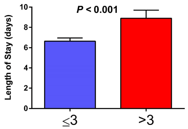
A total of 200 (F = 85) patients where included in the study. We found a significant correlation between the ZHS and length of stay (r .385, P < .001). Moreover, HMR analysis revealed that the ZHS was an independent predictor of length of stay in the patient population. Moreover, t-test revealed that patients with ZHS scores of > 3 stayed significantly (P < 0.001) longer (M ± SD; 8.9 ± 6.6) hospitalized than those with ZHS of ≤ 3 (5.6 ± 3.6), see figure 1. A total of six specific criteria were valuable for determining hypoxic state in these patients with DMII. Statistically, no criterion proved to be superior to another (p > 0.05), however, the presence of more than three of any of these criteria was associated with an increased length of hospital stay. Multiple regression reveled a an increase in length of hospital stay of up to three days more was found in patients with more than three criteria independent of gender (Table 2).
Discussion
In this study, we sought to investigate the clinical signs and symptoms of hypoxia, as well as other objective information currently gathered as part of inpatient medical care (vitals and laboratory studies), to create a standardized hypoxic assessment scale which does not require additional hospital resources or extensive and/or invasive medical testing. The novel results of this study revealed an association between DMII and ZHS in this population suggesting that the developed instrument to measure clinical sings of hypoxia could be useful for recognizing over all disease severity of hospitalized patients and length of stay.
Tissue hypoxia seems to play an integral part in diabetic eteo pathogenesis, with hypoxia complications affecting the cardiovascular system [9], accelerating the progression of diabetic retinopathy [3], diabetic feet [10], and other detrimental effects. Thus tissue oxygenation has been a modality for alleviating these detrimental effects [7]. Thus a standardized modality for quick assessment of the manifestations of tissue hypoxia could be of clinical value. Accordingly, in the present study it was determined that six specific criteria were valuable for determining hypoxic state in these patients with DMII. Statistically, no criterion proved to be superior to another, however, the presence of more than three of any of these criteria was associated with an increased length of hospital stay a finding that merits further investigation and replication. The data revealed that females had shorter hospital courses in general when compared to males, however, the increase in length of hospital stay of up to three days more was found in patients with more than three criteria independent of gender. Therefore, the hypoxic criteria predicted increased length of stay which might hold important clinical implications in the standardization of medical care when assessing for hypoxic state and implementing medical treatment.
These parameters can be easily identified and extracted from the data routinely collected as part of the inpatient medical care. We are proposing to implement these six criteria in a Yes/No fashion in order to recognize hypoxic state. Results of these criteria can be used to predict length of stay and increase the level of medical care in order to improve medical outcomes and decrease hospital length of stay. Early activation of medical management of hypoxic states will improve hospital course, save human and pharmaceutical resources in the hospital, and shorten length of hospital stay. As a result, overall cost of hospital care will decrease.
Conclusions
Using the results of the present scale, we propose that early implementation of short-term oxygen therapy in patients with > 3 clinical signs of hypoxia could potentially result in decreased LOS and improved patient and hospital outcomes. We propose nasal cannula at 2LPM for 2 hours twice daily (70/7.0/2-2-2).Moreover, developing a hypoxic scale can provide an additional tool to assess level of hypoxia in other medical conditions where hypoxia can occur, i.e metabolic diseases, toxicology, pulmonary diseases, and so on. We propose the use of the ZHSin other possible medical hypoxic states in order to improve medical management and collect data to corroborate the practical significance of this research.
- Florkowski C. HbA1c as a Diagnostic Test for Diabetes Mellitus - Reviewing the Evidence. Clin Biochem Rev. 2013; 34: 75-83. https://goo.gl/AzdxCe
- Bouche C, Serdy S, Kahn CR, Goldfine AB. The cellular fate of glucose and its relevance in type 2 diabetes. Endocr Rev. 2004; 25: 807-30. https://goo.gl/wq656c
- Nyengaard J R, Ido Y, Kilo C, Williamson JR. Interactions between hyperglycemia and hypoxia: implications for diabetic retinopathy. Diabetes. 2004; 53: 2931-8. https://goo.gl/VDiWvu
- Braslavskaia N ShteingardtIu. [Tissue oxygenation and tissue hypoxia in diabetes mellitus]. Probl Endokrinol (Mosk). 1976; 22: 13-7. https://goo.gl/mdzGEQ
- Pokorski M, Pozdzik M, Antosiewicz J, Dymecka A, Mazzatenta A, Di Giulio C. Hypoxic Ventilatory Reactivity in Experimental Diabetes. Adv Exp Med Biol. 2015; 860: 123-32. https://goo.gl/14AG5T
- Marschner J P, N Rietbrock. Oxygen release kinetics in healthy subjects and diabetic patients. I: The role of 2,3-diphosphoglycerate. Int J Clin Pharmacol Ther. 1994; 32: 533-5. https://goo.gl/Gm3Uea
- Vera Cruz P, Guerreiro F, Ribeiro MJ, Guarino MP, Conde SV. Hyperbaric Oxygen Therapy Improves Glucose Homeostasis in Type 2 Diabetes Patients: A Likely Involvement of the Carotid Bodies. Adv Exp Med Biol. 2015; 860: 221-5. https://goo.gl/3r3wNW
- Ditzel J. The problems of tissue oxygenation in diabetes mellitus. III. The "three-in-one concept" for the development of diabetic microangiopathy and a rational approach to its prophylaxis. Acta Med Scand Suppl. 1975; 578: 69-83. https://goo.gl/PA2hFz
- Mansor L S, Mehta K, Aksentijevic D, Carr CA, Lund T, Cole MA, et al., Increased oxidative metabolism following hypoxia in the type 2 diabetic heart, despite normal hypoxia signalling and metabolic adaptation. J Physiol. 2016; 594: 307-20. https://goo.gl/4xK4LB
- Vogelberg K H, M Konig. Hypoxia of diabetic feet with abnormal arterial blood flow. Clin Investig. 1993; 71: 466-70. https://goo.gl/VdujYU
Sign up for Article Alerts