Pulmonary Artery Obstruction Index and RV/LV Ratio in Patients with Pulmonary Embolism as Severity Index Risk Factor for Obstructive Sleep Apnea
Doaa M. Magdy1*, Ahmed Metwally1, Mohamed Adam1, Marwa Makboul2 and Shimaa Farghaly2
1Department of Chest Diseases, Faculty of Medicine, Assuit University
2Department of Radio diagnosis, Faculty of Medicine, Assiut University
*Address for Correspondence: Doaa M. Magdy, Department of Chest Diseases, Faculty of Medicine, Assuit University, Egypt, Tel: + 010-062-610-10; E-mail: [email protected]
Submitted: 08 April 2019; Approved: 10 May 2019; Published: 13 May 2019
Citation this article: Magdy DM, Metwally A, Adam M, Makboul M, Farghaly S. Pulmonary Artery Obstruction Index and RV/LV Ratio in Patients with Pulmonary Embolism as Severity Index Risk Factor for Obstructive Sleep Apnea. Int J Sleep Disord. 2019;2(1): 005-009.
Copyright: © 2019 Magdy DM, et al. This is an open access article distributed under the Creative Commons Attribution License, which permits unrestricted use, distribution, and reproduction in any medium, provided the original work is properly cited
Keywords: Pulmonary embolism; Polysomnography
Download Fulltext PDF
Introduction: There is growing evidence that Obstructive Sleep Apnea (OSA) is a risk factor for Pulmonary Embolism (PE). This association represents a major public health burden.
Aims and Objectives: To investigate Computed Tomography Obstruction Index (CTOI) and the Right Ventricular (RV) to Left Ventricular (LV) diameter ratio with OSA severity.
Materials and Methods: 46 Patients with (PE) were evaluated for OSA. Pulmonary Artery Obstruction Index (PAOI) and RV/LV diameter ratio was measured by pulmonary angiography. Pulmonary Embolism Severity Index (PESI) was determined. Epworth Sleepiness Scale (ESS) and Polysomnography (PSG) was performed for all patients. Based on the PAOI, patients divided into (< 15%, 15-50%, > 50%)
Results: No differences were found regarding age, neck and waist circumference. Patients with a higher PAOI had a significant increase in BMI, more often provoked PE, a higher rate of thrombolysis, a longer duration of hospitalization, higher rate of recurrent VTE, and a higher RV/LV diameter ratio. 55.6% and 38.9% of patients with a higher PAOI presented with snoring and daytime sleepiness.
Regarding PSG, AHI, obstructive and hypopnea index were significantly increased with increase the PAOI. Also, desaturation index and time spent with oxygen less than 90% were significantly increased. A positive correlation was observed between PAOI, RV/LV diameter ratio and AHI. In logistic regression analysis, PAOI >40%, RV/ LV >1 were predictors of sleep apnea.
Conclusion: Patients with greater pulmonary artery obstruction had more sever sleep apnea. PAOI > 40%, RV/LV > 1 were predictors of sleep apnea among patients with acute PE.
Introduction
Pulmonary Embolism (PE) is considered as a major risk for patients suffering from venous thromboembolism and can often be fatal. Recently, there is growing evidence from various cross-sectional and longitudinal studies that obstructive sleep apnea is a risk factor for pulmonary embolism. This association represents a major public health burden, given the high prevalence of both disorders with a higher mortality rates [1,2].
PE occurred as a result of Virchow’s classic triad, namely vascular endothelial impairment, stasis of blood flow, and increased coagulability. OSA could hypothetically affect all three mechanistic pathways. Intermittent hypoxia increases oxidative stress, and inflammatory response that impairs endothelial function. OSA-related hemodynamic alterations may slow intravenous flow, and lastly, increased coagulability, platelet activity, and decreased fibrinolytic capacity. These same pathophysiologic derangements are prothrombotic and could promote the development of venous thromboembolic disease. Being aware of the impact of sleep apnea on pulmonary embolism is important, as it can help doctors identify patients who may be at risk and to instruct their patients on how to treat and reduce their risk of both sleep apnea and pulmonary embolism [3].
Computed Tomography Pulmonary Angiography (CTPA) is considered as a first-line diagnostic strategy in patients suspected with PE [4]. The CT obstruction index provides an objective tool to quantify the obstruction severity of pulmonary arteries [5]. However, no longitudinal studies to date have explored the association’s between severity index of Pulmonary Embolism (PE) and severity of OSA. In this study, the aim is to evaluate Computed Tomography Obstruction Index (CTOI) and the Right Ventricular (RV) to Left Ventricular (LV) diameter ratio with OSA severity.
Patients and Methods
Study design
This prospective cohort study was conducted between November 2018 and March 2019 in Assuit university hospital, sleep lab center. Egypt.
46 patients who had a high clinical suspicion of PE according to Wells score and confirmed by Computed Tomography (CT) pulmonary angiography were enrolled in the study.
After diagnosis and clinical stabilization of the acute PE episode was achieved. The patients were transferred to sleep center to evaluate presence of sleep disorder. The study was accepted by the institutional ethics committees. All patients filled the consent form and information was collected by a check list.
• Inclusion criteria
All patients with PE confirmed by Computed Tomography (CT) pulmonary angiography.
• Exclusion criteria
1. Dissatisfaction of patient for participating in the study
2. pregnancy
3. Patients younger than 18 years old were excluded
All patients were subjected to the following: Baseline demographics were collected from all patients age, gender, co-morbid conditions (active cancer, arterial hypertension, diabetes mellitus, acute or chronic heart failure, chronic pulmonary disease, history of VTE, type of PE (provoked versus unprovoked). Unprovoked PE was defined as PE in the absence predisposing factors (immobilization, major surgery, or active cancer during the last three months [6].
Pulmonary Embolism Severity Index (PESI): The PESI is easily and widely used prognostic score suggested by Aujesky et al. [7] for patients with acute PE, composed of 11 indices. PESI (age, sex, cancer, heart failure, chronic lung disease, heart rate >110 beats/min, systolic pressure < 100 mmHg, respiratory rate ≥ 30 times/min, temperature < 36°C, altered mental status and arterial oxygen saturation < 90%).
CT scanning protocol: CTPA scan were performed using 64-channel Multi-detector CT scanner (Toshiba, Japan) Aquilion machine with 16x1.2mm collimation, 120-140 kV, tube current 150-280 mA, all transverse images were reconstructed to 0.625 mm-slice images. A 100 ml of non-ionic contrast agent Ultravist 370 mg iodine concentration (Iopromide 0.769 g) was injected at a rate 4ml/sec into the left antecubital vein.
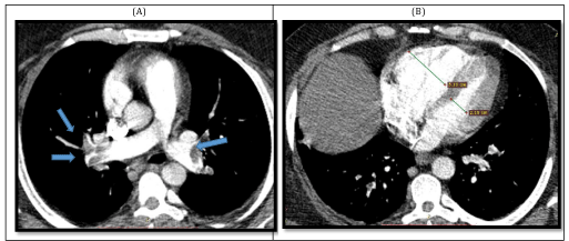
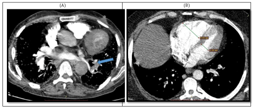
Diagnostic criteria of embolism; was presence of non-enhancing endoluminal clots on CT examination, central emboli defined as emboli within the main arteries, or lobar arteries or both, and peripheral emboli defined as emboli within segmental and/or sub-segmental arteries (Figure 1).
Pulmonary Artery Obstruction Index (PAOI): The percentage of vascular obstruction was calculated according to Qanadli et al. [8]. The arterial tree of each lung have 10 segmental arteries (3 in the upper lobes, 2 in the middle lobe and in the lingula, and 5 in the lower lobes). The presence of an embolus in a segmental artery was scored 1 point. Central or paracentral emboli were scored a value equal to the number of segmental arteries arising distally. Depending on the degree of vascular obstruction a weighting factor was assigned to each value (0 = no thrombus; 1 = partial occlusion; and 2 = total occlusion). Isolated subsegmental embolus was considered as a partially occluded segmental artery and was assigned a value of 1. Thus, The percentage of obstruction = Σ(n.d)/40×100 where n = the number of segmental branches arising distally (minimum, 1; maximum, 20) and d = degree of obstruction (minimum, 0; maximum, 2).
Based on the percentage of vascular obstruction, patients were then divided into three groups (< 15% versus 15-50% versus > 50%) [8].
RV/LV diameter ratio: Right Ventricular (RV) to Left Ventricular (LV) diameter ratio was measured to evaluate determine RV dysfunction through a standard axial view from the maximum ventricular endocardium and the interventricular septum. RV/LV diameter ratio of > 0.9 was defined as right ventricular dysfunction (Figure 2) [9].
Epworth Sleepiness Scale (ESS): All study participants filled out an Epworth Sleepiness Scale (ESS) to assess sleepiness [10].
Nocturnal Polysomnography (PSG): Nocturnal Polysomnography (PSG) was performed in all patients; results were reviewed and rescored according to standard criteria [11]. AHI was calculated by dividing the total number of apneas and hypopneas by the hours of sleep time recorded overnight.
Statistical analysis
Statistical analyses were performed (SPSS for Windows, version 15.0, SPSS Inc., Chicago, Illinois, USA). The data were expressed as the mean± standard deviation and/ or number (percentage). The distribution of the variables was analyzed with the Kolmogorow-Smirnow test. Differences between parametric and non-parametric variables of two groups were assessed by Student t test and Mann-Whitney U test as appropriate. The relationship between the categorical variables was determined by the chi-square test. Pearson correlation analysis was used according to distribution of variables. A P value under 0.05 was considered statistically significant.
Results
Total 46 patients with acute PE confirmed by CTPA, 12, 16, and 18 of patients had a PAOI of < 15%, 15-50%, and > 50%, respectively. No significant differences were found between groups regarding age, sex, neck and waist circumference.
Patients with a higher PAOI had a significant increase in BMI (P = 0.001), more often provoked PE (P = 0.001), a higher rate of thrombolysis (P = 0.000), a longer duration of hospitalization (P = 0.000), higher rate of recurrent VTE, and a higher RV/LV diameter ratio (P = 0.001). 55.6% of patients with a higher PAOI presented with snoring while 38.9% of them presented with excess daytime sleepiness. Also, Epworth Sleepiness Scale was significantly increased.
Regarding PSG records, AHI, obstructive apnea and hypopnea index were significantly increased with increase the PAOI. Also, desaturation index and time spent with oxygen were significantly increased in patients with higher PAOI.
The mean percentage of pulmonary artery vascular obstruction was (32.5 ± 21.9) % (range 5%- 70%). There was a high correlation coefficient between mean percentage of PAOI and AHI (r = 0.957, P = 0.000). Also, a positive correlation coefficient was observed between RV/LV diameter ratio and AHI (r = 0.825, P = 0.000) (Table 3). In logistic regression analysis, we found that PAOI > 40%, RV/ LV > 1 were predictors of sleep apnea among patients with acute PE (Table 4). On the other hand neck and waist circumference were not considered predictors.
Discussion
In many studies, Obstructive Sleep Apnea (OSA) has been shown to be an independent risk factor for PE and both share many of the same risk factors and pathogenic mechanism. This is considered as major concern due to high prevalence of OSA and high morbidity and mortality associated with PE. CT obstruction index has been evaluated in many published articles, and can be used as an objective and reproducible tool to stratify the severity of PE [5]. Hence, the current study we aim to investigate severity of PE through CT parameters with severity of OSA.
Several studies have reported a higher prevalence of sleep disordered breathing in patients presented with DVT or PE supporting the association between OSA and PE [12,13]. It is well known that PE is the result of Virchow’s classic risk triad, vascular endothelial impairment, venous stasis, and/or increased coagulability. OSA could hypothetically affect all three mechanistic pathways. Repeated hypoxic episodes interspersed with intervals of re-oxygenation may have systemic implication that impairs endothelial function. OSA-related hemodynamic alterations and sedentarism may slow intravenous flow, and lastly, increased coagulability, platelet activation, and decreased fibrinolytic capacity [14].
The main finding in this study is that patients with a greater PAOI had a significant increase in OSA severity as confirmed by PSG. Moreover, that PAOI > 40%, RV/ LV > 1 were predictors of sleep apnea among patients with acute PE.
In agreement with our results, Toledo et al. studied prevalence of OSA in 120 patients with acute PE and investigated whether OSA is associated with PE severity scores. They reported 45.8% had AHI > 15 events/h and that a major proportion of patients with moderate-severe OSA (AHI > 15) had obstruction index in the third and fourth quartiles as compared to those group with AHI ≤ 15 events/h [15].
Furthermore, our study demonstrated that snoring and Excessive Daytime Sleepiness (EDS) were more prevalent clinical presentation in patients with higher PAOI. Similarly, Epstein et al. reported that snoring complaint was more common in PE subjects with OSA risk [16]. In contrast, Kosoval et al. investigated clinical symptoms suggestive of OSA among patients with acute PE and found that snoring and witnessed apnea was more common in control group [17].
In Conclusion
Patients with greater pulmonary artery obstruction had more sever sleep apnea. PAOI > 40%, RV/ LV >1 were predictors of sleep apnea among patients with acute PE. Snoring and EDS were frequent complaint among patients with higher PAOI. Hence, OSA should be evaluated for all patients presented with PE.
- Lippi G, Mattiuzzi C, Franchini M. Sleep apnea and venous thromboembolism. A systematic review. Thromb Haemost. 2015; 114: 958-963. https://bit.ly/2HbChd1
- Arzt M, Luigart R, Schum C, Lu¨thje L, Stein A, Koper I, et al. “Circulation and Sleep’’ working group of the German Society of Sleep Research and Sleep Medicine (DGSM) Sleep-disordered breathing in deep vein thrombosis and acute pulmonary embolism. Eur Respir J. 2012; 40: 919-924. https://bit.ly/304jZ53
- Linz D, Woehrle H, Bitter T, Fox H, Cowie MR, Bo¨hm M, et al. The importance of sleep-disordered breathing in cardiovascular disease. Clin Res Cardiol. 2015; 104: 705-718. https://bit.ly/2H9YPe8
- Erdur B, Karabulut N, Turkcuer I, Ergin A. Diagnostic work-up of patients with suspected pulmonary embolism: a survey of strategies used by emergency physicians. Diagn Interv Radiol. 2009; 15: 166-171. https://bit.ly/2VWPONG
- İnönü H, Acu B, Pazarli AC, Doruk S, Erkorkmaz U, Altunkaş A. The value of the computed tomographic obstruction index in the identification of massive pulmonary thromboembolism. Diagn Interv Radiol. 2012; 18: 255-260. https://bit.ly/2vLRI58
- Anderson FA Jr, Spencer FA. Risk factors for venous thromboembolism. Circulation. 2003; 107: 9-16. https://bit.ly/2VqidMM
- Aujesky D, Obrosky DS, Stone RA, Auble TE, Perrier A, Cornuz J, et al. Derivation and validation of a prognostic model for pulmonary embolism. Am J Respir Crit Care Med. 2005; 172: 1041-1046. https://bit.ly/2Yp6Y4r
- Qanadli SD, El Hajjam M, Vieillard-Baron A, Joseph T, Mesurolle B, Oliva VL, et al. New CT index to quantify arterial obstruction in pulmonary embolism: comparison with angiographic index and echocardiography. AJR Am J Roentgenol. 2001; 176: 1415-1420. https://bit.ly/2LuwuDy
- Van der Meer RW, Pattynama PM, Van Strijen MJ, van den Berg-Huijsmans AA, Hartmann IJ, Putter H, et al. Right ventricular dysfunction and pulmonary obstruction index at helical CT: prediction of clinical outcome during 3-month follow-up in patients with acute pulmonary embolism. Radiology. 2005; 235: 798-803. https://bit.ly/2Yc0CFc
- Johns M. A new method for measuring day-time sleepiness: the Epworth Sleepiness Scale. Sleep. 1991; 14: 540-541. https://bit.ly/2VmDYNz
- Berry RB, Budhiraja R, Gottlieb DJ, Gozal D, Iber C, Kapur VK, et al. Rules for scoring respiratory events in sleep: Update of the 2007 AASM manual for the scoring of sleep and associated events. Deliberations of the Sleep Apnea definitions task force of the American academy of sleep medicine. J Clin Sleep Med. 2012; 8: 597-619. https://bit.ly/2Q0p0Ha
- Hasegawa R, Shiomi T, Sasanabe R, Otake K, Banno K, Oki Y, et al. Sleep Apnea syndrome in patients with pulmonary thromboembolism. Psychiatry Clin Neurosci. 2000; 54: 342-343. https://bit.ly/2Ha71ev
- Arnulf I, Merino-Andreu M, Arnaud Perrier. Obstructive Sleep Apnea and venous thromboembolism. JAMA. 2002; 287: 2655-2656. https://bit.ly/2PXktFl
- Ryan S, Taylor CT, McNicholas WT. Selective activation of inflammatory pathways by intermittent hypoxia in obstructive sleep apnea syndrome. Circulation. 2005; 112: 2660-2667. https://bit.ly/2VqjJys
- Toledo P, Cerdà M, Alonso-Fernández A, Sanchez-Raga J, Suquía A, Peña M, , et al. Obstructive Sleep Apnoea is a Pulmonary Embolism severity risk factor. 2017; 50: 1399-3003. https://bit.ly/2YkcV2b
- Epstein MD, Segal LN, Ibrahim SM, Friedman N, Bustami R. Snoring and the risk of obstructive Sleep Apnea in patients with pulmonary embolism. Sleep. 2010; 33: 1069-1074. https://bit.ly/2Lygpgg
- Kosoval D, Uyar M, Elbek O, Bayram N, Özsaraç I, Yarar E, et al. Obstructive sleep apnea is prevalent in patients with pulmonary embolism. Clin Invest Med. 2013; 36: E277-E281. https://bit.ly/2VbzhRu
Sign up for Article Alerts