Modelling Mesenchymal Stem Cells in Cancer Therapy
Serakinci N1* and Can Erzik2
1Near East University, Faculty of Art and Sciences, Head of Department of Molecular Biology and Genetics Near East Avenue, 99138, Nicosia, Northern Cyprus
2Marmara University, School of Medicine, Department of Medical Biology, Istanbul Turkey
*Address for Correspondence: Nedime Serakinci, Near East University, Faculty of Medicine, Department of Medical Genetics, Lefkosa, E-mail: [email protected]
Submitted: 30 June 2016; Approved: 21 February 2017; Published: 23 February 2017
Citation this article: Serakinci N, Erzik C. Modelling Mesenchymal Stem Cells in Cancer Therapy. Int J Stem Cell Res. 2017;3(1): 007-011.
Copyright: © 2017 Serakinci N, et al. This is an open access article distributed under the Creative Commons Attribution License, which permits unrestricted use, distribution, and reproduction in any medium, provided the original work is properly cited
Keywords: Mesenchymal stem cells; Cancer therapy; Telomere
Download Fulltext PDF
Stem cells have the ability to perpetuate themselves through self-renewal and generate mature cells of a particular tissue through differentiation. Mesenchymal Stem Cells (MSCs) play an important role in tissue homeostasis supporting tissue regeneration. MSCs are rare pluripotent cells supporting hematopoietic and mesenchymal cell lineages. MSCs have a great potential in cancer therapy, also the stem cell exosome and/or microvesicle-mediated tissue regeneration abilities may be used a potential to the therapeutic applications. In this review, use of hMSCs in stem cell-mediated cancer therapy is discussed.
Introduction
Mesenchymal Stem Cells (MSCs) have distinct characteristics and they can undergo self-renewing divisions as well as give rise to progenitor cells. They are multipotent and can be obtained mainly from bone marrow and adipose tissue. MSCs have the ability to differentiate into diverse tissue types of other lineages within or across germ lines including the mesodermal lineage, such as adipocytes, osteocytes, chondrocytes and cells of other embryonic lineages like glial cells [1]. They secrete several paracrine factors including chemo-attractants for endothelial lineage cells, monocytes, and macrophages, as well as inflammatory factors such as various chemokines and interleukins. Through chemokine signaling, MSCs interact with the extracellular matrix that results in the transcriptional activation of target genes in cancer cells as well as macrophages and lymphocytes. One of the main characteristics of hMSCs is their homing abilities to the primary tumor site and metastatic sites. Chemokines and their receptors were proposed to be involved in hMSC migration and homing [2,3]. Moreover, recent studies have shown that hMSCs have antiapoptotic characteristics Yang, et al. [4] in Bone Marrow-derived MSCs (BM-MSCs), which decrease oxidative stress, apoptosis and hippocampal damage in the brain [5]. Similar to the BM-MSCs, MSC-derived exosomes were shown to have similar functions in suppressing and repairing inflammatory responses which lead to tissue damage and modulating the immune system, however, these findings remain to be controversial [6]. MSCs may interact with tumor cells to promote tumor growth directly or indirectly through autocrine/paracrine mechanisms. MSCs are considered to be the source of Tumor-Associated Fibroblasts (TAFs), which are important components of tumor stroma. Therefore, MSCs play an important role in orchestrating the tumor microenvironment through angiogenesis and modulation of both immune system and tumor stromal architecture [7]. It is possible that MSCs provide a specific microenvironment or a niche for cancer stem cells. Therefore, investigating the interaction between stem cells and their specific microenvironment/niche cells will enhance the understanding of cancer development, especially metastasis [7-11]. Several studies including animal models and pre-clinical investigations report using MSCs in cancer treatments. Although BM-MSCs are the most common choice in these studies, they are not very practical, since harvesting bone marrow is an invasive procedure and it yields a small number of cells. Besides, the differentiation potential and the lifespan of BM-MSCs decrease with the advanced donor’s age [11-16]. Although BM-MSCs is the gold standard for the in-vitro experiments as well as clinical applications Batsali, et al. [17], other alternative sources of MSCs, such as adipose tissue and umbilical cord blood, have gained more importance in the recent years. Adipose tissue-derived MSCs are obtained from the subcutaneous tissue. They have similar expansion potential, differentiation capacity, and MSC immunophenotype as the MSCs derived from the bone marrow [14]. Umbilical cord, obtained after the removal of placenta, is rich in hematopoietic stem cells [18,19]. One of the differences between the MSCs obtained from bone marrow and umbilical cord is that umbilical cord-derived MSCs have a higher expansion rate compared to both bone marrow and adipose tissue-derived MSCs [14,20] that could be due to the higher telomerase activity of the umbilical cord-derived MSCs [21].Therapeutic use of hMSCs in cancer therapy
To date, cancer therapy is still one of the most challenging treatments. One reason is that cancer has a dysregulated cellular self-renewal capacity. Gene and viral cancer therapies have shown improved outcomes, however, there is still a great need for development. Cancer therapy directed at tumor cells is very difficult. However, the fundamental issue in cancer research is the identification of the cell type capable of sustaining the outgrowth of the neoplastic clone within solid tumors. Therefore therapies specifically directed at the cancer stem cells have gained importance to reduce and stop the metastatic tumors. Recent studies have shown that use of stem cells obtained from adult the tissue may be a novel vehicle for stem cell-mediated cancer therapy with improved anti-tumor effects.
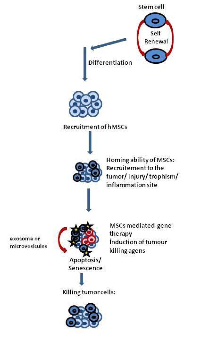
Up to date, various agents have been used with stem cells as vehicles to reduce the tumor size or extend the survival of the organism [22,23]. All these vehicles and agents showed different success rates. In the last decade, hMSCs have been proposed as a great tool in different therapeutic applications. MSCs serve as a powerful cell-based delivery vehicle for the site-specific release of anticancer drugs due to their homing abilities, easy acquisition, hypoimmunogenic properties, fast in vitro expansion and feasibility of autologous transplantation properties [24]. The use of self-derived hMSCs has an advantage over using other vehicles for delivery since they would have significantly a lower risk of being rejected as foreign objects by the immune system. Once these stem cells carrying biological agents are injected into the patient’s bloodstream, they can migrate to the tumor site and release the anti-carcinogenic agent.
The use of MSCs transduced with TRAIL showed induction of apoptosis and a subsequent reduction of tumor cell viability in colorectal carcinoma, gliomas, lung, breast, squamous and cervical cancer [25,26]. Targeted delivery was also proven to be successful using hMSCs in a xenogenic mouse model. The growth of malignant cells in lungs of mice was inhibited both in-vitro and in-vivo following local delivery of MSCs transduced with interferon-β (IFN-β). However, this inhibition was not achieved during the systematic or non-local delivery of the cells to the tumor site [27,28]. In the prostate stroma of a castrate resistant mouse model, where MSCs were used to deliver Frizzled Related Protein-2 (SFRP2) to antagonize the Wnt-mediated cancer progression, decreased tumor growth, increased apoptosis, and tumor necrosis have been observed [29]. In mice with both solid and metastatic tumors, it was shown that intratumoral injection of MSCs expressing modified interleukin-12 (MSCs/IL-12M) caused strong tumor-specific T-cell responses and anti-metastatic effects as well as inhibitory effects of solid tumor growth. These effects were proven to be stronger than interleukin-12 expressing adenovirus [30]. In 2011, Serakinci, et al. [10] showed the homing, engrafting and proliferation abilities of hMSCs in a human xenograft model in an ovarian cancer cell line transplanted into immune compromised mice [10]. Human BM-MSCs were shown to secrete interferon-b (IFNb) and diminish melanoma, breast carcinoma, and lung metastases [25]. Similarly, MSCs derived from amniotic fluid was capable of transporting IFNb to the site of neoplasia of a bladder tumor model and inhibit the tumor growth as well as prolonged survival of mice [25].
Limiting factors and difficulties of MSC use in cancer therapy
Difficulties already start with the isolation process, since only 1 in every 105 cells obtained in isolation is expected to be an MSCs. MSCs have low grafting efficiency as well as potency. The limited division potential of hMSCs also restrains their therapeutic applications especially considering that a high number of cells is required for therapy in humans. This raises the need for large scale MSC expansion [22].
Here, the telomere dynamics play an important role in stem cell function in particular during the expansion of stem cell population. Telomere homeostasis and telomerase play a critical role in tumor progression and it is well known that the cancer cells rely on telomerase for its survival. One of the main functions of telomeres is to protect the chromosome ends as being detected as DNA double strand breaks by DNA repair machinery.
Serakinci, et al. [12] established an immortalized human MSCs (hMSC-telo1) cell line by introducing a retrovirus carrying the hTERT gene, which codes for the catalytic subunit of telomerase reverse transcriptase. hTERT, together with hTERC, the telomerase RNA, function to lengthen the end of DNA strand, namely the telomeric repeat sequence and allows the stem cell to maintain its characteristics with an expanded life span This gene manipulation allowed cells to bypasses the naturally built-in cellular controls which govern the delicate balance between cell proliferation, senescence, and carcinogenesis. Although this is very promising in the use of MSCs for therapeutic applications, manipulation of telomere-telomerase activity in order to extend the proliferative capacity of stem cell populations may increase the risk for stem cell susceptibility to carcinogenesis. The transduced cell line presented variations indicative of neoplastic development, such as contact inhibition, anchorage independence and in-vivo tumor formation in Severe Combined Immunodeficiency (SCID) mice [11]. These can be due to critically shortened telomeres leading to senescence that can be considered as a barrier against cancer formation via a telomere-mediated checkpoint. Dysfunctional telomeres have the ability to disturb the genomic stability via Break-Fusion-Break cycles causing excessive genomic instability and aberrations, which might lead to rapid cell proliferation, loss of contact inhibition, a gradual increase in telomerase activity [31,32]. These phenotypic and genotypic alterations were also reported in adipose-derived hMSCs [33] and bone marrow-derived mouse MSCs [34].
One of the options to resolve the issue of neoplastic transformation of hMSCs could be the self-destruction of the vehicle after the drug delivery. This kind of treatment has been applied in tumor-selective viruses that mediate oncolytic effects on tumors and destroy targeted cancer cells. This kind of viruses has been engineered at the telomerase promoter sequence with tumor-specific transcriptional response elements. These therapies target the telomerase-positive cells and combine the genetically engineered vehicle stem cell and suicide gene therapies [35-37]. Serakinci and colleagues [38] have reported that hMSC-telo1 cells do not necessarily give rise to spontaneous transformation. The neoplastic transformation was observed in the telomerase introduced hMSCs when they were subjected to 2.5 Gy of gamma irradiation followed by long-term culturing. Thus, the neoplastic transformation was suggested to occur due to DNA damage caused by irradiation and telomere damage leading to temporary cell cycle arrest [12,38]. Telomerase may help in the production of a large number of cells, but it may have an impact on neoplastic transformation. Therefore these cells require close monitoring before and after the application and treatment.
In addition to the malignant transformations observed in cells with manipulated telomere-telomerase activity, unmanipulated BM-MSCs were also shown to produce a sub-population of cells with high levels of telomerase activity, chromosomal aneuploidy, translocations and capacity in the formation of tumors in multiple organs of NOD/SCID mice [22,39]. Malignant transformations were also reported in rodent models, in mouse, and in hMSC populations [11,33,22,39]. The underlying molecular mechanism in this spontaneous transformation was suggested to occur after hMSC by-passing the senescence by repressing p16 expression, acquisition of telomerase activity, deletion of the Ink4a/Arf locus and hyperphosphorylation of Rb [33]. This raised concerns whether hMSCs can lead to spontaneous malignant transformation when forced to extensive expansion. On the contrary to these studies, analysis of hMSCs with comparative genomic hybridization, karyotyping and subtelomeric fluorescence in situ hybridization showed that there is no evidence of spontaneous hMSC transformation during long-term culture [40,41]; however, maintaining a normal karyotype will not eliminate the possibility of epigenetic changes to occur. Indeed, telomerase-immortalized hMSCs were shown to accumulate genetic and epigenetic variations leading to spontaneous transformation [11,42]. Exogenously administered hMSCs may get engaged to developing tumors, after being infused systemically in animal models for glioma, colon carcinoma, gastric cancer, ovarian carcinoma, Kaposi’s sarcoma and melanoma [43,44]. Although these studies support the possibility of neoplastic transformation of hMSCs during in-vitro expansion, it is still controversial and the molecular pathogenesis underlying such mechanism is not fully established yet.
Moreover, a number of studies investigating the use of MSCs for graft-versus-host disease showed no signs of tumor formation [43,45,46]. A recent small size phase 2 clinical study was reported for the use of MSCs for the treatment of Crohn’s disease that showed promising results. Although all these results are promising, in order to establish a definite role of MSC in the prevention of tumorigenesis, comprehensive information on the regulation of adult stem cell growth and monitoring the outcome of clinical applications are necessary.
Conclusions and future directions
Lately, hMSCs have become a great therapeutic target for many diseases due to their homing abilities once reconstructed to inflammation or tumor site. MSCs can be acquired from the patients’ own body and use of these cells lowers the risks of rejection. In addition to their tumor homing properties, MSCs are also easily transduced by integrating vectors due to their high levels of amphotropic receptors and offer long-term gene expression without alteration of the phenotype. Gene and viral-based therapies have shown enhancements in cancer treatment and a number of anticancer genes have been successfully engineered into MSCs, which then promoted anticancer effects in various carcinoma models. However, since one of the most apparent characteristics of cancer is the continued cell growth that is associated with telomerase activity, there may be an increased risk of cancer development with the use of genetically modified cells in cancer therapies. Therefore, use of targeted treatment with self-suicide vehicles may be a better approach to improve the cancer therapy and reduce the risk of developing a secondary tumor.
The stem cell exosome/microvesicle-mediated regeneration, cell migration and homing abilities of the hMSCs had an important role in the treatment of cancer and other diseases. Several anticancer genes have been successfully engineered into MSCs. Lentivirus-, retrovirus-, plasmid- and adenovirus-mediated MSC deliveries showed promising results in animal studies with prolonged life of the animals, reduced complications, and/or tumor growth. All these promising results have drawn the attention to the potential of MSCs in cell-based therapies. Current literature and discussion indicate that hMSC use in cancer therapy would benefit from the combination of the gene therapy with stem cell therapy approach.
It is worth mentioning that use of genetically modified MSCs and their target specificities need further exploring. Therefore, clinical studies play a crucial role in determining the potential of MSC usage in cancer in combination with conventional therapy strategies.
In conclusion, modeling of hMSCs is a promising approach for cancer therapy. The use of hMSCs with suicidal gene therapies is a hopeful approach and the combinatory effect of gene therapy with stem cell therapy may be the strategy forward.
- Jiang Y, Jahagirdar BN, Reinhardt RL, Schwartz RE, Keene CD, Ortiz-Gonzalez XR, et al. Pluripotency of mesenchymal stem cells derived from adult marrow. Nature. 2002; 418: 41-49. https://goo.gl/ZkvAjK
- Kortesidis A, Zannettino A, Isenmann S, Shi S, Lapidot T, Gronthos S. Stromal-derived factor-1 promotes the growth, survival, and development of human bone marrow stromal stem cells. Blood. 2005; 105: 3793-3801. https://goo.gl/tuYQdr
- Wynn RF, Hart CA, Corradi-Perini C, O'Neill L, Evans CA, Wraith JE, et al. A small proportion of mesenchymal stem cells strongly expresses functionally active CXCR4 receptor capable of promoting migration to bone marrow. Blood. 2004; 104: 2643-2645. https://goo.gl/X3Zd3L
- Yang C, Deqiang Lei, Weixiang Ouyang, Jinghua Ren, Huiyu Li, Jingqiong Hu, et al. Conditioned media from human adipose tissue-derived mesenchymal stem cells and umbilical cord-derived mesenchymal stem cells efficiently induced the apoptosis and differentiation in human glioma cell lines in vitro. BioMed Research International. 2014. https://goo.gl/tdGmn3
- Calió ML, Marinho DS, Ko GM, Ribeiro RR, Carbonel AF, Oyama LM, et al. Transplantation of bone marrow mesenchymal stem cells decreases oxidative stress, apoptosis, and hippocampal damage in brain of a spontaneous stroke model. Free radical biology & medicine. 2014; 70: 141-154. https://goo.gl/ne9NeQ
- Yu B, Zhang X, Li X. Exosomes derived from mesenchymal stem cells. International Journal of Molecular Sciences. 2014; 15: 4142-4157. https://goo.gl/Gx1KyE
- Honoki K, Tsujiuchi T. Senescence bypass in mesenchymal stem cells: a potential pathogenesis and implications of pro-senescence therapy in sarcomas. Expert review of anticancer therapy. 2013; 13: 983-996. https://goo.gl/s4MEyD
- Sohni A, Verfaillie CM. Mesenchymal stem cells migration homing and tracking. Stem Cells International. 2013. https://goo.gl/TmZdbx
- D’Souza A, Lacy M, Gertz M, Kumar S, Buadi F, Hayman S, et al. Long-term outcomes after autologous stem cell transplantation for patients with POEMS syndrome (osteosclerotic myeloma): A single-center experience. Blood. 2012; 120: 56-62. https://goo.gl/XWDDA9
- Serakinci N, Christensen R, Fahrioglu U, Sorensen FB, Dagnæs-Hansen F, Hajek M, et al. Mesenchymal stem cells as therapeutic delivery vehicles targeting tumor stroma. Cancer biotherapy & radiopharmaceuticals. 2011; 26: 767-773. https://goo.gl/ucgPL5
- Serakinci N, Guldberg P, Burns JS, Abdallah B, Schrødder H, Jensen T, et al. Adult human mesenchymal stem cell as a target for neoplastic transformation. Oncogene. 2004; 23: 5095-5098. https://goo.gl/w7CLnM
- Serakinci N, Christensen R, Graakjaer J, Cairney CJ, Keith WN, Alsner J, et al. Ectopically hTERT expressing adult human mesenchymal stem cells are less radiosensitive than their telomerase negative counterpart. Experimental Cell Research. 2007; 313: 1056-1067. https://goo.gl/XMrznI
- Bentzon JF, Stenderup K, Hansen FD, Schroder HD, Abdallah BM, Jensen TG, et al. Tissue distribution and engraftment of human mesenchymal stem cells immortalized by human telomerase reverse transcriptase gene. Biochemical and Biophysical Research Communications. 2005; 330: 633-640. https://goo.gl/bsNFpl
- Kern S, Eichler H, Stoeve J, Klüter H, Bieback K. Comparative analysis of mesenchymal stem cells from bone marrow, umbilical cord blood, or adipose tissue. Stem cells. 2006; 24: 1294-1301. https://goo.gl/mFUqZY
- Mueller SM, Glowacki J. Age-related decline in the osteogenic potential of human bone marrow cells cultured in three-dimensional collagen sponges. Journal of cellular biochemistry. 2001; 82: 583-590. https://goo.gl/6w9bl3
- Vellasamy S, Sandrasaigaran P, Vidyadaran S, George E, Ramasamy R. Isolation and characterisation of mesenchymal stem cells derived from human placenta tissue. World journal of stem cells. 2012; 4: 53-61. https://goo.gl/UaFfL9
- Batsali AK, Kastrinaki MC, Papadaki HA, Pontikoglou C. Mesenchymal stem cells derived from Wharton’s Jelly of the umbilical cord: biological properties and emerging clinical applications. Current stem cell research & therapy. 2013; 8: 144-155. https://goo.gl/Ui03GQ
- Rubinstein P, Dobrila L, Rosenfield RE, Adamson JW, Migliaccio G, Migliaccio AR, et al. Processing and cryopreservation of placental/umbilical cord blood for unrelated bone marrow reconstitution. Proceedings of the National Academy of Sciences of the United States of America. 1995; 92: 10119-10122. https://goo.gl/KJZTcQ
- Wyrsch A, Dalle Carbonare V, Jansen W, Chklovskaia E, Nissen C, Surbek D, et al. Umbilical cord blood from preterm human fetuses is rich in committed and primitive hematopoietic progenitors with high proliferative and self-renewal capacity. Experimental Hematology. 1999; 27: 1338-1345. https://goo.gl/TNaucL
- Goodwin H, Bicknese AR, Chien SN, Bogucki BD, Quinn CO, Wall DA. Multilineage differentiation activity by cells isolated from umbilical cord blood: Expression of bone, fat, and neural markers. Biology of Blood and Marrow Transplantation. 2001; 7: 581-588. https://goo.gl/49mmUI
- Chang Y, Tseng CP, Hsu LF, Hsieh TB, Hwang SM. Characterization of two populations of mesenchymal progenitor cells in umbilical cord blood. Cell Biology International. 2006; 30: 495-499. https://goo.gl/OfIjsV
- Momin EN, Mohyeldin A, Zaidi HA, Vela G, Quiñones-Hinojosa A. Mesenchymal stem cells: new approaches for the treatment of neurological diseases. Current stem cell research & therapy. 2010; 5: 326-344. https://goo.gl/gxqcmk
- Aboody KS, Najbauer J, Danks MK. Stem and progenitor cell-mediated tumor selective gene therapy. Gene therapy. 2008; 15: 739-752. https://goo.gl/tt5CWO
- Gao Z, Zhang L, Hu J, Sun Y. Mesenchymal stem cells: A potential targeted-delivery vehicle for anti-cancer drug, loaded nanoparticles. Nanomedicine: Nanotechnology, Biology, and Medicine. 2013; 9: 174-184. https://goo.gl/Ty3YTT
- Shah K. Encapsulated stem cells for cancer therapy. Biomatter. 2013; 3: e24278. https://goo.gl/dxtXVZ
- Menon LG, Kelly K, Yang HW, Kim SK, Black PM, Carroll RS. Human bone marrow-derived mesenchymal stromal cells expressing S-TRAIL as a cellular delivery vehicle for human glioma therapy. Stem Cells. 2009; 27: 2320-2330. https://goo.gl/W0ChFf
- Studeny M, Marini FC, Champlin RE, Zompetta C, Fidler IJ, Andreeff M. Bone marrow-derived mesenchymal stem cells as vehicles for interferon-beta delivery into tumors. Cancer research. 2002; 62: 3603-3608. https://goo.gl/ZwUD3t
- Studeny M, Marini FC, Dembinski JL, Zompetta C, Cabreira-Hansen M, Bekele BN, et al. Mesenchymal stem cells: Potential precursors for tumor stroma and targeted-delivery vehicles for anticancer agents. Journal of the National Cancer Institute. 2004; 96: 1593-1603. https://goo.gl/fNwbAe
- Placencio VR, Li X, Sherrill TP, Fritz G, Bhowmick NA. Bone marrow derived mesenchymal stem cells incorporate into the prostate during regrowth. PLoS One. 2010; 5: e12920. https://goo.gl/gu05ss
- Seo SH, Kim KS, Park SH, Suh YS, Kim SJ, Jeun SS, et al. The effects of mesenchymal stem cells injected via different routes on modified IL-12-mediated antitumor activity. Gene therapy. 2011; 18: 488-495. https://goo.gl/hSBzYb
- Serakinci N, Graakjaer J, Kolvraa S. Telomere stability and telomerase in mesenchymal stem cells. Biochimie. 2008; 90: 33-40. https://goo.gl/5r2Sen
- Furlani D, Ugurlucan M, Ong L, Bieback K, Pittermann E, Westien I, et al. Is the intravascular administration of mesenchymal stem cells safe? Mesenchymal stem cells and intravital microscopy. Microvascular Research. 2009; 77: 370-376. https://goo.gl/ZN6yyH
- Rubio D, Garcia S, De la Cueva T, Paz MF, Lloyd AC, Bernad A, et al. Human mesenchymal stem cell transformation is associated with a mesenchymal-epithelial transition. Experimental Cell Research. 2008; 314: 691-698. https://goo.gl/R606S2
- Miura Y, Gao Z, Miura M, Seo BM, Sonoyama W, Chen W, et al. Mesenchymal stem cell-organized bone marrow elements: an alternative hematopoietic progenitor resource. Stem cells. 2006; 24: 2428-2436. https://goo.gl/Ss3eJl
- Abdul-Ghani R, Ohana P, Matouk I, Ayesh S, Ayesh B, Laster M, et al. Use of transcriptional regulatory sequences of telomerase (hTER and hTERT) for selective killing of cancer cells. Molecular therapy : the journal of the American Society of Gene Therapy. 2000; 2: 539-544. https://goo.gl/ygFxn4
- Komata T, Koga S, Hirohata S, Takakura M, Germano IM, Inoue M, et al. A novel treatment of human malignant gliomas in vitro and in vivo: FADD gene transfer under the control of the human telomerase reverse transcriptase gene promoter. Int J Oncol. 2001; 19: 1015-1020. https://goo.gl/tQ0nec
- Plumb JA, Bilsland A, Kakani R, Zhao J, Glasspool RM, Knox RJ, et al. Telomerase-specific suicide gene therapy vectors expressing bacterial nitroreductase sensitize human cancer cells to the pro-drug CB1954. Oncogene. 2001; 20: 7797-7803. https://goo.gl/meuR66
- Christensen R, Alsner J, Brandt Sorensen F, Dagnaes-Hansen F, Kolvraa S, Serakinci N. Transformation of human mesenchymal stem cells in radiation carcinogenesis: long-term effect of ionizing radiation. Regenerative medicine 2008; 3: 849-861. https://goo.gl/QwnnQu
- Wang Y, Huso DL, Harrington J, Kellner J, Jeong DK, Turney J, et al. Outgrowth of a transformed cell population derived from normal human BM mesenchymal stem cell culture. Cytotherapy. 2005; 7: 509-519. https://goo.gl/tBdCAh
- Bernardo ME, Zaffaroni N, Novara F, Cometa AM, Avanzini MA, Moretta A, et al. Human bone marrow-derived mesenchymal stem cells do not undergo transformation after long-term in vitro culture and do not exhibit telomere maintenance mechanisms. Cancer Research. 2007; 67: 9142-9149. https://goo.gl/TxejGk
- Meza-Zepeda LA, Noer A, Dahl JA, Micci F, Myklebost O, Collas P. High-resolution analysis of genetic stability of human adipose tissue stem cells cultured to senescence. Journal of cellular and molecular medicine. 2008; 12: 553-563. https://goo.gl/44l3hZ
- Burns JS, Abdallah BM, Guldberg P, Rygaard J, Schrøder HD, Kassem M. Tumorigenic heterogeneity in cancer stem cells evolved from long-term cultures of telomerase-immortalized human mesenchymal stem cells. Cancer Research. 2005; 65: 3126-3135. https://goo.gl/Cnbd8z
- Correa P, Houghton J. Carcinogenesis of Helicobacter pylori. Gastroenterology. 2007; 133: 659-672. https://goo.gl/Fyx8xK
- Lazennec G, Jorgensen C. Concise review: adult multipotent stromal cells and cancer: risk or benefit? Stem cells. 2008; 26: 1387-1394. https://goo.gl/lF7NK7
- Barkholt L, Flory E, Jekerle V, Lucas-Samuel S, Ahnert P, Bisset L, et al. Risk of tumorigenicity in mesenchymal stromal cell-based therapies - Bridging scientific observations and regulatory viewpoints. Cytotherapy. 2013; 15: 753–759. https://goo.gl/lXOjXv
- Resnick IB, Barkats C, Shapira MY, Stepensky P, Bloom AI, Shimoni A, et al. Treatment of severe steroid resistant acute GVHD with mesenchymal stromal cells (MSC). American journal of blood research. 2013; 3: 225-238. https://goo.gl/svjCF0
Sign up for Article Alerts