Inhibition of Porcine Reproductive and Respiratory Syndrome Virus by Nitric Oxide Induced Through Toll-Like Receptor Activation in Porcine Alveolar Macrophages?
Huan Liu1, Jia Liu2, Ning Ding3, Kun Li3, Peng Wang1, Hui Han1 and Qinfu Wang1*
1Institute of Immunology, College of Life Science and Technology, Dalian University, Dalian, Liaoning 116622, China
2Animal Husbandry Service of Dalian, Dalian 116037
3College of Medicine, Dalian University, Dalian, Liaoning, 116622
*Address for Correspondence: Qinfu Wang, Institute of Immunology, College of Life Science and Technology, Dalian University, Dalian, Liaoning 116622, China, Tel: +86-411-87402134; E-mail: [email protected]
Submitted: 14 September 2017; Approved: 25 October 2017; Published: 26 October 2017
Citation this article: Liu H, Liu J, Ding N, Li K, Wang P, et al. Inhibition of Porcine Reproductive and Respiratory Syndrome Virus by Nitric Oxide Induced Through Toll-Like Receptor Activation in Porcine Alveolar Macrophages. Adv J Toxicol Curr Res. 2017;1(2): 063-068.
Copyright: © 2017 Liu H, et al. This is an open access article distributed under the Creative Commons Attribution License, which permits unrestricted use, distribution, and reproduction in any medium, provided the original work is properly cited
Keywords: Nitric Oxide; Reactive Oxygen Species; Porcine Reproductive and Respiratory Syndrome Virus (PRRSV); Porcine Alveolar Macrophages (PAM); Cytokine
Download Fulltext PDF
Porcine Reproductive and Respiratory Syndrome Virus (PRRSV) induces respiratory distress in piglets and causes reproductive failure in sows. In this work, the anti- PRRSV effect of Nitric Oxide (NO) induced through toll-like receptor activation in Porcine Alveolar Macrophages (PAMs) was investigated. Our results show the ratio of arginase-1 (Arg1)/Nitric Oxide Synthase (iNOS) was decreased in PAMs stimulated by a toll-like receptor ligand, polyinosinic polycytidylic acid (poly I:C), leading to the release of NO and the decrease of PRRSV copy numbers. In order to examine whether NO prohibits PRRSV infection in PAMs, we added a NO donor, S-Nitroso-N-Acetylpenicillamine (SNAP), to stimulate the release of NO in PAMs. Our results show that PRRSV copies were further decreased, indicating the NO level is negatively related with PRRSV propagation. The cytokine level in the poly I:C treated PAMs after PRRSV infection was further investigated. The results show the TNFα and IL-6 levels were decreased, but the IL-1β level was significantly increased after PRRSV infection, indicating IL-1β may be more relevant in anti-PRRSV than TNFα and IL-6. In conclusion, our results suggest that poly I:C activates the innate immunity by releasing NO, which in turn inhibits PRRSV infection.
Introduction
Porcine Reproductive and Respiratory Syndrome (PRRS) is one of the most economically significant diseases affecting the swine industry worldwide and causes great economic losses each year. The major characteristics of PRRS include reproductive failure and respiratory diseases, which usually lead to compromised lung-defense mechanisms and secondary complications [1]. Porcine Reproductive and Respiratory Syndrome Virus (PRRSV) primarily infects Porcine Alveolar Macrophages (PAMs) and Dendritic Cells (DCs) [2]. One characteristic of a pathologic change in pigs infected with PRRSV is the dramatic decrease of the macrophage population [3,4]. PRRSV infection can delay and down-modulate adaptive immune functions, leading to reduced efficacy of vaccination [5].
A safe and effective PRRSV vaccine is the best solution for the ultimate control of the PRRS pandemic. Vaccines for PRRS in China are predominately modified live products [6]. The anti-PRRSV protective immunity does not begin sooner than four weeks post-infection [7] which explains why our endeavor to prevent the PRRS epidemic by inoculating inactivated or attenuated vaccines is not always successful. Mechanisms other than acquired immunity may be primarily involved in producing an environment that is resistant to PRRSV infection, limiting viral replication enough to reduce the extended period of viremia, and/or limiting or abolishing persistent infection [7]. If an enhanced innate immunity can be induced in uninfected individuals, it would markedly improve the efficiency against PRRSV infection in conjunction with T-cell-directed vaccines. Toll-Like Receptors (TLRs) play a key role in the activation of innate immunities to produce Interferons (IFNs) and pro-inflammatory cytokines [8]. In this paper, the anti-PRRSV effect of a TLR ligand, polyinosinic polycytidylic acid (poly I:C) in PAM cells was investigated. Our results show the elevated Nitric Oxide (NO) level was negatively related to PRRSV copy numbers, indicating NO plays an important role in regulating PRRSV infection.
Materials and Methods
Virus and cells
A PRRSV wild type strain TJ-F10 was kindly provided by Dr. Hua Wu from Sinovet Biotechnology Co. Ltd., Beijing, China. The virus was passaged ten times in Marc-145 cells with a titer of 108 TCID50/mL. Marc-145 cells were grown in Dulbecco’s Modified Eagle’s Medium (DMEM; Invitrogen, Carlsbad, CA, USA) supplemented with 10% Fetal Bovine Serum (FBS; Invitrogen), 100 U/mL penicillin G, 100 mg/mL Na streptomycin sulfate, and 2 mM l-glutamine.
Pigs
Three to four week-old weaned pigs (conventional Large White-Duroc crossbred) were procured from a swine herd seronegative for PRRSV, porcine respiratory coronavirus, transmissible gastroenteritis virus, and porcine circovirus 2 virus. The experimental protocol was approved by the ethics committee of Dalian University, Dalian, China. All procedures were performed in accordance with the current Chinese legislations on the care and use of laboratory animals.
Alveolar macrophages
Pigs were euthanized by exsanguination. The trachea was ligated to prevent total pulmonary collapse, and the heart and lungs were removed from the thorax. PAMs were collected from lungs by bronchoalveolar lavage. Bronchoalveolar Lavage Fluid (BALF) preparations are almost exclusively composed of alveolar macrophages [9]. The PAMs were re-suspended in RPMI-1640 with 10% Fetal Bovine Serum (FBS), 2 mM L-glutamine, 1 µg/mL fungizone, 100 U/mL penicillin and 100 µg/mL streptomycin. The PAM cultures were incubated in Petri dishes at 37°C in a humidified 5% CO2 atmosphere for 2 h. The medium and non-adherent cells were removed by aspiration and washed once with complete RPMI-1640 medium. The adhesive cells were digested by trypsin, washed three times, counted and scored for viability by trypan blue dye exclusion. The cells were plated at a density of 1×10⁵ cells/well in 96-well culture plates or 1×10⁶ cells/well in 6-well culture plates.
Lung interstitial macrophages
The lung tissue was chopped into pieces of less than 1 mm3. To remove remaining alveolar macrophages and blood cells, the tissue was washed with PBS until the filtrate appeared clear. The tissue was then digested using 0.025% collagenase IA (Sigma, St. Louis, MO, USA) at 37°C in a shaking water bath for 60 min. The product of the digestion was filtered by a 200 mesh stainless steel filter, centrifuged at 1500 rpm for 10 min, and re-suspended in RPMI-1640 medium with 10% FBS [10]. After removing the non-adherent cells, the adhesive IM cells were collected, scored for viability and plated for testing.
Nitric oxide assay
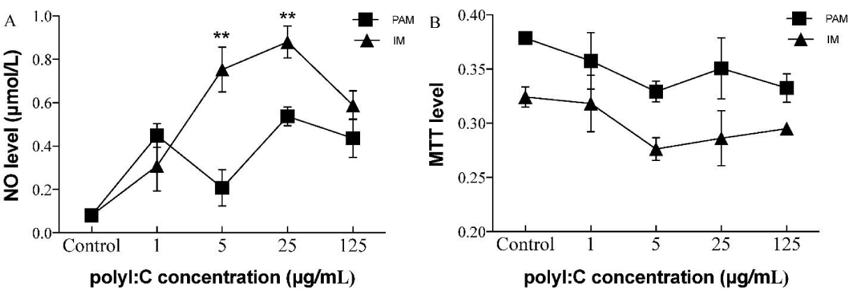
The PAM and IM cells were plated at a density of 1×10⁵ cells/well in 96-well plates for 24 h, and stimulated with different doses of poly I:C (1, 5, 25, 125 and 250 μg/mL) for 24 h. The culture media were collected for the NO assay. The nitric oxide concentration was measured by detection of its stable oxidative metabolite, nitrite. In brief, 80 μL culture media were mixed with an equal volume of Griess reagent (0.1% naphthylethylenediamine dihydrochloride and 1% sulfanil-amide in 5% phosphoric acid) and shaken at room temperature for 5 min. The absorbance at 550 nm was measured, and the concentration was determined using a calibration curve constructed with sodium nitrite standards [11].
Cell viability assay
Cell viability was evaluated based on the conversion of yellow MTT (3-[4,5-dimethylthiazol-2-yl]-2,5-diphenyl tetrazolium bromide) to purple formazan.
Nitric oxide regulated the mRNA expression of Arg1 and iNOS in PAMs and IMs
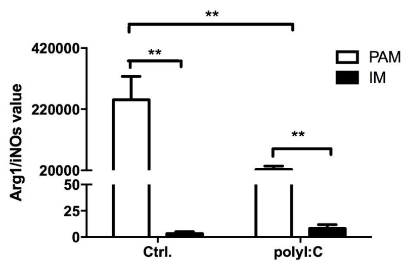
To determine if the poly I:C treatment induces polarization of PAM and IM cells, the mRNA expressions of Arg1 and iNOS were measured and the Arg1/iNOS ratio was calculated. The Arg1/iNOS ratio represents the NO production inhibitory level, which is higher in PAMs than IMs. Our results show the poly I:C treatment significantly changed the Arg I/iNOS ratio in PAMs (Figure 2). In the poly I:C pretreated PAMs, the Arg1/iNOS ratio decreased, indicating the NO level elevated.
Dynamic changes of NO by different doses of SNAP in PAMs pretreated with poly I:C
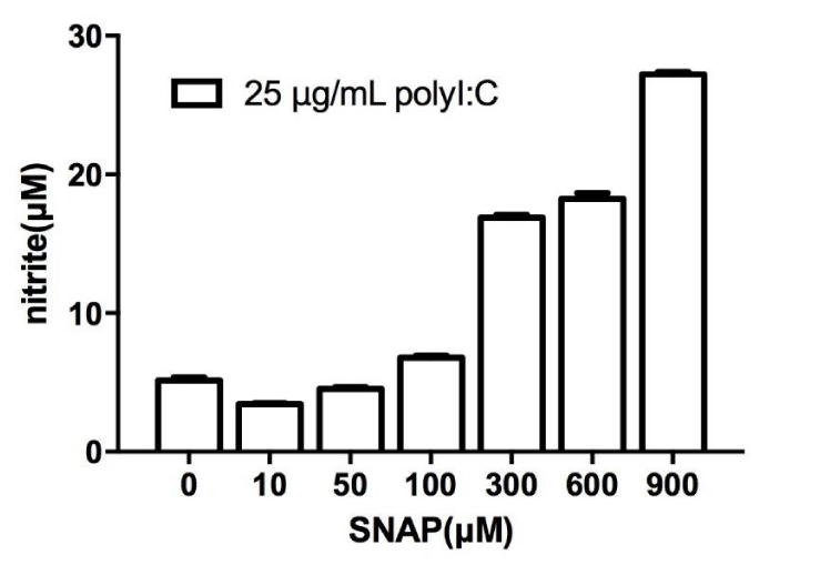
In order to determine whether NO plays an important role against PRRSV infection, we stimulated the PAMs with SNAP in different concentrations (10, 50, 100, 300, 600 and 900 μM). The results show the NO level was elevated in a SNAP concentration-dependent manner (Figure 3).
Correlation of the NO level with PRRSV copy numbers in the poly I:C treated PAMs
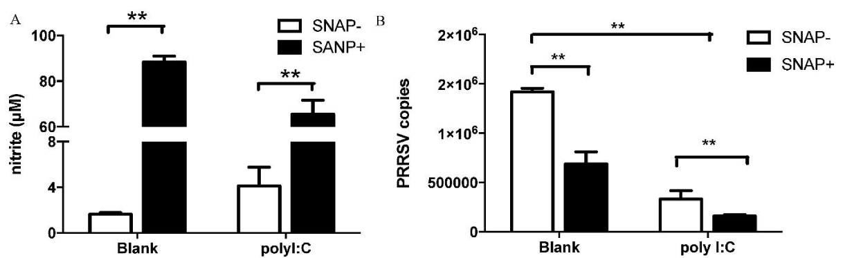
Our results show that PAM cells were more sensitive to PRRSV infection than IM cells. The mRNA level of PRRSV in the PAM cells is 10 fold to 100 fold higher than those in the IM cells when infected with PRRSV at 0.2, 1 and 5 MOI, respectively [12]. Activation of TLR3 signaling was reported to reduce viral multiplication in PAM cells [13]. To understand the effects of poly I:C against PRRSV infection in PAM cells, the copy number of the wild type strain PRRSV (TJ-F10) was measured using quantitative RT-PCR in the PAMs infected with TJ-F10 at MOI of 1. The results show that the NO level was increased significantly in the PAMs pretreated with poly I:C (Figure 4A), which led to decreased expression of PRRSV (Figure 4B). When SNAP was added, the NO level was further increased in both blank and PAMs pre-treated with poly I:C. However, the PRRSV copies did not change. The copy number of PRRSV in poly I:C pre-treated PAMs was lowered than that in the untreated PAMs.
Correlation of the ROS level with PRRSV copies in the poly I:C treated PAMs
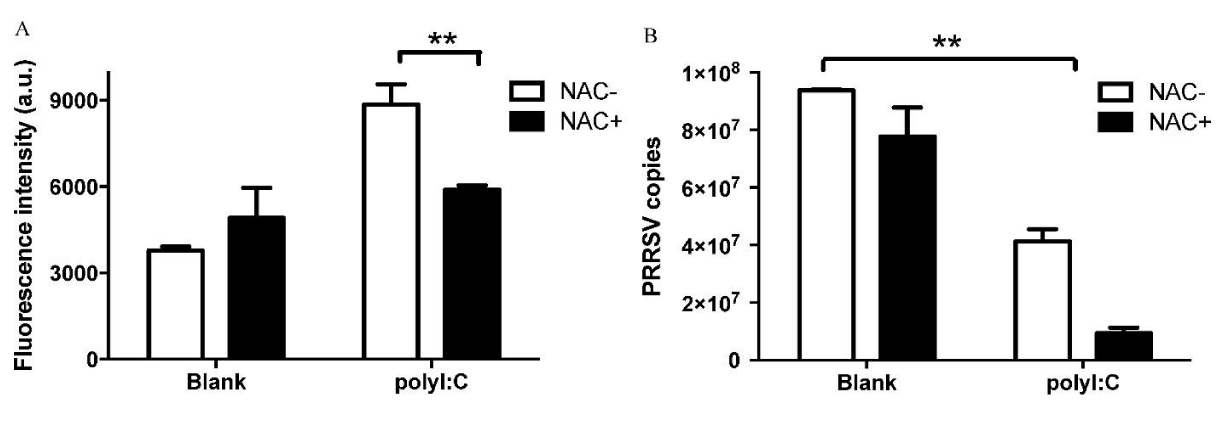
The results showed that the ROS level was decreased by NAC in the poly I:C treated group. However, the PRRSV was not increased, indicating the ROS level is not relevant to the anti-PRRSV effect of poly I:C.
Expression of cytokines in the poly I:C treated PAMs before and after PRRSV infection
The proinflammatory cytokines interleukin-1β (IL-1β), tumor necrosis factor-α (TNFα), and IL-6 are important mediators of several respiratory diseases. IL-1β and TNFα are among the first cytokines that are produced in the lungs during an infection inducing a cascade of secondary cytokines, such as IL-6. Our results showed the expression of all three cytokines was changed upon PRRSV infection, indicating these cytokines may play an important role in the infection of PRRSV. The IL-1β level was elevated in the poly I:C treated group; among all three groups, the IL-6 and TNFα levels were decreased after PRRSV infection, but the IL-1β level was increased significantly after PRRSV infected, indicating IL-1β may be more sensitive in the PRRSV infected PAMs than TNFα and IL-6.
Discussion
Nitric oxide is a key early signal involved in various pathophysiologic processes including infection and immunity [14]. Nitric oxide in mammalian macrophages is known to play a significant role in intracellular defense against a number of microbial infections [15]. Nitric oxide inhibits viral protein and/or RNA synthesis by modification of molecules essential for viral replication, such as ribonucleotide reductase and viral protease [16]. Nitric oxide has been shown to inhibit Japanese encephalitis virus, vesicular stomatitis virus, mouse hepatitis virus, Epstein-Barr virus, influenza virus, herpes simplex virus type 1, vaccinia virus and SARS coronavirus [17]. To investigate the inhibition effect of NO on PRRSV, the dose of poly I:C was optimized, and the dynamic changes of the NO levels were investigated in PAMs and IMs stimulated by various doses of poly I:C. Our results demonstrated that the NO secretion increased I a dose-dependent manner in the PAM and IM cells treated with low doses of poly I:C (≤ 25μg/ml). However, the NO secretion declined sharply when the concentrations of poly I:C were higher than 125μg/ml. Our data show no significant difference in viability among these samples treated with different doses of poly I:C. The decline of the NO level at high poly I:C doses needs further investigation.
Arg1 is used as a marker for M2 phenotype macrophages, which are considered as anti-inflammatory cells. iNOS serves as a marker for M1 macrophage [18,19]. iNOS competes with Arg1 for the same substrate and produces NO. iNOS uses L-Arg to produce NO and Arg1 uses L-Arg to produce L-ornithine and urea. The competition for the same substrate by these two enzymes acts as a way to control the production of NO [20]. The relative level of Arg1 to iNOS is used as an index for the ratio between M2 and M1 functional macrophages [21]. TLR3 is an important element in controlling viral replication. TLR3 recognizes dsRNA, a molecular pattern associated with viral infection. Poly I:C, a synthetic analog of dsRNA, is the ligand of choice for TLR3.
NO production is associated with the resistance to microbial killing in different cell lines [22]. To determine if poly I:C treatment induce polarization of PAM and IM cells, the mRNA expressions of Arg1 and iNOS were measured and the Arg1/iNOS ratio was calculated [23]. Our results showed the poly I:C treatment significantly decreased the Arg1/iNOS ratio.
The reason for usage of PAMs, not IMs, to study the anti-PRRSV effect is that PAM is a main target of PRRSV infection [12]. Additionally, studies using primary rat or mouse macrophages suggest that PAMs are more effective than IMs in producing cytokines involved in antimicrobial defense [10]. The PAM cells appear to be a non-specific first line of defense against inhaled pathogens, whereas IM cells show a more pronounced regulatory function [24]. These dissimilarities should be taken into consideration in future studies on the roles of swine lung macrophages in inflammatory response.
The infection of PRRSV was inhibited in the PAMs pre-treated with the optimal dose of poly I:C, When the TLR3 expression level in the target cells decreases, the PRRSV replication increases [25]. In order to understand whether NO plays an important role against PRRSV infection, we add 900 μM SNAP in the poly I: C treated PAM cells. Our result shows the NO level was elevated and accordingly the PRRSV copy number decreased, indicating No plays an important anti-PRRSV role.
We further investigate the role of ROS in the anti-PRRSV process. The ROS level was decreased by NAC. However, the PRRSV copies did not change, indicating the anti-PRRSV effect induced by poly I:C is not related with ROS.
In addition to the effect of NO on the inhibition of PRRSV infection, we also investigated the molecular mechanisms against PRRSV infection in PAMs. Poly I:C, selectively activates the MyD88-dependent and -independent pathways via TLR4 and TLR3, stimulating the production of pro-inflammatory mediators such as Nitric Oxide (NO), Tumor Necrosis Factor-α (TNF-α), and interleukins. We detected the TNFα, IL-1β, and IL-6 levels in the cell culture medium of PAMs. The results showed TNFα and IL-1β were elevated in the poly I:C treated PAMs. PRRSV infection in PAMs led to the increase of IL-1β and decrease of TNFα and IL-6. We presumed that IL-1β is relevant to the anti-PRRSV effect. Although pro-inflammatory cytokines usually exhibit antiviral effects, the increase of these cytokines may also be a risk factor in accelerating development of PRRS. For example, pigs inoculated with PRRSV-LPS developed severe respiratory disease [26]. Therefore, protective effects of TLR-mediated innate immunity on PRRSV infection would be limited to the primary site of infection. In addition, even though some TLR ligands potentially suppress PRRSV replication in vitro, their effects in vivo need to be further investigated.
Conclusion
In this study, the dynamic change of the NO level was investigated in the ploy I:C treated PAMs and IMs. We found the PRRSV infection was inhibited in the poly I:C pretreated PAMs, indicating that TLR3 activation by poly I:C could be a potential application for the prevention of PRRSV infection. The finding highlights the feasibility of using poly I:C and combination immunotherapy to construct new strategies for the therapy of the PRRSV infection. The molecular mechanism of anti-PRRSV effects of NO was investigated. Our results show the proinflammatory cytokine, IL-1β, may be relevant to the anti-PRRSV effect, compared with TNFα and IL-6. Further research needs to examine more closely the links between cytokines and PRRSV infection.
Acknowledgements
This study was supported by the National Natural Science Foundation of China (No. 81172777) and Dalian Science and Technological Project (No. 2014B11NC096).
- Fan B, Liu X, Bai J, Li Y, Zhang Q, Jiang P. The 15N and 46R Residues of Highly Pathogenic Porcine Reproductive and Respiratory Syndrome Virus Nucleocapsid Protein Enhance Regulatory T Lymphocytes Proliferation. PLoS One. 2015; 10: 138772. https://goo.gl/VCSXfh
- Fu Y, Quan R, Zhang H, Hou J, Tang J, Feng WH. Porcine Reproductive and Respiratory Syndrome Virus Induces Interleukin-15 through the NF-kappa B Signaling Pathway. J Virol. 2012; 86: 7625-7636. https://goo.gl/dt3yvM
- Labarque G, Van Gucht S, Nauwynck H, Van Reeth K, Pensaert M. Apoptosis in the lungs of pigs infected with porcine reproductive and respiratory syndrome virus and associations with the production of apoptogenic cytokines. Vet Res. 2003; 34: 249-260. https://goo.gl/wxbd3H
- Rodriguez-Gomez IM, Barranco I, Amarilla SP, Garcia-Nicolas O, Salguero FJ, Carrasco L et al. Activation of extrinsic- and Daxx-mediated pathways in lymphoid tissue of PRRSV-infected pigs. Vet Microbiol. 2014; 172: 186-194. https://goo.gl/Gkfbgi
- Wongyanin P, Buranapraditkul S, Yoo D, Thanawongnuwech R, Roth JA, Suradhat S. Role of porcine reproductive and respiratory syndrome virus nucleocapsid protein in induction of interleukin-10 and regulatory T-lymphocytes (Treg). J Gen Virol. 2012; 93: 1236-1246. https://goo.gl/vGor7E
- Galliher-Beckley A, Li X, Bates JT, Madera R, Waters A, Nietfeld J, et al. Pigs immunized with Chinese highly pathogenic PRRS virus modified live vaccine are protected from challenge with North American PRRSV strain NADC-20. Vaccine. 2015; 33: 3518-3525. https://goo.gl/c5a7Qn
- Michael P. Murtaugh. PRRS immunology: what are we missing? American Association Of Swine Veterinarians. 2004. https://goo.gl/8dSj3Q
- Takeda K, Akira S. Toll-like receptors in innate immunity. Int Immunol. 2005; 17: 1-14. https://goo.gl/dHf2Mi
- Ait-Ali T, Wilson AD, Westcott DG, Clapperton M, Waterfall M, Mellencamp MA, et al. Innate immune responses to replication of porcine reproductive and respiratory syndrome virus in isolated Swine alveolar macrophages. Viral Immunol. 2007; 20: 105-118. https://goo.gl/EdQnUo
- Hoppstadter J, Diesel B, Zarbock R, Breinig T, Monz D, Koch M, et al. Differential cell reaction upon Toll-like receptor 4 and 9 activation in human alveolar and lung interstitial macrophages. Respir Res. 2010; 11: 124. https://goo.gl/h5Kebz
- Su YW, Chiou WF, Chao SH, Lee MH, Chen CC, Tsai YC. Ligustilide prevents LPS-induced iNOS expression in RAW 264.7 macrophages by preventing ROS production and down-regulating the MAPK, NF-kappaB and AP-1 signaling pathways. Int Immunopharmacol. 2011; 11: 1166-1172. https://goo.gl/wX1EaW
- Yaping Hu, Meichen Wang, Xuemei Zhang, Peng Wang, Huan Liu, Qinfu Wang. Heterogeneity of swine lung macrophages inoculated by porcine reproductive and respiratory syndrome virus. Food Agr Immunol. 2016; 27: 724-733. https://goo.gl/g4DLb4
- Sang Y, Ross CR, Rowland RR, Blecha F. Toll-like Receptor 3 Activation Decreases Porcine Arterivirus Infection. Viral Immunol. 2008; 21: 303-313. https://goo.gl/D2Gppj
- Wink DA, Hines HB, Cheng RY, Switzer CH, Flores-Santana W, Vitek MP, et al. Nitric oxide and redox mechanisms in the immune response. J Leukoc Biol. 2011; 89: 873-891. https://goo.gl/YraH7u
- Miller LC, Neill JD, Harhay GP, Lager KM, Laegreid WW, Kehrli ME Jr. In-depth global analysis of transcript abundance levels in porcine alveolar macrophages following infection with porcine reproductive and respiratory syndrome virus. Adv Virol. 2010; 2010: 864181. https://goo.gl/jxN97w
- Miller LC, Lager KM, Kehrli ME Jr. Role of Toll-like receptors in activation of porcine alveolar macrophages by porcine reproductive and respiratory syndrome virus. Clin Vaccine Immunol. 2009 ; 16: 360-365. https://goo.gl/AGBT1i
- Klingstrom J, Akerstrom S, Hardestam J, Stoltz M, Simon M, Falk KI, et al. Nitric oxide and peroxynitrite have different antiviral effects against hantavirus replication and free mature virions. Eur J Immunol. 2006; 36: 2649-2657. https://goo.gl/rQ9Krt
- Italiani P, Boraschi D. From Monocytes to M1/M2 Macrophages: Phenotypical vs. Functional Differentiation. Front Immunol. 2014; 5: 514. https://goo.gl/5p1k9G
- Sica A, Mantovani A. Macrophage plasticity and polarization: in vivo veritas. J Clin Invest. 2012; 122: 787-795. https://goo.gl/sxhBXx
- Nelson SM, Lei X, Prabhu KS. Selenium levels affect the IL-4-induced expression of alternative activation markers in murine macrophages. J Nutr. 2011; 141: 1754-1761. https://goo.gl/5q6B76
- Chiang CS, Chen FH, Hong JH, Jiang PS, Huang HL, Wang CC, et al. Functional phenotype of macrophages depends on assay procedures. Int Immunol. 2008; 20: 215-222. https://goo.gl/KbPmLQ
- Utaisincharoen P, Anuntagool N, Arjcharoen S, Limposuwan K, Chaisuriya P, Sirisinha S. Induction of iNOS expression and antimicrobial activity by interferon (IFN)-beta is distinct from IFN-gamma in Burkholderia pseudomallei-infected mouse macrophages. Clin Exp Immunol. 2004; 136: 277-283. https://goo.gl/A39ta1
- Davis MJ, Tsang TM, Qiu Y, Dayrit JK, Freij JB, Huffnagle GB, et al. Macrophage M1/M2 polarization dynamically adapts to changes in cytokine microenvironments in Cryptococcus neoformans infection. MBio. 2013; 4: 264-313. https://goo.gl/TU1MpN
- Boorsma CE, Draijer C, Melgert BN. Mediators of inflammation. Mediators Inflamm. 2013; 2013: 769214. https://goo.gl/qMeCe4
- Darwich L, Diaz I, Mateu E. Certainties, doubts and hypotheses in porcine reproductive and respiratory syndrome virus immunobiology. Virus Res. 2010; 154: 123-132. https://goo.gl/34oect
- van Gucht S, van Reeth K, Pensaert M. Interaction between porcine reproductive-respiratory syndrome virus and bacterial endotoxin in the lungs of pigs: potentiation of cytokine production and respiratory disease. J Clin Microbiol. 2003; 41: 960-966. https://goo.gl/RL8LjJ
Sign up for Article Alerts