Near Complete Airway Obstruction Secondary to Tracheal Stent Migration?
Christine M Carr1, Russell S Allinder2 and Diann M Krywko1*
1Department of Emergency Medicine, Medical University of South Carolina
2Department of Emergency Medicine, Roper St. Francis Healthcare System
*Address for Correspondence: Diann M Krywko, Department of Emergency Medicine, Medical University of South Carolina, Charleston, SC, Home: 1880 Hall Point Rd. Mount Pleasant, SC 29466, United States, E-mail: [email protected]
Submitted: 13 December 2020; Approved: 22 December 2020; Published: 25 December 2020
Citation this article: Carr CM, Allinder RS, Krywko DM. Near Complete Airway Obstruction Secondary to Tracheal Stent Migration. Int J Case Rep Short Rev, 2020 Dec 25; 6(12): 085-00. doi: 10.37871/ijcrsr.id92
Copyright: © 2020 Carr CM, et al. This is an open access article distributed under the Creative Commons Attribution License, which permits unrestricted use, distribution, and reproduction in any medium, provided the original work is properly cited
Download Fulltext PDF
Case Report
A 73-year-old male presented to the Emergency Department (ED) with acute onset of severe shortness of breath for six hours, not relieved by rest. His medical history included systolic cardiac dysfunction with failure, coronary artery disease, cardiac bypass, hiatal hernia, transient ischemic attacks, in addition to a remote tracheal injury from a motor vehicle accident as a teenager complicated by vocal cord paralysis resulting in tracheostomy dependence for four years. The patient noted progressive worsening stridor and respiratory difficulties six months prior to ED presentation. Twenty-four hours prior to arrival, the patient underwent a rigid bronchoscopy for subglottic stenosis tracheal stent placement. He denied chest pain, hemoptysis, unilateral leg swelling, cough, fever, chills or other symptoms.
On physical examination, the patient was in respiratory extremis. Vital signs were: temperature 37°C, blood pressure 136/90 mmHg, pulse 111 beats per minute, respiratory rate 22 breaths per minute, and 92% room air oxygen saturation. He was noted to have a hoarse voice, inspiratory stridor, accessory muscle usage, and a markedly prolonged expiratory time. No adventitious lung sounds were appreciated, abdomen was unremarkable, and extremities were without edema or circumferential size abnormality. Heart sounds revealed no murmurs, gallops or rubs, and pulses were palpable and normal throughout all extremities.
The patient was placed on a cardiac monitor, 4 liters of oxygen via Nasal Cannula (NC) was applied, and Intravenous (IV) access obtained. Cardiac monitoring showed normal sinus tachycardia. A portable chest radiograph was obtained. Though difficult to observe on imaging, the radiograph revealed inferior migration of the recently placed tracheal stent with the distal portion pointing towards the left main bronchus (Figure 1).
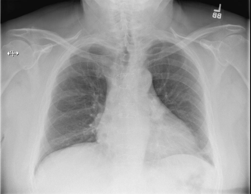
Ten milligram (mg) IV dexamethasone, 0.5 milliliter (mL) nebulized racemic epinephrine for presumed post-procedural edema, and 4% nebulized lidocaine in anticipation of an awake fiberoptic ED intubation was administered. Otolaryngology (ENT) and anesthesia were simultaneously consulted for fiberoptic nasopharyngoscopy with the anticipated potential need for surgical airway. The patient exhibited decreased stridor after the IV dexamethasone and nebulized epinephrine. The ENT physician performed nasopharyngoscopy at the bedside and visualized severe arytenoid edema with a significantly narrowed airway. At this time, an awake fiberoptic intubation was recommended. The patient was administered 0.2 mg IV glycopyrolate, a muscarinic anti-cholinergic medication used to decrease secretions, and 100% O2 by fnon-rebreather face mask with 2L nasal cannula in anticipation of an emergent difficult airway in the ED.
ENT service elected to re-perform nasopharyngoscopy and recommended aborting the awake intubation in ED immediately, with transfer of the patient to Operating Room (OR) to perform a controlled intubation with sophisticated surgical back up if needed. The patient was safely transported from the ED to the OR where he was successfully orally intubated with 6.0 Endotracheal Tube (ETT). The following day, he was taken back to the OR. Bronchoscopy was performed, exchanging an 8.0 ETT over catheter in order to remove the tracheal stent. The patient was extubated on post-procedure day two without further complications.
To this author’s knowledge, this specific complication of tracheal stent placement and management have not yet been reported in emergency medicine literature.
Discussion
In most emergent airway management cases, orotracheal intubation and tracheotomy are often considered definitive airway management. Ventilation should improve immediately once the airway is secured. However, if traditional airway management does not correct the ventilation problem, further etiologies and alternative interventions must be considered.
There are occasions where the area of airway obstruction is distal to the endotracheal or tracheotomy tube, such as a stent or other foreign body migrating into the lower trachea or carina. In a case such as this, orotracheal intubation and tracheotomy would be rendered ineffective. It is important to recognize the presence of a distal obstruction and to employ the appropriate intervention(s). Prompt tracheobronchoscopy must be considered as the next step in the algorithm.
Sign up for Article Alerts