Fahr Syndrome: Two Observations in Togo?
B. N’timon1*, M. Dagbe1, V.K. Kumako2, A.Amadou3, M.Tchaou3, L.Sonhaye3, P.Gbande3, A.Segbedji4, T.Tchamdja5, L.Djalogue5, L. Agoda-Koussema3 and K.Adjenou33
1Department of Radiology, University of Kara, Togo
2Department of Neurology, University of Kara, Togo
3Department of Radiology, University of Lomé, Togo
4Department of Pediatrics, University of Kara, Togo
55Department of internal medicine, University of Kara, Togo
*Address for Correspondence: Bidamin N’timon, Department of Radiology, University of Kara (Togo), France, E-mail: [email protected]
Submitted: 17 November 2017; Approved: 07 December 2017; Published: 09 December 2017
Citation this article: N’timon B, Dagbe M, Kumako VK, Amadou A, Tchaou M, et al. Fahr Syndrome: Two Observations in Togo. Sci J Neurol Neurosurg. 2017;3(4): 079-082.
Copyright: © 2017 N’timon B, et al. This is an open access article distributed under the Creative Commons Attribution License, which permits unrestricted use, distribution, and reproduction in any medium, provided the original work is properly cited
Keywords: Fahr Syndrome; CT-Scan; Calcifications; Basal Ganglia; Hypoparathyroidism; Togo
Download Fulltext PDF
Fahr syndrome is a rare anatomo-clinical entity. It combines bilateral and symmetrical calcification of Basal Ganglia (BG) and may be associated with dysparathyroidism. We report two cases of symptomatic Fahr syndrome in two Togolese. The first case concerns a 16-year-old boy with generalized tonic-clonic convulsions. The second case concerns a 39-year-old male adult with aphasia, behavioral disorders and dysphagia. Cerebral computed tomography showed bilateral and symmetrical intracerebral calcification of BG in both patients and a phospho-calcium balance revealed signs of hypoparathyroidism. Correction of phospho-calcium disorders had improved clinical manifestations.
Introduction
Fahr syndrome (SF) associates various neuropsychic disorders with basal ganglia calcifications [1,2]. Phosphocalcic metabolic disorders, mainly hypoparathyroidism, are often described. The polymorphism of the clinical signs also characterizes this affection which is rare [3,4]. In the light of two observations, we are discussing the clinical aspects, imaging and biology.
Cases Reports
Patient 1
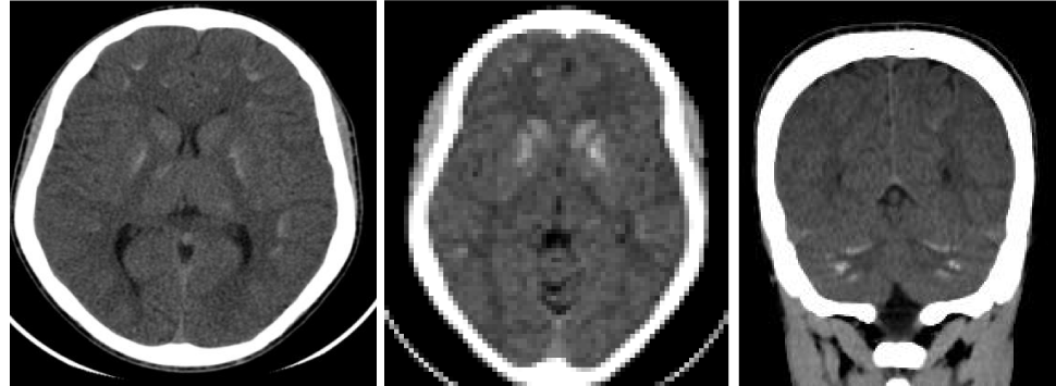
Mr E.F, a 16-year-old male, consulted in the neurology department for generalized tonic-clonic seizures. The first crisis dates back to 09 months ago. He had a personal history of meningitis treated 11 years ago and head trauma with initial loss of consciousness with indefinite duration, unexplored on imaging 10 months ago. There was no family history. At physical examination the general condition was good. His weight was 54 kg for a height of 1.63 meters, a body mass index of 20.32. His temperature was 37 degrees Celsius. The neurological examination and the rest of the physical examination were normal. The hypothesis of an under or extra dural hematoma was raised. Non-injected cranioencephalic CT showed bilateral and symmetrical calcifications of BG and serrated nuclei (Figure 1). The diagnosis of Fahr’s disease or Fahr’s syndrome was made. The patient had been hospitalized and the biological assessment showed hypocalcemia at 62 mg / l (84 < N < 105mg / l), hyperphosphatemia at 101mg / l (25 < N < 45mg / l), hypocalciuria at 11mg / 24h (100 < N < 300 mg / 24 h) and hypophosphaturia at 285 mg / 24 h (400 < N < 1300 / 24h). Magnesium, blood count, blood glucose, and serum creatinemia were normal. The parathyroid hormone level was not measured. The patient was placed on calcivitaminotherapy D and sodium valproate. The follow-up made it possible to note a cessation of seizures two months after the start of treatment. The patient was seen again 6 months and a year later and was still free of crisis. Paraclinically, a gradual normalization of the phosphocalcic balance was noted.
Patient 2
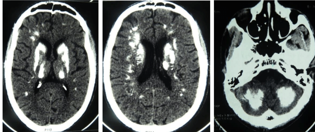
Mr P. K, male, aged 39, consulted in the neurology department of the Kara Teaching Hospital for an aphasia and behavioral disorders that have been progressively settled since about 6 months, associated with a swallowing disorder type of dysphagia that started two weeks ago. He had no particular personal or family history or chronic pathology known according to his family. At the physical examination, his general condition was good. There were spasmodic laughter and crying, signs of dementia with a Mini Mental Status at 16, and bilateral pyramidal syndrome. Otorhinolaryngological (ENT) examination including indirect laryngoscopy was normal. Oesophageal endoscopy and oesophageal transit were normal. Oesophageal manometry, oesophageal radiography and oesophageal biopsies were not performed. The hypothesis of an intracranial expansive process, or a dementia or a psychiatric affection, associated with unidentified esophageal disease was raised. Non - injected cranio - encephalic CT showed massive, bilateral and symmetrical calcifications of BG, white - gray matter junction, and cerebellar hemispheres (Figure 2). The chest X-ray in frontal incidence and parathyroid gland ultrasound were normal. The biological assessment showed a hypocalcemia at 60 mg / l (84 < N < 105 mg / l), a hyperphosphatemia at 105 mg / l (25 < N < 45 mg / l), a low parathyroid hormone at 2 ng / l (14 < N < 63ng /). Serology cysticercosis and HIV serology were negative. The blood count, blood glucose and serum creatinemia were normal. The diagnosis of Fahr syndrome was noticed.
A nasogastric tube had been placed to the patient to feed and calcivitaminotherapy D was initiated. He also had specific rehabilitation for his dysphagia. The follow-up after 03 years noted a partial but significant regression of the neurological and psychiatric clinical signs with a complete regression of the dysphagia in only 01 months after the beginning of the treatment, and a progressive correction of the phosphocalcic balance.
Discussion
Described for the first time by the German neurologist Fahr T in 1930 [5]. Fahr syndrome is synonymous for some authors of Fahr disease or idiopathic calcifications of basal ganglia [6]. It is a rare disease with a prevalence of between 0.49% and 10.02% in a review of the literature in 2013 [6] in which patients selected for CT showed neuropsychiatric manifestations in 03 different studies. It can be asymptomatic and in this case brain calcifications are fortuitously discovered during cerebral CT. It occurs classically between the ages of 30 and 60 [7, 8]. Our first observation is peculiar by discovery at a relatively early age, although pediatric cases are rarely described [9,10]. The clinical manifestations of this syndrome are non specific and are very varied associating at various but more frequent proportions, abnormal movements and neuropsychiatric disorders. The abnormal movements described in order to decrease frequency are: parkinsonism, chorea, tremor, dystonia, atheosis and orofacial dyskinesia [11]. The neuropsychiatric disorders described are: depression, anxiety, cognitive disorders, hallucinations, personality disorders, schizoid psychosis and dementia [7]. Other neurological manifestations are possible but less common, such as extrapyramidal syndrome, focal or generalized seizures, more rarely cerebellar or pyramidal syndrome [3,12,13]. In our first observation, the syndrome was revealed by seizures that is not the most common mode of disclosure [3,12] and in the second observation, aphasia, behavioral disorders, signs of dementia and dysphagia. This last rare symptom makes it a special feature. This dysphagia may have another etiology given that the etiological explorations of dysphagia have not been carried out because of insufficient equipment. However, the favorable evolution after the start of calcivitaminotherapy D and specific rehabilitation measures make us think that it may be related to Fahr syndrome. The pyramidal syndrome, which is a rare manifestation [3,12], was found in patient 2. In terms of medical imaging, the reference examination that makes it possible to diagnose Fahr’s syndrome is cerebral CT without injection of the contrast medium. It shows intracerebral calcifications, bilateral and symmetrical, interesting the BG. The semi-oval center and the sub-cortical gray matter can also be affected [6]. This is the case of our two patients. Similarly, cerebral Magnetic Resonance Imaging (MRI), when performed, reveals in the calcified areas of BG, a T2 hyposignal, a hypo or T1 hypersignal. The cerebellar lesions may be more heterogeneous with the possibility of T1 and T2 hypersignal due to gliosis in calcified areas [8]. The new MRI sequences “SWAN, SWI and VenoBOLD” nowadays allow the diagnosis of micro calcifications with a higher sensitivity compared to other MRI sequences and its sequences should be integrated in MRI protocols for the exploration of neurodegenerative diseases [14]. X-rays of the skull have been described as a diagnostic imaging modality. It shows calcium opacities, punctuate, grouped, symmetrical and located above the saddle turcic, corresponding to the calcifications of BG, while subcortical calcifications and cerebellar appear wavy [8].
These lesions on imaging are associated with biological signs of dysparathyroidism including hypoparathyroidism. The rate of parathyroid hormone was not measured in our first observation because of lack of financial means and the insufficiency of the technical platform. Signs of hyperparathyroidism are rarely described [4, 15].The diagnostic criteria for Fahr’s disease involve [16,13]: neuroimaging of bilateral calcifications of BG, progressive neurological or neuropsychiatric manifestations, onset of clinical manifestations generally occurring between 40 and 50 years of age ( an earlier onset is possible), the absence of evidence of biochemical abnormalities and clinical features suggesting the presence of mitochondrial or metabolic disease or other systemic disorders, the non-infectious, traumatic or toxic etiologies of intracerebral calcifications and the Presence of family history of BG calcification consistent with autosomal dominant inheritance. In case of family history, the diagnosis can be made in the absence of one of the first 2 criteria. If the family history is negative, meeting the first 5 criteria is sufficient for the diagnosis of Fahr’s disease only if the calcifications are typical of Fahr’s disease. In our two observations, no other case was found in both families. Fahr syndrome should not be confused with other conditions that can cause intracerebral calcification, especially in our tropical environment, infections primarily neurocysticercosis followed by toxoplasmosis, syphilis and finally rubella which has become very rare currently. Other conditions that can cause brain calcification are: endocrinopathies (hypothyroidism, hypogonadism), systemic pathologies (systemic scleroderma, systemic lupus erythematosus, celiac disease), various diseases (chronic renal failure, vitamin D intoxication, mitochondrial cytopathies) and primary or secondary calcified brain tumors. In these conditions, intracerebral calcifications are not bilateral, symmetrical or localized to BG, as seen in patients with Fahr syndrome [17].
Conclusion
Fahr syndrome has no clinical specificity. The diagnosis is confirmed by CT which shows the bilateral and symmetrical calcifications of the basal ganglia. We must look for these calcifications, the biological signs of a dysthyroidism (hypoparathyroidie in our observations) and who can be responsible for the clinical manifestations and whose correction improves its clinical manifestations. Our first observation is peculiar to the early age of diagnosis and the second to swallowing disorders as a clinical manifestation
Declarations
Ethics approval and consent to participate
This case report was approved by the Department of Radiology of the teaching hospital of Lomé. We obtained the approval from the patients. The patients gave their consent.
Consent for publication
The Department of Pathology of the teaching hospital of Lomé authorized the publication of this manuscript.
Authors Contributions
BN was responsible for the design of the study, undertook the field study, performed data collection, analysis and interpretation, and wrote the manuscript.
TD, MD, PG, AA, TM, LS, SB and AB: participated in the design of the study, supervised the data collection and participated in the data analysis. LKAK is responsible for the overall scientific management of the study, the analysis and interpretation, and preparation of the final manuscript. All authors have read and approved the final manuscript to be submitted for publication.
- Morgane L, Trimarchi F, Benvenga S. Fahr’s disease. Lancet. 2002; 359: 759. https://goo.gl/KbTYwY
- Murat Gulsun, Ali FuatBaykız, SerdarKabata, Hasan Belli. Fahr’s Syndrome –Three cases presenting with psychiatric signs. Eur J Gen Med. 2006; 3: 35-40. https://goo.gl/YBMpPh
- el Maghraoui A, Birouk N, Zaim A, Slassi I, Yahyaoui M, Chkili T. Syndrome de Fahr et dysparathyroidie : trois observations. Press Med. 1995; 24: 1301-4. https://goo.gl/q2pTBT
- Khadir K, Moussaid L, Azzouzi S. Fahr syndrome secondary to hypoparathyroidism with dermatological revelation. Ann Dermatol Venerol. 2004; 131: 979-83. https://goo.gl/efZ6Ao
- Fahr, T. Idiopathic calcification of the cerebral vessels. Zentralbl Allg Pathol. 1930; 50: 129-133.
- Saleem et al. Fahr’s syndrome: literature review of current evidence. Orphanet Journal of Rare Diseases. 2013; 8: 156. https://goo.gl/5PzSME
- Konig P. Psychopathological alterations in cases of symmetrical basal ganglia sclerosis. Biol Psychiatry. 1989; 25: 459-468. https://goo.gl/m7aMo5
- Sobrid SH MJ, Geschwind DH. Familial Idiopathic Basal Ganglia Calcification: GeneReviews™ [Internet]. Seattle (WA): University of Washington, Seattle; 2004. Updated 2007 Sep 20.
- Kahloul N, Chaari W, Boughamoura L, Charfeddine L, Khammeri S, Amri F. Pseudo hypoparathyroidism revealed by Fahr syndrome. Arch Pediatr. 2009; 16: 444-8. https://goo.gl/dcMFna
- Souad R, Ilhame D, Mohamed B, Fawzi B, Ismail R, Rachid A . Confusional Syndrome Revealing Fahr Syndrome with Hyperparathyroidism .Pan Afr Med J. 2013; 14: 123. https://goo.gl/SuxpiB
- Manyam BV, Walters AS, Narla KR. Bilateral striopallido dentate calcinosis: clinical characteristics of patients seen in a registry. Mov Disord. 2001; 16: 258-64. https://goo.gl/YpmhwH
- Rumbach L, Buffler M, Callard M. Fahr's disease and Fahr's syndrome about 10 observations. J Med Strasbourg. 1984; 15: 375-7.
- Lazar M, Ion DA, Streinu-Cercel A, Badarau AI. Fahr’s syndrome: diagnosis issues in patients with unknown family history of disease. Rom J Morphol Embryol. 2009; 50: 425-8. https://goo.gl/Jswn2S
- Sahin N, Solak A, Genc B, Kulu U. Fahr disease: use of susceptibility-weighted imaging for diagnostic dilemma with magnetic resonance imaging. Quant Imaging Med Surg. 2015; 5: 628-632. https://goo.gl/b3iZNb
- Seyahi N, Apaydin S, Sariyar M, Erek E. Intracranial calcification and tumoural calcinosis during vitamin D therapy. Nephrol. 2004; 9: 89-93. https://goo.gl/YT5W2M
- Manyam BV. What is and what is not ‘Fahr’s disease’. ParkinsonismRelat Disord. 2005; 11: 73-80. https://goo.gl/DhvKDQ
- Pfaender M, D'Souza WJ, Trost N, Litewka L, Paine M, Cook M. Visual disturbances representing occipital lobe epilepsy in patients with cerebral calcifications and coeliac disease: a case series. J Neurol Neurosurg Psychiatr. 2004; 75: 1623-5. https://goo.gl/5ay2oi
Sign up for Article Alerts