Beyond the Biopsy: a Case of Erdheim-Chester Disease with Concurrent Lung Cancer
Nadia Solomon1*, Arne Landwehr1, Zerwa Farooq2, Garfield Gutzmore3, Horace Tang3, Vincent Rizzo2 and Sandeep Singh Tuli4
1St. George’s University School of Medicine, True Blue, Grenada
2Department of Medicine, Icahn School of Medicine at Mount Sinai, Queens Hospital Center, Jamaica, NY, USA
3Department of Hematology & Oncology, Icahn School of Medicine at Mount Sinai, Queens Hospital Center, Jamaica, NY, USA
4Department of Radiology, Icahn School of Medicine at Mount Sinai, Queens Hospital Center, Jamaica, NY, USA
*Address for Correspondence: Nadia Solomon, School of Medicine, St. George’s University, True Blue, Grenada, Tel: +609-439-9160; ORCID ID: 0000-0003-0728-0095; E-mail: [email protected]
Submitted: 30 March 2019; Approved: 19 April 2019; Published: 22 April 2019
Citation this article: Landwehr A, Solomon N, Sanelli-Russo S, Rizzo V. Beyond the Biopsy: a Case of Erdheim-Chester Disease with Concurrent Lung Cancer. American J Rare Dis Diagn Ther. 2019;2(1): 001-004.
Copyright: © 2019 Solomon N, et al. This is an open access article distributed under the Creative Commons Attribution License, which permits unrestricted use, distribution, and reproduction in any medium, provided the original work is properly cited
Keywords: Erdheim-Chester disease; Histiocytosis; Diagnostics
Download Fulltext PDF
Introduction: Erdheim-Chester Disease (ECD) is a rare disorder characterized by tissue infiltration by pathologic histiocytes, leading to widespread inflammation and damage. Clinical presentation can range from asymptomatic infiltration of bone to multiple organ system dysfunction.
Methods: We present a case of a patient with several unusual imaging findings that led to a differential diagnosis of ECD; however, a biopsy of a mediastinal mass suspected to be due to hystiocyte infiltration instead revealed primary lung cancer. Ultimately, ECD could not be ruled out and the patient was referred to dermatology for a facial xanthogranuloma biopsy.
Results: Biopsy of right cheek xanthogranuloma revealed a dermal nodular infiltrate of histiocytes with foamy cytoplasm mixed with lymphocytes and rare Touton-type giant cells, consistent with ECD.
Conclusions: ECD should be considered in patients with signs of malignancy and diffuse tissue thickening and fibrosis. Concurrent ECD and adenocarcinoma is highly unusual: in this case, adenocarcinoma was confirmed by biopsy but was not consistent with the majority of imaging findings. This case demonstrates the importance of a thorough investigation and the consideration that not all findings may be attributable to a single disease process.
Abbreviations
ECD: Erdheim-Chester Disease; CT: Computed Tomography; MRI: Magnetic Resonance Imaging; CNS: Central Nervous System
Introduction
Erdheim-Chester Disease (ECD) is a rare, non-Langerhans’ cell histiocytosis, with only several hundred cases appearing in the literature [1,2]. Originally described as “lipoid granulomatosis,” [3] ECD is characterized by tissue infiltration by pathologic, foamy histiocytes, leading to inflammation and damage [1,4]. More than 50% of patients demonstrate mutations in the proto-oncogene BRAF [3,4], leading to uncontrolled clonal expansion [4]. Clinical manifestations range from asymptomatic infiltration of bone to multiple organ system dysfunction [1-3].
We present a case of ECD in a patient with an additional malignancy initially distracting from diagnosis. Findings of multiple imaging studies are discussed, emphasizing the importance of a thorough workup.
Case Presentation
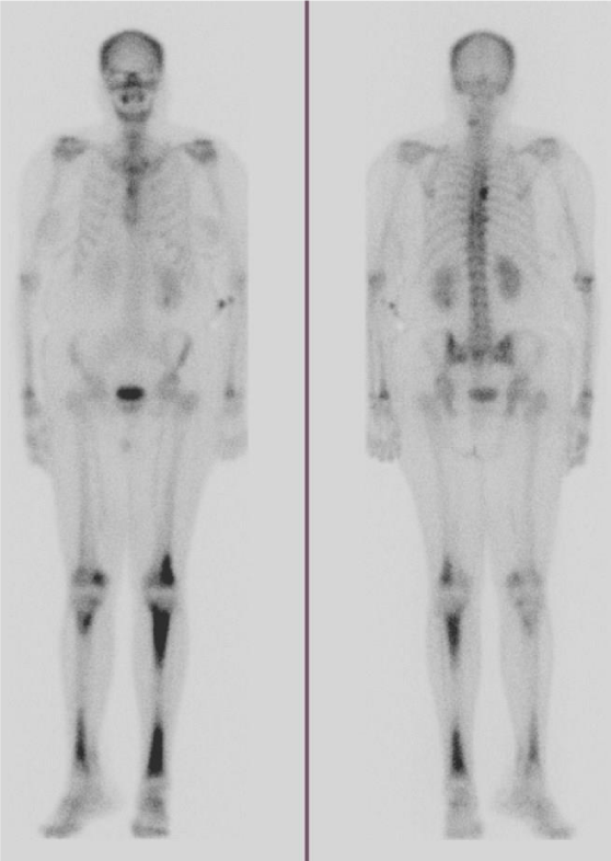
A 56 year-old Trinidadian female presented to the emergency department with intermittent left-sided chest pain for 3 days, which was exacerbated by exertion and associated with shortness-of-breath. Additional symptoms included headache, fatigue, and 15-20 pound weight loss with bilateral lower extremity pain over the past year. Numerous facial xanthogranulomas were present. Chest x-ray revealed an enlarged cardiac silhouette, suspected pulmonary vascular congestion, and subcentimeter calcifications due to old granulomatous disease. Computed Tomography (CT) of the chest revealed small pericardial and left pleural effusions, and a large left upper-lobe paramediastinal mass with extensive adenopathy [Figure 1], warranting metastatic workup. Subsequent CT abdomen revealed perinephric soft tissue surrounding both kidneys. CT head without and with intravenous contrast revealed metastases to the right frontal cortex and right posterior cerebellum with mild mass effect of the right lateral ventricle. Magnetic Resonance Imaging (MRI) of the brain confirmed these findings, demonstrating enhancing lesions in the right cerebellum and right frontal lobe adjacent to the frontal operculum. A total body bone scan revealed abnormal uptake in the seventh vertebral body and increased uptake in both lower extremities [Figure 2].
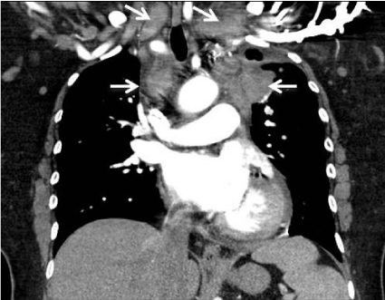
CT-guided biopsy of the superior mediastinal lymph nodes revealed poorly differentiated adenocarcinoma with necrosis positive for cytokeratins AE1/ AE3 and TTF1, compatible with lung primary. Findings and suspicions were discussed with the patient, and she was referred to dermatology. Biopsy of right cheek xanthogranuloma revealed a dermal nodular infiltrate of histiocytes with foamy cytoplasm mixed with lymphocytes and rare Touton-type giant cells, consistent with ECD.
Discussion
Although definitive diagnosis of ECD relies on identification of CD68 (+), CD1a (-) histiocytes on biopsy, radiological findings can suggest the diagnosis [2]. Because ECD affects multiple organs and organ systems, however, presentation at diagnosis and associated imaging findings can be highly variable. In this case, demonstration of perinephric soft tissue encapsulating the kidneys on CT and giving them a “hairy” appearance [2] was the first sign alerting clinicians to the potential presence of ECD. CT findings of pericardial and pleural effusions [2], combined with findings of increased uptake in the lower extremities on bone scan [1], further supported this diagnosis. As Central Nervous System (CNS) involvement, often characterized by multiple lesions, has been reported in >50% of patients [1,2,5], the two brain lesions were also considered consistent with the working diagnosis. It was therefore somewhat surprising when the mediastinal lesion was biopsy proven as adenocarcinoma, with ECD proven later on via xanthogranuloma biopsy. Given these findings, it is unclear if the CNS lesions are due to ECD or metastatic spread of adenocarcinoma.
Conclusion
ECD should be considered in patients with signs of malignancy and diffuse tissue thickening and fibrosis. Simultaneous occurrence of ECD and adenocarcinoma is highly unusual: in this case, presence of adenocarcinoma was confirmed but could not explain the majority of imaging findings. Ultimately, this case demonstrates the importance of a thorough workup: in a disease like ECD with such diverse features, it is important to consider that not all findings may be attributable to a single disease process.
- Cives M, Simone V, Rizzo FM, Dicuonzo F, Cristallo Lacalamita M, Ingravallo G, et al. Erdheim-Chester disease: a systematic review. Crit Rev Oncol Hematol. 2015; 95: 1-11. https://bit.ly/2Xolpou
- Mazor RD, Manevich-Mazor M, Shoenfeld Y. Erdheim-Chester Disease: a comprehensive review of the literature. Orphanet J Rare Dis. 2013; 8: 137. https://bit.ly/2UloXpT
- Nikpanah M, Kim L, Mirmomen SM, Symons R, Papageorgiou I, Gahl WA, et al. Abdominal involvement in Erdheim-Chester Disease (ECD): MRI and CT imaging findings and their association with BRAF(V600E) mutation. Eur Radiol. 2018; 28: 3751-3759. https://bit.ly/2GvO31x
- Durham BH. Molecular characterization of the histiocytoses: Neoplasia of dendritic cells and macrophages. Semin Cell Dev Biol. 2019; 86: 62-76. https://bit.ly/2VayggS
- Parks NE, Goyal G, Go RS, Mandrekar J and Tobin WO. Neuroradiologic manifestations of Erdheim-Chester disease. Neurol Clin Pract. 2018; 8: 15-20. https://bit.ly/2GoBILe
Sign up for Article Alerts