Revisitation of Autoimmune Addison’s Disease: known and Open Pathophysiologic and Clinical Aspects
Annamaria De Bellis*, Giuseppe Bellastella, Maria Ida Maiorino, Vlenia Pernice, Miriam Longo, Carmen Annunziata, Antonio Bellastella and Katherine Esposito
Department of Advanced Medical and Surgical Sciences, University of Campania “Luigi Vanvitelli”, Naples, Italy
*Address for Correspondence: Annamaria De Bellis, Department of Advanced Medical and Surgical Sciences, University of Campania “Luigi Vanvitelli”, Naples, Italy, Tel/Fax: 0039-081-566-5245; E-mail: [email protected]
Submitted: 30 January 2019; Approved: 21 March 2019; Published: 22 March 2019
Citation this article: Bellis AD, Bellastella G, Maiorino MI, Pernice V, Longo M, et al. Revisitation of Autoimmune Addison’s Disease: known and Open Pathophysiologic and Clinical Aspects. Int J Clin Endocrinol. 2019;3(1): 001-013.
Copyright: © 2019 Bellis AD, et al. This is an open access article distributed under the Creative Commons Attribution License, which permits unrestricted use, distribution, and reproduction in any medium, provided the original work is properly cited
Download Fulltext PDF
Addison’s Disease (AD) or primary adrenal insufficiency has been thought a rare disease for a long time, but recent epidemiological studies have reported a rising prevalence in developed countries. Among the causes of apparently idiopathic forms, autoimmunity plays a relevant role. This review will be focused on several aspects of autoimmune AD, which may manifest either as an isolated disorder or associated with other autoimmune diseases among the autoimmune polyglandular syndromes. HLA plays a key role in determining T cell responses to antigens, and various HLA alleles have been shown to be associated with many T cell-mediated autoimmune disorders, but the mechanism by which the adrenal cortex is destroyed in AD is still discussed. Cytotoxic T lymphocytes are thought to be the most important effector cells in mediating the autoimmune tissue destruction, because Adrenal Cortex Autoantibodies (ACA) and/or autoantibodies against 21 idroxylase (21-OHAb) do not seem to be directly involved in the pathogenesis, being considered only good marker of the disease both in clinical and in preclinical stage. In fact, subclinical autoimmune AD can evolve trough 5 functional stages from stage 0 (only presence of autoantibodies) to stage 4 (clinically overt disease). All the five stages are characterized by the presence of these antibodies but only when they are present at high titre in subclinical stages are associated with the progression towards clinically overt autoimmune AD, whereas a spontaneous remission of subclinical adrenal dysfunction with their disappearance may occur when they are present at low titres. Treatment of AD is based on the use of hydrocortisone or cortisone for symptomatic patients; fludrocortisone should be used as substitute for mineral-corticosteroids. In some cases, an early replacement therapy has been shown to be helpful to interrupt the progression towards the clinical stage with disappearance of these autoantibodies and recovery of adrenal function. In addition, a life-threatening adrenal crisis in patients with chronic adrenal insufficiency under established replacement therapy may occur. Clinical medicine must pay attention to these situations because an untreated Addisonian crisis is a medical emergency that requires hospitalization, and if not caught early can be fatal.
Introduction
Thomas Addison first described the disease which now bears his name in an article in the London Medical Gazzette, entitled “Disease: chronic suprarenal insufficiency, usually due to tuberculosis of suprarenal capsule” [1]. However, his work was criticized by other specialists of the time that did not agree about the existence of this condition, until an eminent French physician, Armand Trousseau, finally recognized adrenal failure and first called it Addison’s Disease (AD) [2]. In 1912, Harvey William Cushing discovered that adrenal function was regulated by pituitary gland [3] and in 1948 Harris demonstrated the control of pituitary by hypothalamus [4]. Since 1938, it was introduced a replacement therapy with corticosterone, synthesized with deoxycorticosterone, for AD in humans [5-7]. AD, or primary adrenal insufficiency, is an endocrine disorder characterized by impaired secretions of steroid hormones by adrenal gland (glucocorticoids and mineralocorticoids) with consequent high levels of both Adrenocorticotropic Hormone (ACTH) and plasmatic renin activity. Less frequent is the form of secondary adrenocortical insufficiency, where a deficiency of pituitary ACTH leads to insufficient cortisol production. Tertiary adrenal insufficiency can be caused by diseases affecting the hypothalamus and the secretion of Corticotropic-Releasing Hormone (CRH). Nowadays, chronic primary adrenal insufficiency has a prevalence of 93-140 per million and an incidence of 4.7-6.2 per million/year in European populations [8-10]. These recent numbers are higher than those reported during the 1960s and 1970s [11,12] despite a continuous decline in tuberculous adrenalitis (the most common cause of AD until the discovery of rifampicin antibiotic), suggesting an increasing incidence of idiopathic adrenalitis. The prevalence of idiopathic form of AD has been estimated at 30-60/ million in European and North American populations, and its prevalence is highest in the fourth decade of life [13]. Moreover, prevalence of AD in Germany has increased between 2008 and 2012, with higher numbers in women than men [14].
Considerable evidence suggests that idiopathic adrenal atrophy is an autoimmune disease. As most of the autoimmune diseases [15], autoimmune AD mostly affects women, with at least 65% of patients being female [11-13]. We will further analyse autoimmune aspects of AD in detail. In patients with AD, autoimmune comorbidities are rather frequent. Approximately 60% of these patients develop an Autoimmune Polyglandular Syndrome (APS) comprising autoimmune AD and at least one other autoimmune disease like autoimmune thyroid disorders, type 1 diabetes, autoimmune oophoritis leading to primary ovarian failure, celiac disease, atrophic gastritis, vitiligo, and/or autoimmune alopecia [14].
Secondary adrenal insufficiency has an estimated prevalence of 150-280 per million, affecting more frequently women than men (age at diagnosis peaks in the fourth and in the sixth decade of life, respectively) [13]. Clinical features of adrenal insufficiency can include severe fatigue and weakness, loss of weight, increased pigmentation of the skin (only in primary adrenal insufficiency due to high ACTH levels), low blood pressure, nausea, vomiting, painful muscles and joints. The results of either undiagnosed Addison’s disease, or pathological insult suddenly affecting adrenal function, or an intercurrent problem (e.g. infection, trauma) in the setting of known Addison’s disease, may be the Addisonian crisis (Table 1). Additionally, this situation may develop in those patients under glucocorticoids treatment who have suddenly ceased taking their medication [16]. Moreover, rarer conditions favouring this crisis have been recently reported in literature. In particular, an Addisonian crisis has been described in a young boy several years after hematopoietic stem cell transplantation [17] and another case in a patient after the first infusion of zoledronic acid [18]. Clinical medicine must pay attention to these situations because an Addisonian crisis is a medical emergency that requires hospitalization, and if not caught early can be fatal.
Etiology
The causes of primary adrenal insufficiency can be grouped according to the type of hormone secretion impairment. According to Ten et al. [19], the causes can be divided into three categories: 1) adrenal dysgenesis; 2) impaired steroidogenesis; 3) adrenal destruction or atrophy. Table 2 summarizes the factors involved in the mentioned categories; whereas, table 3 describes the causes of secondary adrenal insufficiency. This minireview aims to focus on autoimmune adrenal insufficiency.
Recent studies have shown the correlation between Addison’s disease and adverse effects of check-point-inhibitors [20]. Immune Checkpoint Inhibitors (ICPIs) are a new class of antineoplastic agents used to treat melanoma, lung cancer, and increasingly, other malignancies. These drugs include anti-Cytotoxic T-Cell Antigen 4 Antibodies (anti-CTLA-4 Abs), Programmed Death 1 (PD-1) and Programmed Death Ligand 1 (PD-L1) inhibitors. ICPIs target the immune system at regulatory checkpoints, allowing disinhibition of the immune system and identification and destruction of cancer cells. As a result of this nonspecific and dysregulated activation of the immune system, autoimmune conditions, including several endocrinopathies (hypophysitis, thyroiditis) have been described as an off-target effect [21]. The mechanism of development of autoimmune adrenal insufficiency in patients treated with these agents, as ipilimumab or nivolumab, has not yet been clarified, mainly due to a few studies so far reported. However, it is interesting to note that antibodies directed against steroidogenic enzymes such as 21-hydroxylase, typically observed in patients with autoimmune adrenalitis, were absent in reported cases of AD. However, it is difficult to determine whether the development of adrenal insufficiency post-ICPS follows a different mechanism than traditional autoimmune AD [20].
Clinical Presentation
The clinical manifestations of primary adrenal insufficiency result from deficiency of all adrenocortical hormones (aldosterone, cortisol, androgens). The majority of the symptoms are early non-specific and can delay diagnosis and treatment of the disease. Symptoms of the disease can include severe fatigue and weakness, loss of weight, low blood pressure, nausea, vomiting, painful muscles and joints. Hypoglycaemia can be the presenting sign in children with adrenal insufficiency. A specific sign of chronic, but not acute, primary adrenal insufficiency is hyperpigmentation, which predominantly affects areas of skin subjected to pressure (elbows, knuckles, palmar creases, lips, buccal mucosa). The preclinical time course of adrenal insufficiency can span many years after detection of early metabolic changes by screening, even in the presence of high specific autoantibody titres and significantly raised corticotropin concentrations [22].
In autoimmune adrenal insufficiency, the zona glomerulosa is generally the first zone affected by immune-mediated destruction, possibly because it is thinner than fasciculata and reticularis zona and more vulnerable to autoimmune attack. This feature might explain the first step of adrenal failure, which is characterised by low aldosterone concentrations and high plasma renin activity. Subsequently, a progressive decline in glucocorticoid secretion appears, initially with inadequate response to stressful stimuli and then by a phase of overt failure with low basal cortisol concentrations [23].
The clinical manifestations of secondary or tertiary adrenal insufficiency result from glucocorticoid deficiency only (secretion of aldosterone is preserved); however, they can also include signs of the primary underlying disorder. Characteristic of secondary and tertiary forms is the absence of hyperpigmentation, because corticotropin levels are not increased. There might also be present symptoms and signs of deficiency of other anterior pituitary hormones.
In addiction, a life-threatening adrenal crisis can be the first presentation of adrenal insufficiency or it may occur in patients with chronic adrenal insufficiency, even under established replacement therapy [24]. The incidence of adrenal crises is 8.3% patient-years in patients with chronic adrenal insufficiency. Clinical features include vomiting, abdominal pain, myalgia, joint pains, severe hypotension, and hypovolaemic shock associated with hyponatremia, hyperkalemia, or hypoglycaemia. The acute presentation can be triggered by a physiological stress, such as surgery, trauma, an intercurrent infection such as gastrointestinal infection, fever, major pain, strenuous physical activity, pregnancy or emotional stress. The stress-related cortisol response is reduced in chronic adrenal insufficiency and has to be mimicked by the patient with a transient steroid dose increase greater than the standard replacement dosage [24]. Moreover, this situation may develop in those patients who have spontaneously reduced the glucocorticoids dosage. It is a medical emergency, and if untreated it can be fatal.
Criteria for Organ-Specific Autoimmunity in Addison’s Disease
In 1957 Witebsky et al. proposed the criteria to define a disease as autoimmune [25], these criteria subsequently revisited by Rose and Bona focused on the question whether autoimmunity was the real cause of the disease, rather than a consequence or a normal accompaniment. On the basis of this approach the definition of autoimmunity was based on direct, indirect or circumstantial criteria [26]. Direct evidence requires transmissibility of the characteristic disease lesions from human to human, or from human to animal. This type of evidence, which is mediated by autoantibodies or autoreactive T-cells and applicable to some autoimmune diseases (such as Graves’ disease, myasthenia gravis, pemphigus vulgaris) is lacking for AD. Adrenal Cell Antibodies (ACA) are present in autoimmune AD but they incapable of transmitting disease from mother to newborn [27]. However, the recently described concordance for multiple autoimmune manifestations in monozygotic twins with autoimmune AD, deposes for the heritability of autoimmune AD and for strong genetic influence on disease specificity in organ-specific autoimmunity [27]. Indirect evidence is based on the reproduction of autoimmune disease in experimental animals [25]; however, spontaneous animal models of AD are lacking.
Circumstantial evidence is the listing of markers descriptive of autoimmune disease. In autoimmune AD, these markers are represented by autoantibodies (ACA, autoantibodies to adrenal antigens and other autoantibodies), by demonstration of a cell mediated immunity to adrenal cortex antigens, by lymphoplasmacytic nature of the bioptical lesions, and by association with other autoimmune diseases. Moreover, sufficient data are available in the genetic aspects of autoimmune Addison’s disease [28-31] as well as genes classically thought to influence autoimmunity, such as those of the MHC locus and cytotoxic T lymphocyte antigen 4 (CTLA-4) [32] and the gene AIRE in autoimmune polyendocrine syndrome type 1 [33].
Lymphocytic infiltration in the affected gland is a usual finding of the autoimmune diseases. In some autoimmune endocrine diseases this infiltration can be proven relatively easily due to the easy approach to the gland. This is certain true in the case of autoimmune thyroid diseases, but it is much more difficult in many other autoimmune diseases including AD, in which histopathological observations are only obtained by autoptical examination [34]. Thus, the research of respective organ-specific autoantibodies may help to diagnose an autoimmune adrenalitis.
After many years of autoimmune AD, adrenal gland will appear extremely small, with a complete loss of adrenocortical tissue and preservation of medulla. However, a diffuse lymphocytic infiltration with disappearance of the epithelial cells of the adrenal cortex has often been observed also in patients at the initial phase of adrenal failure for autoimmune AD [34,35].
Autoantigens in the autoimmune Addison’s disease
Identification and characterization of specific autoantigens are integral parts of research for several reasons: I) to understand the pathogenesis of autoimmune diseases; II) to study B and T cell responses; III) to characterize autoantibodies; IV) to develop methods for measurement of autoantibodies. Autoimmune endocrine diseases are the most frequent organ-specific autoimmune diseases and many of their autoantigens have been identified and studied in detail. Many organ- specific autoantigens are generally protein with well-defined functions and activities, such as enzymes or receptors. The expression of these antigens in the affected organs is often specific as thyroid peroxidase; but some of these antigens do not fulfill strictly the criteria of organ specificity, as Glutamic Acid Decarboxilase (GAD) or the calcium sensing receptor [36].
Regarding autoimmune AD, Chen S et al. identified a 55kDa adrenal microsomal protein by an immunoprecipitation technique [37]. Autoantibodies reacted to this protein, appeared to be adrenal specific, since the antigen was not precipitated from placenta or thyroid microsomes. In subsequent years, sera from patients with AD reacted against members of the cytochrome p450 family of enzymes involved in steroid synthesis. In particular, three steroidogenic enzymes: 1) 21 hydroxylase [p450c21], 2) 17 alpha hydroxylase [p450c17] 3) cholesterol side chain-cleaving enzyme [p450scc]) were shown to be autoantigens in adrenal autoimmune disease [38-41]. While p450c21 is an adrenal specific enzyme, the antigens p450c17 and p450scc are found in all steroid-producing cells such as gonadal (both antigens) and placenta tissue (only p450scc). In immunofluorescence studies, both p450c21 and p450scc antigens were present in all three layers of the adrenal cortex while antigen p450c17 was not present in the cells of the zona glomerulosa [40].
Detection and characteristics of adrenal cortex autoantibodies
Anderson et al. in the later 1950, first detected ACA by complement fixation, in some patients with idiopathic Addison’s disease [42]. Subsequently these antibodies were detected by indirect Immunofluorescence (IF) in many patients with idiopathic AD. Occasionally, ACA have been detected also in few patients with Addison’s disease due to tuberculosis [1]. IF was and is still considered the gold standard method for detection ACA in routine use [43,44].
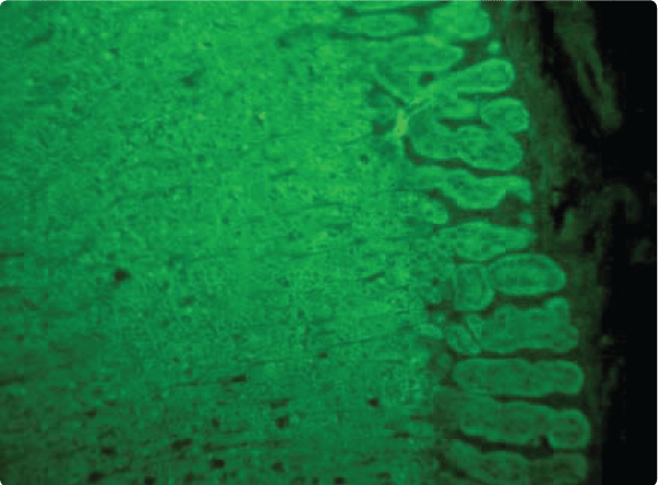
The choice of substrate, using unfixed frozen sections of adrenal gland, have an important role in identifying these antibodies. Ideal tissues are constituted by human fresh adrenal gland immediately frozen in order to avoid cytoplasm antigen alterations [45]; bovine and monkey adrenal glands have also been used in the immunofluorescence test [44,45]. Human adrenal glands from adult subjects aged > 50 year or old baboon adrenal glands are not considered suitable because of the presence of lipofuscin pigment that could interfere with the evaluation of antibodies. Adrenal cortex antibodies react to cells of all three layers of the adrenal cortex (Figure 1) even if sometimes, especially in preclinical stages of the Addison’s disease, they immunostained cells of one or two of three cortical layers [46].
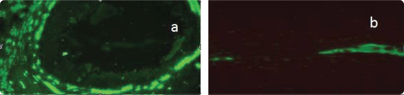
It has been shown that ACA are IgG (IgG1, IgG 2 and IgG4 subtypes) and sometimes complement fixing antibodies when present at high titers [47]. They react to cytoplasmatic antigens distinct from adrenal hormones as well as occurring for other organ specific antibodies as Islet Cell Antibodies (ICA), Vasopressin Cell Antibodies (AVPcAb) and Antipituitary Antibodies (APA) which react to cytoplasmatic antigens distinct from the hormones of the respective glands. Sometimes in sera of patients with autoimmune AD positive for ACA, Steroid-producing cell Antibodies (StcAb) may be also detected by immunofluorescence [47,48]. In these cases, both kind of antibodies immunostained not only adrenal cells but also cytoplasmatic antigens of other steroid-producing cells such as those present in the testis (Figure 2) the ovary and the placenta. However, preabsorption tests with homogenates of the steroid producing cells may contribute to differentiate the antibodies, because ACA recognize exclusively hormone-secreting cells from the adrenal gland [49]. Among the enzymes involved in adrenal steroid synthesis, the enzyme steroid 21 hydroxilase has been identified as the major autoantigen in autoimmune AD.
The identification and molecular cloning of steroid 21OH allowed to develop high sensitive and specific radio binding assay for 21OH antibodies (21OHAb) [37,50]. Direct evidence that 21OH is the major autoantigen recognized by ACA emerges from absorption studies [38]. The authors showed that after incubation with purified human recombinant 21OH, sera previously positive for ACA and 21OHAb did not show any more reaction to the respective antigens or resulted positive at lowest titre. It is now clear that 17OH and P450scc are the major component of StcAb measured by immunofluorescence. Using an immunoprecipitation assay, 35S-labeled cloned antigens were used to test this reactivity and good correlation between StcAb and the antibodies to these specific enzymes was found [51,52]. At least, it has been postulated that autoantibodies to the ACTH receptor with function and growth blocking activities were responsible for hypoadrenalism in patients with idiopathic Addison’s disease [53-55]. However, more recent studies with purified patients’ IgGs did not seem to confirm these earlier observations.
Prevalence and clinical significance of ACA/21OHAb, StcA/17OHAb sccAb
The presence of ACA and 21OHAb in patients with clinical AD allows an etiological diagnosis of autoimmune disease [49,56,57], even if ACA or 21OHAb have also been found occasionally at low titers in sera of patients with AD secondary to tuberculosis [56,57]. A good correlation between ACA and 21OHAb detection has been shown by some studies, but other studies found some discrepancies [28,36]. The prevalence of ACA/21OHAb in clinical AD depends on duration time of disease and on different clinical forms of autoimmune AD. Some organ specific antibodies as ICA in type 1 diabetes mellitus and Antipituitary Antibodies (APA) in Lymphocytic Hypophysitis (LYH) are present in an early clinical phase of the diseases but subsequently could disappear [58-61] while ACA/21OHAb in AD and AVPcAb in autoimmune central diabetes insipidus persist over time during the clinical course of the disease [57,60]. In particular, when ACA/21OHAb are identified at the onset of clinical AD, both antibodies are present in 92-100% of cases belonging to Autoimmune Polyglandular Syndromes (APS) while in patients with isolated AD the presence of ACA and 21OHAb are present in 76-80% of cases, respectively [28,62,63]. It’s possible that an earlier diagnosis in patients with preexisting autoimmune endocrinopathies could be a reason for that difference. The time-lag in diagnosis may be explained by the fact that isolated AD often develops slowly over months that patients may not notice physical changes in the beginning unless the patient suffers from an acute adrenal crisis. In addition, most symptoms are unspecific and are not directly leading to the diagnosis of AD. Therefore, diagnosing isolated AD is still difficult.
An early diagnosis and start of replacement therapy seem to have a positive effect on the quality of life [64]. Women are often diagnosed later than men. A possible explanation might be that women have pain experience levels higher than men [65].
At the onset of clinical AD, both antibodies are present at very high titers/levels, whereas their titer significantly decreases in patients with longer disease duration (personal unpublished data). In fact, ACA may persist more than 20 years after the onset of disease especially in patients with APS including AAD [57]; instead StcAb may be present at high titers in patients with AD of recent onset but they disappear during the follow-up of disease [66,67]. These antibodies have been found above all in females with premature ovarian failure, and then in some kind of APS [47,48]. In the majority of cases StcAb positive patients are positive also for 17OHAb and or P450sccAb [40].
ACA/21OHAb have also been found in 0.2-0.8% of normal controls and in 1-3% of first-degree relatives of patients with autoimmune AD. Both antibodies can be observed in 1,2-1.3% of patients with other endocrine autoimmune diseases without clinical phase of diseases [28,62]. The prevalence of ACA/21OHAb is higher in some autoimmune diseases as idiopathic hypoparathyroidism, autoimmune thyroid diseases, type 1 diabetes mellitus and autoimmune premature ovarian failure than in other diseases as celiac disease, autoimmune chronic hepatitis, vitiligo, autoimmune CDI and LYH. In particular, the presence of ACA/21OHAb in patients with idiopathic hypoparathyroidism and/or candidiasis indicates clinical or potential APS type 1, while in patients with autoimmune thyroid diseases and/or type 1 diabetes mellitus indicates potential APS type 2 (see next paragraphs). Since 1988, in our Endocrine Diseases Laboratory, adrenal cortex antibodies and other organ specific autoantibodies have been evaluated in sera of a large cohort of autoimmune endocrine diseases (also diagnosed in other Endocrine Units). In particular, ACA have been found in several patients with autoimmune endocrine diseases without clinical AD [44]. Subsequently ACA positive patients have been tested for 21OHAb and 90/103 resulted positive for these antibodies [63,68]. The majority of ACA/21OHAb positive patients had autoimmune thyroid diseases and/or type 1 diabetes mellitus but some of them had other autoimmune diseases as celiac disease and autoimmune Central Diabetes Insipidus (CDI) and LYH [63].
Adrenal autoantibodies: pathogenetic role or only markers of disease?
The inaccessibility of the adrenal glands has been a major bar to progress in understanding the pathogenesis of autoimmune AD. Until recently, knowledge has been acquired either by the evidence that a particular region of 21OH could be an immunodominant T cell epitope in AD or by the suggestion of possible pathogenetic role of the CXCL10 in endocrine autoimmune diseases and in particular in AD [69].
The mechanism by which the adrenal cortex is destroyed in autoimmune AD is not yet clear even if TH1 lymphocytes are thought to be the most important cells in mediating the autoimmune adrenal gland destruction. Evidence for a direct pathogenetic role of antibodies directed to the 21-OH in adrenal failure is weak. Some authors indicated that the central and the C-terminal regions of the 21-OH sequence are important for autoantibody binding [69]. Using a panel of mouse monoclonal antibodies to human recombinant 21-OH, it has been possible the identification of three different short aminoacidic sequences in the 5-terminal part of 21-OH which are important for binding 21-OH. The C-terminal region of the 21-OH molecule is important also for enzyme activity. Antibody preparation from Addisonian patients caused a marked-dose dependent inhibition of 21-OH enzyme activity (thus blocking the conversion of progesterone to deoxy-progesterone) in vitro. However, studies of adrenal steroid synthesis markers under ACTH stimulation in patients with high levels of 21-OHAb did not confirm an inhibitory effect of 21-OHAb on 21-OH enzyme activity in vivo. Since the autoantigens targeted in AD are intracellular proteins localized in the endoplasmic reticulum, it is unknown whether the corresponding autoantibodies and their effector function play a pathogenic role. According to some studies, these adrenal antigens are also represented on plasma membrane as result of stress, for example during a viral infection [69]. Therefore, the question is: are these autoantibodies the cause of the tissue destruction or just a result of it? Antibodies may destroy cells by activating the cytolytic complement cascade, thus leading to deposition of immune complexes in the respective gland with consequent organ damage. Moreover, antibodies may bind the antigen and Natural Killer Cells (NKC) can cause antibody dependent cellular cytotoxicity, through FC receptor binding. However, autoimmune AD has been recently shown to be associated with significantly decreased NKC cytotoxicity, which impairs anti-viral immune defense with increased rate of respiratory infections and ultimately mortality in affected patients [70]. It has been shown that the identify of IgG subclasses of antibodies may indicate which T helper (Th) cell subset is involved in autoimmune response. In humans, IgG1, and IgG3 indicate Th1 responses while IgG4 is seen when a Th2 response predominates. Boe et al. [71] showed that autoantibodies against 21 hydroxilase and side-chain cleavage enzyme in AD are predominantly of the IgG1 subclass suggesting that CD4 T cell response in autoimmune AD is dominated by Th1 cells. In particular, even if autoantibodies to adrenocortical antigens may not be directly pathogenetic, they may modulate T cell response to the antigens they bind. Efficient complement and Fc receptor-mediated uptake of antigen-antibody complexes by dendritic cells (APC) may lead the stimulatory presentation (increase of IL12) of adrenal autoantigens on CD4 Th1 cells with subsequent increase of INFγ. These cells could have a principal pathogenetic role in autoimmune AD even if little is known about the T-cells mediated destruction of the adrenal cortex. Cell-mediated immune reaction to adrenal antigens were first described by Nerup and Bendixen in 1969 [72] and subsequently several studies have confirmed and extended their initial results. In particular, Freeman and Weetnam [73] were able to demonstrate in autoimmune AD T-cell proliferation when cells were stimulated with adrenal proteins fractionated according to molecular weight. However, individual antigens were not identified. Moreover, an increased expression of circulating Ia-positive T-cells among patients with recent onset of AD compared with controls have been reported [74]. Recently Husebye et al. [75] demonstrated that when SJL and BALB/c mice were immunized with recombinant 21-OH and with 21OH derived peptides, a significative T-cell proliferation (CD4 T lymphocytes restriction) in lymph nodes of these animals could be present. Moreover, the authors demonstrated that T-cell proliferation (CD4 cells) could be determined by 21-OH peptide derived from a part [76]. This important result suggests that 21-OH [77] determined the immunogenicity of the proteins and then the proliferative response observed after immunization with both 21-OH and 21-OH derived peptide [76] were restricted to CD4 positive cells, a strong indication of MHC class 2 presentation. This 21-OH derived peptide could bind to DR4 molecules that (especially the DR4-subtype DR4 0404 together with DR3) are the two main susceptibility haplotypes for AAD. The presentation of peptides from exogenous antigens has been regarded as the main function of MHC class II molecules in humans and mice [78]. However, as 21OH is an intracellular enzyme, the role of MHC class II in the pathogenesis of autoimmune AD remains unclear. However, many cell types (endothelial cells, B cells, dendritic cells, epithelial cells) appear to be capable of presenting cytoplasmatic antigens via MHC class II molecules and then CD4 T-lymphocytes can be activated by cytosolic antigens in this context. The adrenal cortex and particularly the zona reticularis, has also been found to express MHC class II molecules in normal conditions. In autoimmune AD an increased expression of these molecules is observed, facilitating the activation of T lymphocytes against the adrenal cells. It has been postulated that HLA DR expression could be the result of previous infectious episodes and of the INFγ produced by activated B lymphocytes [79]. This would enhance the expression of HLA DR molecules on cells and could permit to T cells to react with the autoantigen. However, the consequence of T cells interaction with HLA on the target (adrenocortical cells) will be determined by the costimulation with other membrane proteins. In the absence of costimulation or in case of expression of B7-2 on the target, interaction with CTLA-4 will inhibit rather than amplify the T cell response. In this context it is of interest that genetic CTLA-4 polymorphism have been found to be associated with the development of AD [32].
Recently, the “pivotal” role of CD4 T helper 1 lymphocytes (Th1) responses in pathogenesis of autoimmune endocrine diseases as autoimmune AD has been demonstrated. Th1 cells produce cytokines, such as IL-2, INF-gamma which determine the activation of macrophages (INF-gamma-activate scavenger macrophages), the production of the complement fixing antibodies and also cytotoxicity. Moreover, INF-gamma induces production of CxCr3 binding chemokines as CXCL10 by different cell types (Th1 cells, epithelial cells and endothelial cells). These chemokines could have a role in determining Th1 response amplification in inflamed tissues of autoimmune diseases. Rotondi et al. [69] recently showed that the serum levels of CXCL10 are increased in patients with both overt and subclinical autoimmune AD compared with patients with not autoimmune adrenal failure and with healthy subjects. Moreover, these authors evidenced in vitro that human zona fasciculata cells from normal adrenal glands could be able to product CXCL10 after stimulation with INF-gamma or INF-gamma plus TNF-alfa. According to the last experimental evidence, Rotondi et al. [61] proposed that in autoimmune AD patients Th1 cells and inflamed adrenal cells not only can attract and requite Th1 cells in adrenal gland but they can also up regulate INF-gamma secretion by Th1 cells and by the same inflamed adrenal cells. For this reason, these authors support the active role played by adrenal cells in determining the activation of the autoimmune process in autoimmune AD. High levels of CXCL10 in sera of patients with AD could be markers of activation of TH1 lymphocytes that are thought to be the most important effector cells in mediating the autoimmune adrenal destruction. At least Kriegel et al. [80] showed that patients with autoimmune polyendocrine syndrome type 2 (in which Addison disease is one of the components) but not those with isolated Addison disease were found to have CD4+ CD25+ regulatory T cells with defective suppressor capacity, suggesting an impaired regulatory function in the pathogenesis of autoimmune AD.
Natural History of Autoimmune AD
Betterle et al. [57] studied the natural history of autoimmune AD in patients positive for adrenal autoantibodies with other organ specific autoimmune diseases. In particular, on the basis of their different adrenocortical function, ACA positive patients can be included into 5 functional stages ranging from a normal function to clinically overt AD. In particular:
-stage 0, with normal adrenal function (potential stage);
-stage 1, with normal ACTH values, normal basal and ACTH-stimulated cortisol levels, high PRA, low or normal aldosterone levels and no clinical signs or symptoms of AD;
-stage 2, similar to stage 1 but with low cortisol response to ACTH;
-stage 3, similar to stage 2, but with high ACTH levels;
-stage4, clinically overt AD.
The zona glomerulosa (which produces the mineralcorticoids) is first involved because probably it is more sensitive to a generalized autoimmune attack to the adrenal cortex. The zona fasciculate (which produces glucocorticoids) is affected in a second phase because it is protected by autocrine production of corticosteroids for a longer period of time [28]. However, the same group of authors recently described a non-classical presentation of AD in a 19-year-old female with Hashimoto thyroiditis but positive for ACA and 21-OHAb [81]. She had increased basal ACTH levels with normal basal cortisol not responding to ACTH associated with normal levels of PRA and aldosterone, indicating that the immune attack in this case involved firstly the zona fasciculata.
It has been evidenced that reciprocal relationship between immune response and neuroendocrine system could be present in autoimmune diseases [82]. In particular, the involvement of the Hypothalamic-Pituitary-Adrenal axis (HPA) seems crucial in the development and maintenance or interruption of inflammatory autoimmune conditions. During the inflammatory process some cytokines (TNF-alfa, IL1beta, IL6), released from sites of inflammatory and/or immune process, activate HPA and then endocrine (adrenal gland) and autocrine (immune cells) secretion of corticosteroids increases. The release of glucocorticoids is the most powerful endogenous mechanism to suppress the inflammatory process in autoimmune diseases [76]. The active role of adrenocortical cell itself, as an active player in the autoimmune process, has been also stressed by a recent paper by Hellesen et al. [83]. It is possible hypothesize that during natural history of autoimmune AD the relationship between immune process and HPA is normal in early stages of the disease. Subsequently, if any type of perturbing situation (physical and psychical stress, infections, surgery) or the pituitary involvement by the autoimmune process [59] occur, the response of HPA may be impaired, leading to the progression of the autoimmune adrenalitis with deterioration of adrenal function until to clinically overt stage of AD. The autoimmune diseases are caused by a failure of immune cell tolerance, and the abnormal response of immune-neuroendocrine system may contribute to the break of tolerance [34].
Many studies evidenced that both ACA and 21OHb are good positive markers of clinical autoimmune AD in patients with other autoimmune endocrine diseases [57,84] but the rate of progression to clinical AD varies greatly, depending on some factors as patients age, autoantibody titers and adrenal function status. Concerning this, it has been reported that the presence of ACA and 21OHAb is a marker of rapid progression toward clinical AD in children [46,50], and of low progression in adults with other endocrine autoimmune diseases [85]. Longitudinal studies by us [44,56] and others [57] on a large population of organ-specific autoimmune disease patients without clinical AD to evaluate the time-related behavior of adrenal autoantibodies, showed that the levels of these autoantibodies were correlated with the degree of adrenal dysfunction with a good concordance between ACA and 21OH behavior, thus concluding that they could be considered good predictive markers of subsequent deterioration of adrenal function only when present at high titers. In particular, at start of the study all patients at stage 2 or 3 had high levels of these antibodies and showed further increase during the follow up with progression of adrenal function impairment until clinically overt AD in all of them. On the contrary ACA/21OHAb, present at low levels only in patients with potential AD (stage 0) or with early stage of subclinical AD (stage 1), disappeared during the follow up accompanied by spontaneous remission of adrenocortical failure in some of them [44,56]. Recently, a study by Rotondi et al. showed elevated levels of circulatory-interferon gamma-inducible chemokine 10 (CXCL10) not only in patients with clinical autoimmune AD (isolated or present in APS) but also in ACA/21OHAb positive patients with subclinical AD, without statistically significant differences in serum CXCL10 levels between overt and subclinical AD and between patients at different stages of subclinical autoimmune AD (patients in stage 1 vs patients in stage 2-3) [69]. Thus, in natural history of AAD, the finding of high levels of ACA/21OHAb more than of CXCL10, strongly signals the destruction phase of the autoimmune process with the deterioration of adrenal function leading to clinically overt AD.
In a recent study Coco et al. evaluated the risk for developing autoimmune AD in many patients with other autoimmune diseases but positive for ACA during a long-term follow up. These authors confirmed that the risk of future clinical AD was higher in children than in adults, in patients with more advanced stages of subclinical AD (stage 2-3) than in patients with potential AD (stage 0) or with early stage of subclinical AD (stage 1) and in patients with high rather than low medium ACA titers. Moreover, the risk of future AD was found to be higher in patients with hypoparathyroidism than in patients with other autoimmune diseases at start of the study [86].
Addison’s Disease: alone or as a Part of Autoimmune Polyglandular Syndromes
Autoimmune Addison’s disease seldom develops as isolated disease. In fact, only in about 50% of cases, the disease manifests itself clinically as a single disease, whereas in the other 50% of patients several glands and organ are involved by the immune process [28]. In 1926, Schmidt reported the presence of a simultaneous lymphocytic infiltration of the thyroid gland and the adrenal cortex in two patients with AD [87]. This description was the first description of an Autoimmune Polyglandular Syndrome (APS). In 1964, Carpenter et al. pointed out that insulin-dependent diabetes mellitus was also frequently present in patients with “Schmidt syndrome” [88]. In 1980 Neufeld and Blizzard [89] proposed a classification of APSs, in which AD was one of the major components (APS type1, 2 and 4) (Table 4). Moreover, APS may be divided in complete and incomplete forms, according to the clinical presence of the disease or only of the associated autoantibodies (potential/subclinical stage). Serum autoantibodies are important useful specific diagnostic markers of disease and have an important role about the predictive value of a future disease, in particular in people at high risk (relatives of patients with autoimmune endocrine diseases, patients affected by autoimmune endocrine diseases and susceptible to other endocrine disease). Progression from the potential to the clinical stage varies in length of time and autoantibodies are usually present along all stages [44,57].
APS type 1
It is characterized by the association of two of these diseases: chronic mucocutaneous candidiasis, hypoparathyroidism, Addison’s disease. It is a very rare disease which is more frequent in pediatric age even if it is so frequent in Finland (1 case every 250000 inhabitants). It is also defined as APECED syndrome (Autoimmune Polyendocrinopathy, Candidiasis, Ectodermal Distrophy). Autoimmune diseases have usually a multifactorial ethiopathogenesis, because they are secondary to mutations of a great number of genes and to environmental factors. Instead, APS type 1 is one of a few numbers of autoimmune diseases which is caused by the mutation of only one gene. It is transmitted in recessive autosomal way and is related to punctiform mutations of AIRE (autoimmune regulator) gene, located on chromosome 21. This gene codifies for nuclear transcriptional factors, whose function is unknown but seems to play a crucial role in the stabilization of the mechanisms of immune tolerance. Instead of other forms of APS, it is not associated with an increased expression of HLA antigens. Together with other major clinical expressions which define this disease, there are other minor clinical expressions, most of them autoimmune, which may appear in these patients (and in those with other APS) during their lives.
APS type 2
APS type 2 is characterized by the association of Addison’s disease (always present), autoimmune thyroid diseases (Grave’s disease and Hashimoto’s thyroiditis), diabetes mellitus type 1.
Female sex is mainly affected by this disease and its first expression is during the adult age. It is easy to find it in parents and descendants of patients affected by APS type 2. In fact, it is transmitted in autosomal dominant way with a variable penetration and it is associated with HLA B8, HLA DR-3 and DR-4 (when diabetes mellitus is present). In APS type 2 may also be present other minor endocrine and not endocrine clinical expressions; the most common of them are: chronic atrophic gastritis, pernicious anaemia, vitiligo, myasthenia gravis, hypophysitis, adult celiac disease, idiopathic thrombocytopenic purpura.
When only one of the three endocrine autoimmune diseases of APS type 2 are present, positiveness for other organ-specific autoantibodies may be observed, characterizing incomplete forms of the disease. In this case, subclinical alteration of the target organ may be observed; these alterations may become evident after some time. Following controls may allow to identify and treat subclinic dysfunctions precociously.
APS type 3
APS type 3 is the most common of the APS but it does not include AD. It is characterized by the association of:
-Autoimmune thyroid diseases (Grave’s disease or Hashimoto’s thyroiditis) which are always present;
-Other organ autoimmune diseases, except Addison’s disease and hypoparathyroidism.
It is often found in more members of the same family. On the basis of the association between autoimmune thyroid diseases and other diseases, APS-3 has been divided into 4 subgroups.
Also, for APS type 3 have been also described incomplete forms, in hidden and in subclinical stage, as for another APS.
APS type 4
This type includes all the combinations which are not present in the previous groups.
Diagnosis and Treatment of Primary Adrenal Insufficiency
General diagnostic procedures
Diagnosis of AD may be sometimes delayed especially in the first stages of the disease or during a prolonged time span of latent adrenal insufficiency [90].
Endocrine Society published in 2016 clinical practice evidence-based guideline with several recommendations to diagnose and treat primary adrenal insufficiency [91]. According to this guideline, diagnostic tests are recommended for the exclusion of primary adrenal insufficiency in all patients with indicative clinical symptoms or signs (for clinical characteristics see our first paragraph-Introduction). In particular, a low diagnostic (and therapeutic) threshold is recommended in acutely ill patients, in patients with predisposing factors and in pregnant women with unexplained persistent nausea, fatigue and hypotension. In these patients a timely therapy with hydrocortisone is recommended prior to the availability of the results of diagnostic tests. Short corticotropin test (250 mcg) is considered the “gold standard” diagnostic tool to establish the diagnosis. Initial measurement of morning plasma ACTH and cortisol levels should be performed if a corticotropin test is not possible in first instance. The authors suggest to perform always a validated assay of 21OH-Ab for the diagnosis of underlying cause. Other causes should be sought in Ab negative individuals [91]. Recently, a multicentre study involving 272 patients with AD suggested that the most consistent biochemical finding of untreated AD was low sodium, more than marked hyperkalaemia, independent of the degree of glucocorticoid deficiency, often associated with increased TSH levels. Thus, the authors conclude affirming that unexplained low sodium and elevated TSH should prompt consideration of an undiagnosed autoimmune AD and on clinical suspicion bring about assay of cortisol and ACTH and search for the presence of 21OH-Ab. Anticipating in these cases additional abnormalities in routine blood tests may delay diagnosis and consequently a timely replacement therapy [92].
Differential diagnosis of different forms of Addison’s disease: role of adrenal autoantibodies evaluation and of other methodological tools
As described in the introduction section, the clinical spectrum of AD has expanded considerable in recent decades, due to the identification and characterization of several novel causative genetic disorders [56]. Despite the recent development of both sophisticated diagnosing imaging technique and novel laboratory technologies that now offer a large spectrum of immunological, biochemical and genetic diagnostic tools, the discrimination of the different etiological forms of AD sometimes remains problematic, also because the disease in some cases may appear under false pretences. Concerning this, a recent paper described a case of a 15-year-old boy who presented with severe systemic inflammation, perimyocarditis and cardiogenic shock, in whom eosinophilic granulomatosis with polyangiitis was initially suspected and later diagnosed with autoimmune adrenalitis, thus belonging to a polyglandular autoimmune syndrome triggered by a large widespread autoimmune mediated process. The authors conclude affirming that autoimmune adrenal insufficiency should be considered in all cases of severe features of pericarditis and perymyocarditis, as timely identification and prompt treatment may be life-saving [93]. Wass et al. [94] suggested in their recent paper that Addison’s disease has to be also considered in differential diagnosis of eating disorders in children and young people. Moreover, cancer immunotherapy has proven its efficacy in a large variety of tumour, but the immune-related side effects have to be considered a new issue in this therapy. Among immune-related endocrine side effects so far reported, adrenal insufficiency is rarely reported, but a recent paper has pointed out a pembrolizumab-induced Addison’s disease as rare but persistent and potentially lethal side-effect of this immunotherapy [95].
As previously described the identification and molecular cloning of the 21OH, has allowed to develop high sensitive and specific radio binding techniques for 21OHAb. Moreover, since good correlation between 21OHAb and ACA has been evidenced, these antibodies can be used in diagnostic flow-chart for the discrimination of patients with clinical autoimmune AD. However, the detection of 21OHAb or ACA in patients with adrenal insufficiency does not always permit the inambiguous diagnosis of AD because adrenal antibodies have sporadically been found in patients with unequivocal post-tubercolosis adrenalitis. For this reason, Italian Society of Endocrinology recently established a specific study group for the etiological classification of AD. In particular, Falorni et al. [96] compared the results of four independent laboratories that determined the levels of ACA or 21OHAb on coded serum samples from 222 AD patients with various disease duration. On the basis of this investigation these authors developed a comprehensive diagnostic flowchart of immunological, biochemical and adrenal imaging for use in routine clinical practice, for etiological diagnosis of AD. For the first step of this flowchart it could be evaluated the possible presence of clinical signs of rare genetic forms of AA. In particular, the presence of delayed puberty with hypogonadotropic hypogonadism in man with AD could be suggestive of X-linked congenital Adrenal Hypoplasia (AHC) due to mutations in DAX-1 a nuclear hormone receptor gene localized on the short arm of X chromosome. Cases of adrenal insufficiency associated with achalasia, alacrimia and sometimes with neurological dysfunction, polyneuropathy, deafness, are suggestive for triple A syndrome.
All the genetic forms of AD do not benefit from the existence of biochemical markers, thus requiring genotyping. The presence of pigmentary retinopathy, ocular myopathy, heart block, ataksia and endocrinopathies associated with AD is suggestive of Keans Sayre Syndrome but the diagnosis has to be confirmed by the demonstration of a mitochondrial DNA deletion. When these genetic signs are absent ACA and 21OHAb evaluation should be performed. The simultaneous presence of both 21OHAb and ACA is the gold standard for diagnosing the autoimmune forms of AD, but also the presence at high but not at low titre of only one of these adrenal autoantibodies is considered sufficient for the diagnosis of AD. When the two adrenal antibodies are absent or present at low titres, a clinical history of lung, bone, pelvic -peritoneal or genitourinary TBC associated with adrenal enlargement or calcification at ultrasound, CT scan, or MRI, could be suggestive for diagnosis of TBC-AD. Increased plasma VLCFA levels allow to formulate diagnosis of Adreno-Leuco-Dystrophy (ALD). Other possible causes of AD, such as post-sepsis or AIDS AD, have to be also taken into consideration in the differential diagnosis. In adrenal antibody-negative subjects or with low levels of one of the two antibodies, with normal pattern or plasma VLCFAs, normal adrenal imaging results, no clinical history of TBC and other infectious diseases, and no signs or symptoms suggestive of a genetic form of AD, the re-evaluation of ACA/21OHAb could be necessary. The presence of positiveness for one or two antibodies allows the diagnosis of autoimmune AD. Moreover, the absence of detectable ACA or 21OHAb in patients with long duration AD but with other autoimmune diseases cannot exclude the possibility of an autoimmune origin of the disease because adrenal autoantibodies could have been present in the past but may have disappeared by the time of the analysis. At least of this flowchart less than 3% of our 222 patients with AD remained truly idiopathic. From this study, it emerges that a clear diagnosis of etiological forms of AD could derive only from the combined use of immunological, clinical, radiological and biochemical data. [97,98]
Treatment of Addison’s disease
Adrenal insufficiency is potentially life-threatening. The treatment should be started as soon as the diagnosis is confirmed, or immediately if the patient presents with adrenal crisis. For this reason, an early diagnosis and replacement therapy seem to have a positive effect on the quality of life.
In fact, the most important factor affecting patients’ quality of life is the latency between the first symptoms and the diagnosis even years later from the onset of the disease [99].
Patients should be educated about the variations of stress doses and equipped with a steroid card and hydrocortisone preparation for parenteral emergency administration. Follow-up should aim at monitoring appropriate replacement dose and to search for the possible association of other autoimmune diseases [91]. A very important part of the management of chronic adrenal insufficiency is education of the patient and his or her family. They should be advised to have supplies of hydrocortisone injections and how and when to administer them.
Treatment of AD is based on the use of hydrocortisone or cortisone for symptomatic patients that is usually given in two or three daily doses, with a half to two-thirds of the daily dose administered in the morning to mimic the physiological cortisol secretion pattern. This may be also obtained administering oral complex compounds, whose daily release mimics circadian cortisol rhythm. Findings of studies indicate that daily cortisol production rates vary between 5 mg/ m2 and 10 mg/ m2 [100-103]. Considering this, replacement therapy should provide the oral administration of 15-25 mg hydrocortisone or 20-35 mg cortisone acetate. It is important to use the smallest dose that relieves the patient’s symptoms. In children the use of hydrocortisone (8 mg/ m2/ d) is recommended.
A timely start of corticosteroid replacement therapy is essential, because an early administration of this therapy in the first stages of AD, may lead in some cases a recovery at normal adrenal function with disappearance of adrenal antibodies [44]. A once-daily dual-release hydrocortisone tablet has also been synthetized to obtain a more physiological circadian-based serum cortisol exposure-time profile. Compared with the conventional approach, treatment with this preparation improved cardiovascular risk factors, glucose metabolism, and quality of life [102]. During minor stress or surgical procedures, the dose of glucocorticoid can be increased to up to three times the usual maintenance dose. During major stress or surgery, doses of glucocorticoid up to ten times the daily production rate might be needed to avoid an adrenal crisis [103]. The problems connected with over replacement therapy include the appearance of symptoms and signs of an iatrogenic Cushing’s syndrome.
In primary adrenal insufficiency, mineralocorticoid replacement therapy is necessary to prevent sodium loss, intravascular volume depletion, and hyperkalaemia. It is given in the form of fludrocortisone in a dose of 0.05–0.20 mg daily, in the morning. The dose of fludrocortisone is titrated individually on the basis of blood pressure, serum sodium and potassium concentrations, and plasma renin activity concentrations. In secondary or tertiary adrenal insufficiency, mineralocorticoid replacement is not necessary, but replacement of other anterior pituitary deficits might be. In women, the adrenal cortex is the main source of androgen production in the form of dehydroepiandrosterone and its sulphate. Treatment with dehydroepiandrosterone improves mood and general wellbeing in adult patients and in children and adolescents with adrenal insufficiency.
Dehydroepiandrosterone replacement should be considered in patients whose wellbeing is greatly impaired despite optimum glucocorticoid and mineralocorticoid replacement. A single oral morning dose of 25–50 mg is sufficient to maintain serum concentrations within the normal range. Oral DHEA treatment has a bimodal effect on naturally occurring regulatory T cells and lymphocyte FoxP3 expression. Oral DHEA replacement restored normal levels of regulatory T cells and led to increased FoxP3 expression. These effects were probably responsible for a suppression of constitutive cytokine expression following DHEA withdrawal. Oral DHEA replacement therapy has been suggested by some authors to improve some basic and clinical aspects of adrenal insufficiency [104,105] but the use of this steroid in AD is still discussed.
Adrenal crisis contributes to the increased mortality in AD patients. Since an increase in cortisol secretion is an important adaptive mechanism during stress, the crisis usually occurs in case of a relative cortisol deficit during stressful events. A quick and sufficient treatment by parenteral Glucocorticoid (GC) administration is essential [106].
Initial management in adrenal crisis is to treat hypotension and to reverse the electrolyte abnormalities and cortisol deficiency. Treatment consists of immediate intravenous administration of 100 mg hydrocortisone and rapid rehydration with normal saline infusion under continuous cardiac monitoring, followed by 100-200 mg hydrocortisone in glucose 5% per 24 h by continuous intravenous infusion; alternatively, hydrocortisone can be given by intravenous or intra muscular injection every 6 h at a dose of 50-100 mg depending on age and body surface area. With daily hydrocortisone doses of 50 mg or more, mineralocorticoid replacement in primary adrenal insufficiency can be stopped or reduced because this dose is equivalent to 0.1 mg fludrocortisone. Once the patient’s condition is stable, intravenous glucocorticoid treatment can be decreased over the next few days and an oral maintenance dose can be instituted.
Equipment of patients with an emergency card and set (GC ampoules) and education in dose adjustment and self-injection of GC recommended by the guidelines. The experts considered 30 minutes as a time limit for “card injection time” in the case of autoimmune AD.
The time since onset of symptoms for GC injection is shorter in case of self-injection against injection by medical personnel. This shows that patients benefit from the possibility of self-injection [107].
Conclusion
An increasing prevalence of Addison’s disease has been reported in developed countries. Autoimmune adrenalitis is a disorder in which the adrenal cortex is destroyed, resulting in the loss of mineralocorticoid, glucocorticoid, and adrenal androgen hormone production. Addison disease can be part of the autoimmune polyglandular syndromes, or it may present as an isolated disorder. The mechanism by which the adrenal cortex is destroyed in autoimmune AD is still discussed. Cytotoxic T lymphocytes are believed to be the main effector cells in the destruction of autoimmune tissue, while adrenal autoantibodies (ACA) and /or 21 Hydroxylases Autoantibodies (21-OHAb) are good markers of the disease both in clinical and preclinical stage. This article focuses on the diagnosis and treatment of Addison disease, in particular with a focus on the treatment of autoimmune adrenalitis not only in preclinical stage but also in subclinical phase, because an early replacement therapy could be useful for stopping the progression to the clinical stage with the disappearance of autoantibodies and the recovery of adrenal function.
Acknowledgements
This work was supported in part by grants from University Research Grants 2015, Department of Medical, Surgical, Neurological, Metabolic and Geriatric Sciences University of Campania “Luigi Vanvitelli”, Naples, Italy. In addition to the authors, the Doctor Paolo Cirillo contributed to the evaluation of adrenocortical antibodies in patients with autoimmune Addison’s disease.
- Addison T. Disease: chronic suprarenal insufficiency, usually due to tuberculosis of suprarenal capsule. London Medical Gazzette. 1849; 43: 517-518.
- Trousseau A. Bronze Addison’s disease. Arch Gen Med. 1856; 8: 478-485.
- Cushing HW. The pituitary body and its disorders. Philadelphia JB Lippincott. 1912
- Harris GW. Electrical stimulation of the hypothalamus and the mechanism of neural control of the adenohypophysis. J Physiol. 1948; 107: 418-429. https://goo.gl/4tyH5m
- De Fremery P, Laqueur E, Reichstein T, Spanhoff RW, Uyldert JE. Corticosterone, a cristalized compound with the biological activity of the adrenocortical hormone. Nature. 1937; 139: 26.
- Simpson SL. The use of syntetic desoxycorticosterone acetate in Addison’s disease. Lancet. 1938; 2: 557-558. https://goo.gl/X55hBS
- Steiger M, Reichstein T. Desoxycorticosterone (21-oxyprogesterone aust-3-oxy-atio cholensaure). Helv Chem Acta. 1937; 20: 1164-1179. https://goo.gl/HsjaGK
- Mason AS, Meade TW, Lee JA, Morris JN. Epidemiological and clinical picture of Addison's disease. Lancet. 1968; 2: 744-747. https://goo.gl/JZm7oz
- Nerup J. Addison's disease-clinical studies. A report of 108 cases. Acta Endocrinol (Copenh). 1974; 76: 127-141. https://goo.gl/2A9qYF
- Kong MF, Jeffcoate W. Eighty-six cases of Addison's disease. Clin Endocrinol (Oxf). 1994; 41: 757-761. https://goo.gl/RCtyFY
- Willis AC, Vince FP, The prevalence of Addison's disease in Coventry, UK. Postgrad Med J. 1997; 73: 286-288. https://goo.gl/DES34j
- Laureti S, Vecchi L, Santeusanio F, Falorni A. Is the prevalence of Addison's disease underestimated? J Clin Endocrinol Metab. 1999; 84: 1762. https://goo.gl/rm97xG
- Lovas K, Husebye ES. High prevalence and increasing incidence of Addison's disease in western Norway. Clin Endocrinol (Oxf). 2002; 56: 787-791. https://goo.gl/eFnDfo
- Meyer G, Neumann K, Badenhoop K, Linder R. Increasing prevalence of Addison's disease in German females: health insurance data 2008-2012. Eur J Endocrinol. 2014; 170: 367-373. https://goo.gl/MhfTdQ
- Erichsen MM, Lovas K, Skinningsrud B, Wolff AB, Undlien DE, Svarteberg J, et al. Clinical, immunological, and genetic features of autoimmune primary adrenal insufficiency: observations from a Norwegian registry. J Clin Endocrinol Metab. 2009; 94: 4882-4890. https://goo.gl/tgmRFP
- Hahner S. Acute adrenal crisis and mortality in adrenal insufficiency: still a concern in 2018. Ann Endocrinol. 2018; 79: 164-166. https://goo.gl/NMFMT5
- Penger T, Albrecht A, Marx M, Stachel D, Metzler M, Dorr HG. Adrenal crisis in a 14-year-old boy 12 years after hematopoietic stem cell transplantation. Endocrinol Diabetes Metab Case Rep. 2018; 6: 18-34. https://goo.gl/FXVUz4
- Smrecnik M, KavcicTrsinar Z, Kocjan T. Adrenal crisis after first infusion of zoledronic acid: a case report. Osteoporosis Int. 2018; 29: 1675-1678. https://goo.gl/FWj7J9
- Ten S, New M, Maclaren N. Addison’s Disease 2001. J Clin Endocrinol Metab. 2001; 86: 2909-2922. https://goo.gl/xZmmDM
- Shariff A I, D'Alessio D A. Primary adrenal insufficiency from immune checkpoint inhibitors. AACE Clinical Case Reports. 2018; 4: e232-e234. https://goo.gl/SyMG3i
- Abdel Rahman O, ElHalawani H, Fouad M. Risk of endocrine complications in cancer patients treated with immune check point inhibitors: a meta-analysis. Future Oncol. 2016; 12: 413-425. https://goo.gl/Xa2YbN
- Baker PR, Nanduri P, Gottlieb P A, Yu L, Klingensmith G J, Eisenbarth G S, Barker J M. Predicting the onset of Addison's disease: ACTH, renin, cortisol and 21-hydroxylase autoantibodies. Clin Endocrinol (Oxf). 2012; 76: 617-624. https://goo.gl/eZwQq7
- Betterle C, Scalici C, Presotto F, Pedini B, Moro L, Rigon F. The natural history of adrenal function in autoimmune patients with adrenal autoantibodies. J Endocrinol. 1998; 117: 467-475. https://goo.gl/LdxycV
- Hahner S, Spinnler C, Fassnacht M, Burger Stritt S, Lang K, Milovanovic D, et al. High incidence of adrenal crisis in educated patients with chronic adrenal insufficiency: a prospective study. J Clin Endocrinol Metab. 2015; 100: 407-416.
- Witebsky E, Rose N R, Terplan K, Paine J R, Egan R. W. Chronic thyroditis and autoimmunization. J Am Med Assoc. 1957; 164: 1439-1447. https://goo.gl/zxvrzt
- Rose NR, Bona C. Defining criteria for autoimmune diseases (Witebsky’s postulates revisited). Immunol Today. 1993; 14: 426-430. https://goo.gl/72nBgE
- Skov J, Hoijer J, Magnusson P K E, Ludvigsson J F, Camper O, Bensing S. Heritability of Addison’s disease and prevalence of associated autoimmunity in a cohort of 112,100 Swedish twins. Endocrine. 2017; 58: 521-527. https://goo.gl/6RP7wT
- Betterle C, Dal Pra C, Mantero F, Zanchetta R. Autoimmune adrenal insufficiency and autoimmune polyendocrine syndromes: autoantibodies, autoantigens, and their applicability in diagnosis and disease prediction. Endocr Rev. 2002; 23: 327-364. https://goo.gl/Rzmh5h
- Drexhage H A. Autoimmune adrenal Failure from “Autoimmune Endocrinopathies” Human Press. Inc. 1999; 309-336.
- Winqvist O, Soderberg A, Kampe O. The autoimmune basis of adrenocortical destruction in Addison's disease. Mol Med Today. 1996; 2: 282-289. https://goo.gl/VALfFQ
- Bigazzi PE, Dekker M, Volpe R. Autoimmunity of the adrenals. Autoimmunity and Endocrine Disease. 1985; 345-374.
- Vaidya B, Imrie H, Geatch DR, Perros P, Ball SG, Baylis PH, et al. Association analysis of the Cytotoxic T Lymphocyte Antigen-4 (CTLA-4) and Autoimmune Regulator-1 (AIRE-1) genes in sporadic autoimmune Addison's disease. J Clin Endocrinol Metab. 2000; 85: 688-691. https://goo.gl/as9EY3
- Ahonen P, Myllarniemi S, Sipila I, Perheentupa J. Clinical variation of Autoimmune Polyendocrinopathy-Candidiasis-Ectodermal Dystrophy (APECED) in a series of 68 patients N Engl J Med. 1990; 322: 1829-1836. https://goo.gl/L7vPUw
- Hayashi Y, Hiyoshi T, Takemura T, Kurashima C, Hirokawa K. Focal lymphocytic infiltration in the adrenal cortex of the elderly: immunohistological analysis of infiltrating lymphocytes. Clin Exp Immunol. 1989; 77: 101-105. https://goo.gl/kUQx3U
- Petri M, Nerup J. Addison’s Adrenalitis. Acta Pathol Microbiol Scand. 1971; 79: 381-388.
- Blizzard RM, Kyle M. Studies of the adrenal antigens and antibodies in Addison’s disease. J Clin Invest. 1963; 42: 1653-1660. https://goo.gl/iCwzEY
- Chen S, Sawicka J, Prentice L, Sanders JF, Tanaka H, Petersen V, et al. Analysis of autoantibody epitopes on steroid 21-hydroxylase using a panel of monoclonal antibodies. J Clin Endocrinol Metab. 1998; 83: 2977-2986. https://goo.gl/hs5x5K
- Winqvist O, Karlsson FA, Kämpe O. 21-Hydroxylase, a major autoantigen in idiopathic Addison's disease. Lancet. 1992; 339: 1559-1562. https://goo.gl/uSpsAf
- Krohn K, Uibo R, Aavik E, Peterson P, Savilahti K. Identification by molecular cloning of an autoantigen associated with Addison's disease as steroid 17 alpha-hydroxylase. Lancet. 1992; 339: 770-773. https://goo.gl/4p38VX
- Winqvist O, Gustafsson J, Rorsman F, Karlsson FA, Kämpe O. Two different cytochrome P450 enzymes are the adrenal antigens in autoimmune polyendocrine syndrome type I and Addison's disease. J Clin Invest. 1993; 92: 2377-2385. https://goo.gl/fU3xXj
- Uibo R, Perheentupa J, Ovod V, Krohn KJ. Characterization of adrenal autoantigens recognized by sera from patients with Autoimmune Polyglandular Syndrome (APS) type I. J Autoimmun. 1994; 7: 399-411. https://goo.gl/v2Mw8Y
- Anderson JR, Goudie RB, Gray KG, Timbury GC. Auto-antibodies in Addison's disease. Lancet. 1957; 272: 1123-1124. https://goo.gl/VJeo3n
- Kosowicz J, Gryczynska M, Bottazzo GF. A radioimmunoassay for the detection of adrenal autoantibodies. Clin Exp Immunol. 1986; 63: 671-679. https://goo.gl/ZRVWQy
- A De Bellis, A Bizzarro, R Rossi, V A Paglionico, T Criscuolo, G Lombardi, et al. Remission of subclinical adrenocortical failure in subjects with adrenal autoantibodies. J Clin Endocrinol Metab. 1993; 76: 1002-1007. https://goo.gl/qs5vFv
- Laureti S, De Bellis A, Muccitelli VI, Calcinaro F, Bizzarro A, Rossi R, et al. Levels of adrenocortical autoantibodies correlate with the degree of adrenal dysfunction in subjects with, preclinical Addison’s disease. J Clin Endocrinol Metab. 1998; 83: 3507-3511. https://goo.gl/cZ7zY5
- Betterle C, Volpato M, Rees Smith B, Furmaniak J, Chen S, Zanchetta R, et al. II. Adrenal cortex and steroid 21-hydroxylase autoantibodies in children with organ-specific autoimmune diseases: markers of high progression to clinical Addison’s disease. J Clin Endocrinol Metab. 1997; 82: 939-942. https://goo.gl/ZDYF2R
- Falorni A, Laureti S, Candeloro P, Perrino S, Coronella C, Bizzarro A, et al. Steroid-cell autoantibodies are preferentially expressed in women with premature ovarian failure who have adrenal autoimmunity. Fertil Steril. 2002; 78: 270-279. https://goo.gl/XTB1D2
- De Bellis A, Bellastella G, Falorni A, Aitella E, Barrasso M, and Maiorino MI, et al. Natural history of autoimmune primary ovarian insuffuciency in patients with Addison’s disease: from normal ovarian function to overt ovarian dysfunction. Eur J Endocrinol. 2017; 177: 329-337. https://goo.gl/ZhEjeL
- Betterle C, Pedini B., Presotto F. Serological markers in Addison’s disease. In: Bhatt R, James VHT, Besser GM, Bottazzo GF, Keen H, Eds. Advances in Thomas Addison’s diseases, Bristol. UK. Journal of Endocrinology. 1994; 2: 67-84.
- Betterle C, Volpato M, Rees Smith B, Furmaniak J, Chen S, Greggio NA, et al. I. Adrenal cortex and steroid 21-hydroxylase autoantibodies in adult patients with organ-specific autoimmune diseases: markers of low progression to clinical Addison's disease. J Clin Endocrinol Metab. 1997; 82: 932-938. https://goo.gl/tcbf3c
- Baker PR, Baschal EE, Fain PR, Triolo TM, Nanduri P, Siebert JC, et al. Haplotype analysis discriminates genetic risk for DR3-associated andocrine autoimmunity and helps define extreme risk for Addison’s disease. J Clin Endocrinol Metab. 2012; 95: E263-70. https://goo.gl/5N9ZBF
- Betterle C, Volpato M, Pedini B, Chen S, Smith BR, Furmaniak J. Adrenal-cortex autoantibodies and steroid-producing cells autoantibodies in patients with Addison's disease: comparison of immunofluorescence and immunoprecipitation assays. J Clin Endocrinol Metab. 1999; 84: 618-622. https://goo.gl/wopbDX
- Keegan CE, Hutz JE, Krause AS, Koehler K, Metherell LA, Boikos S, et al. Novel polymorphisms and lack of mutations in the ACD gene in patients with ACTH resistance syndromes. Clin Endocrinol (Oxf). 2007; 67: 168-174. https://goo.gl/9Gjap6
- Metherell LA, Chan LF, Clark AJ. The genetics of ACTH resistance syndromes. Best Pract Res Clin Endocrinol Metab. 2006; 20: 547-560. https://goo.gl/2ZJfcw
- Clark AJ, Metherell L, Swords FM, Elias LL. The molecular pathogenesis of ACTH insensitivity syndromes. Ann Endocrinol (Paris). 2001; 62: 207-211. https://goo.gl/cxCwfC
- Falorni A, Laureti S, De Bellis A, Zanchetta R, Tiberti C, Arnaldi G, et al. Italian addison network study: update of diagnostic criteria for the etiological classification of primary adrenal insufficiency. J Clin Endocrinol Metab. 2004; 89: 1598-1604. https://goo.gl/FTspjZ
- Betterle C, Scalici C, Presotto F, Pedini B, Moro L, Rigon F, et al. The natural history of adrenal function in autoimmune patients with adrenal autoantibodies. J Endocrinol. 1988; 117: 467-475. https://goo.gl/QgkLCy
- Eisenbarth GS. Type I diabetes mellitus. A chronic autoimmune disease. N Engl J Med. 1986; 314: 1360-1368. https://goo.gl/YT6cPe
- Bellastella A, Bizzarro A, Coronella C, Bellastella G, Sinisi AA, De Bellis A. Lymphocytic hypophysitis: a rare or underestimated disease?. Eur J Endocrinol 2003; 149: 363-376. https://goo.gl/9pY9TX
- De Bellis A, Bizzarro A, Bellastella A. Autoimmune central diabetes insipidus from “Immunoendocrinology in health and disease”. eds. Geenen V & Chrousos G. 2005; 439-459.
- Lethagen AL, Ericsson UB, Hallengren B, Groop L, Tuomi T. Glutamic acid decarboxylase antibody positivity is associated with an impaired insulin response to glucose and arginine in nondiabetic patients with autoimmune thyroiditis. J Clin Endocrinol Metab. 2002; 87: 1177-1183. https://goo.gl/N4SF2c
- Betterle C, Lazzarotto F, Presotto F. Autoimmune polyglandular syndrome type 2: the tip of an iceberg? Clin Exp Immunol. 2004; 137: 225-233. https://goo.gl/qWcb78
- Betterle C, Dalpra C, Greggio N, Volpato M, Zanchetta R. Autoimmunity in isolated Addison's disease and in polyglandular autoimmune diseases type 1, 2 and 4. Ann Endocrinol (Paris). 2001; 62: 193-201. https://goo.gl/EzjsJw
- Bleicken B, Hahner S, Ventz M, Quinkler M. Delayed diagnosis of adrenal insufficiency is common: a cross-sectional study in 216 patients. Am J Med Sci. 2010; 339: 525-531. https://goo.gl/5gw3jc
- Rollman GB, Abdel-Shaheed J, Gillespie JM, Jones KS. Does past pain influence current pain: biological and psychosocial models of sex differences. Eur J Pain. 2004; 8: 427-433. https://goo.gl/mg1uHd
- Reato G, Morlin L, Chen S, Furmaniak J, Smith BR, Masiero S, et al. Premature ovarian failure in patients with autoimmune Addison’s disease: clinical, genetic, and immunological evaluation. J Clin Endocrinol Metab. 2011; 96: 1255-1261. https://goo.gl/KEuPYp
- Bratland E, Skinningsrud B, Undlien DE, Mozes E, Husebye ES. T cell responses to steroid cytochrome P450 21-hydroxylase in patients with autoimmune primary adrenal insufficiency. J Clin Endocrinol Metab. 2009; 94: 5117-5124. https://goo.gl/GNBic2
- Baumann-Antczak A, Wedlock N, Bednarek J, Kiso Y, Krishnan H, Fowler S, et al. Autoimmune Addison's disease and 21-hydroxylase. Lancet. 1992; 340: 429-30. https://goo.gl/Z2VYqd
- Rotondi M, Falorni A, De Bellis A, Laureti S, Ferruzzi P, Romagnani P, et al. Elevated serum interferon-gamma-inducible chemokine-10/CXC chemokine ligand-10 in autoimmune primary adrenal insufficiency and in vitro expression in human adrenal cells primary cultures after stimulation with proinflammatory cytokines. J Clin Endocrinol Metab. 2005; 90: 2357-2363. https://goo.gl/HZWNxD
- Bencos I, Hazeldine J, Chortis V, Hampson P, Taylor AE, Lord JM, et al . Primary adrenal insufficiency is associated with impaired natural killer cell function: a potential link to increased mortality. Eur J Endocrinol. 2017; 176: 471-480. https://goo.gl/vNznHG
- Bøe AS, Bredholt G, Knappskog MP, Hjelmervik TO, Mellgren G, Winqvist O, et al. Autoantibodies against 21-hydroxylase and side-chain cleavage enzyme in autoimmune Addison's disease are mainly immunoglobulin G1. Eur J Endocrinol. 2004; 150: 49-56. https://goo.gl/V6wi7G
- Nerup J, Bendixen G. Anti-adrenal cellular hypersensitivity in Addison's disease. II. Correlation with clinical and serological findings. Clin Exp Immunol. 1969; 5: 341-353. https://goo.gl/x1Ri1v
- Freeman M, Weetman AP. T and B cell reactivity to adrenal antigens in autoimmune Addison’s disease. Clin Exp Immunol. 1992; 88: 275-279. https://goo.gl/3SnvZH
- Rabinowe SL, Jackson RA, Dluhy RG, Williams GH. La-positive T lymphocytes in recently diagnosed idiopathic Addison’s disease. Am J Med. 1984; 77: 597-601. https://goo.gl/nQzmDK
- Husebye ES, Løvås K. Immunology of Addison's disease and premature ovarian failure. Endocrinol Metab Clin North Am. 2009; 38: 389-405. https://goo.gl/2oiSJQ
- Rotondi M, Chiovato L, Romagnani S, Serio M, Romagnani P. Role of chemokines in endocrine autoimmune diseases. Endocr Rev. 2007; 28: 492-520. https://goo.gl/L7hY7g
- Girotra M, Bluestone JA, Herold KC. Immunologic mechanisms causing autoimmune endocrine disease. In: De Groot LJ, Jameson JL, eds. Endocrinology. 5th ed. Philadelphia: Saunders Elsevier. 2006. 769-797.
- Fujii Y, Kato N, Kito J, Asai J, Yokochi T. Experimental autoimmune adrenalitis: a murine model for Addison’s disease. Autoimmunity. 1992; 12: 47-52. https://goo.gl/EtTCtE
- Rotondi M, Lazzeri E, Romagni P, Serio M. Role for interferon-gamma inducible chemokines in endocrine autoimmunity: an expanding field. J Endocrinol Invest. 2003; 26:177-180. https://goo.gl/FowHZJ
- Kriegel MA, Lohmann T, Gabler C, Blank N, Kalden JR, Lorenz H. M. Defective suppressor function of human CD4+ CD25+ regulatory T cells in autoimmune polyglandular syndrome type II. J Exp Med. 2004; 199: 1285-1291. https://goo.gl/EqVp1c
- Manso J, Pezzari R, Scarpa R, Gallo N, Betterle C. The natural history of autoimmune Addison’s disease with non-classic presentation: a case report and review of literature. Clin Chem Lab Med. 2018; 56: 896-900. https://goo.gl/va6umn
- Bellastella G, Rotondi M, Pane E, Costantini S, Colella C, Calemma R, et al. Simultaneous evaluation of the circulating levels of both Th1 and Th2 chemokines in patients with autoimmune Addison's disease. J Endocrinol Invest. 2011; 34: 831-834. https://goo.gl/EnLQyt
- Hellesen A, Bratland E, Husebye E. S. Autoimmune Addison’s disease- an update on pathogenesis. Ann Endocrinol. 2018; 79: 157-163. https://goo.gl/vQYCSQ
- Laureti S, Aubourg P, Calcinaro F, Rocchiccioli F, Casucci G, Angeletti G, et al. Etiological diagnosis of primary adrenal insufficiency using an original flowchart of immune and biochemical markers. J Clin Endocrinol Metab. 1998; 83: 3163-3168. https://goo.gl/AkcPgc
- Betterle C, Volpato M. Adrenal and ovarian autoimmunity. Eur J Endocrinol. 1998; 138:16-25. https://goo.gl/Nf8a4m
- Coco G, Dal Pra C, Presotto F, Albergoni MP, Canova C, Pedini B, et al. Estimated risk for developing autoimmune Addison's disease in patients with adrenal cortex autoantibodies. J Clin Endocrinol Metab. 2006; 91: 1637-1645. https://goo.gl/b2bbZe
- Schmidt MB. Eine biglandulate erkankrung bei morbus Addison’s. Verh Dtsch Ges Pathol. 1926; 21: 212-221.
- Carpenter CC, Solomon N, Silverberg SG, Bledsoe T, Northcutt RC, Klinenberg JR, et al. Schmidt's syndrome (thyroid and adrenal insufficiency). A review of the literature and a report of fifteen new cases including ten instances of coexistent diabetes mellitus. Medicine (Baltimore). 1964; 43: 153-180. https://goo.gl/juH3Qi
- Neufeld M, Maclaren N, Blizzard R. Autoimmune polyglandular syndromes. Pediatr Ann. 1980; 9: 154-162. https://goo.gl/uhW7VN
- Yamamoto T. Latent adrenal insufficiency: Concept, clues to detection and diagnosis. Endocr Pract. 2018; 24: 746-755. https://goo.gl/nDoxtJ
- Bornstein SR, Allolio B, Arlt W, Barthel A, Don Wauchope A, Hammer GD, et al. Diagnosis and treatment of primary adrenal insufficiency: An Endocrine Society Clinical Practice Guideline. J Clin Endocrinol Metab. 2016; 101: 364-389. https://goo.gl/brUQ1c
- Saevik AB, Akerman AK, Grønning K, Nermoen, Valland SF, Finnes TE, et al. Clues for early detection of autoimmune Addison’s disease- myths and realities. J Intern Med. 2018; 283: 190-199. https://goo.gl/zPpKpP
- Lamback EB, Morandi G, Rapti E, Christov G, Brogan PA, Hindmarsh P. Addison’s disease presenting with perimyocarditis. J Pediatr Endocrinol Metab. 2018; 31: 101-106. https://goo.gl/PmzQcc
- Wass J, Newell Price J, Arlt W. Consider Addison’s disease in differential diagnosis of eating disorders in children and young people. BMJ. 2018; 360: 277. https://goo.gl/w11apB
- Hescot S, Haissaguerre M, Pautier P, Kuhn E, Schlumberger M, Berdelou A. Immunotherapy-induced Addison’s disease: A rare, persistent and potentially lethal side-effect. Eur J Cancer. 2018; 97: 57-58. https://goo.gl/1Wqe8b
- Laureti S, Aubourg P, Calcinaro F, Rocchiccioli F, Casucci G, Angeletti G, et al. Etiological diagnosis of primary adrenal insufficiency using an original flowchart of immune and biochemical markers. J Clin Endocrinol Metab. 1998; 83: 163-168. https://goo.gl/sZYsH2
- Betterle C, Garelli S, Presotto F, Furmaniak J. From appearence of adrenal autoantibodies to clinical symptoms of Addison’s disease: natural history. Front Horm Res, Karger. 2016; 46: 133-145. https://goo.gl/kHXuYJ
- Betterle C, Scarpa R, Garelli S, Morlin L, Lazzarotto F, Presotto F, et al. Addison’s disease: a survery on 633 patients in Padova. Eur J Endocrinol. 2013; 169: 773-784. https://goo.gl/cixfRm
- Meyer G, Hackemann A, Penna-Martinez M, Badenhoop K. What affects the quality of life in autoimmune Addison's disease? Horm Metab Res. 2013; 45: 92-95. https://goo.gl/PhvZSn
- Wass JAH, Howell TH, Arlt W Pearce S. Addison’s disease. Potentially life-threatening steroid dependency. Addison’s Disease Self Help Group. 2011.
- Lovas K, Husebye ES. Replacement therapy for Addison’s disease: recent developments. Expert Opin Investig Drugs. 2008; 17: 497-509. https://goo.gl/yCfVVD
- Johannsson G, Nilsson AG, Bergthorsdottir R, Burman P, Dahlqvist P, Ekman B, et al. Improved cortisol exposure-time profile and outcome in patients with adrenal insufficiency: a prospective randomized trial of a novel hydrocortisone dual-release formulation. J Clin Endocrinol Metab. 2012; 97: 473-481. https://goo.gl/YixBgS
- Lovas K, Gjesdal CG, Christensen M, Wolff AB, Almas B, Svartberg J, et al. Glucocorticoid replacement therapy and pharmacogenetics in Addison’s disease: effects on bone. Eur J Endocrinol. 2009; 160: 993-1002. https://goo.gl/QPLVHk
- Rice SP, Agarwal N, Bolusani H, Newcombe R, Scanlon MF, Ludgate M, et al. Effects of dehydroepiandrosterone replacement on vascular function in primary and secondary adrenal insufficiency: a randomized crossover trial. J Clin Endocrinol Metab. 2009; 94: 1966-1972. https://goo.gl/wqdoVQ
- Hunt PJ, Gurnell EM, Huppert FA, Richards C, Prevost AT, Wass JA, et al. Improvement in mood and fatigue after dehydroepiandrosterone replacement in Addison’s disease in a randomized, double blind trial. J Clin Endocrinol Metab. 2008; 85: 4650-4656. https://goo.gl/dhgUzY
- Bornstein SR, Allolio B, Arlt W, Barthel A, Don Wauchope A, Hammer GD, et al. Diagnosis and treatment of primary adrenal insufficiency: an endocrine society clinical practice guideline. J Clin Endocrinol Metab. 2016; 101: 364-389. https://goo.gl/brUQ1c
- urger Stritt S, Kardonski P, Pulzer A, Meyer G, Quinkler M, Hahner S. Management of adrenal emergencies in educated patients with adrenal insufficiency-A prospective study. Clin Endocrinol (Oxf). 2018; 89: 22-29. https://goo.gl/2mQMzJ
Sign up for Article Alerts